. 2006 Apr 19;25(8):1690-9.
doi: 10.1038/sj.emboj.7601074. Epub 2006 Apr 6.

Requirement for SWI/SNF chromatin-remodeling complex in Tat-mediated activation of the HIV-1 promoter

Affiliations

Requirement for SWI/SNF chromatin-remodeling complex in Tat-mediated activation of the HIV-1 promoter

Céline Tréand et al. EMBO J. .

Abstract

Activation of the human immunodeficiency virus type-1 (HIV-1) promoter in infected cells requires the sequential recruitment of several cellular factors to facilitate the formation of a processive elongation complex. The nucleosomal reorganization of the HIV-1 long terminal repeat (LTR) observed upon Tat stimulation suggests that chromatin-remodeling complexes could play a role during this process. Here, we reported that Tat interacts directly with Brm, a DNA-dependent ATPase subunit of the SWI/SNF chromatin-remodeling complex, to activate the HIV-1 LTR. Inhibition of Brm via small interfering RNAs impaired Tat-mediated transactivation of an integrated HIV-1 promoter. Furthermore, Brm is recruited in vivo to the HIV-1 LTR in a Tat-dependent manner. Interestingly, we found that Tat/Brm interaction is regulated by Tat lysine 50 acetylation. These data show the requirement of Tat-mediated recruitment of SWI/SNF chromatin-remodeling complex to HIV-1 promoter in the activation of the LTR.


Figures

Figure 1
Figure 1
Tat associates with Brm in vivo. (A) Tat interacts with the endogenous form of Brm. HeLa nuclear extracts prepared from HeLa S3 cells control or stably expressing Tat-HA-Flag protein (lanes 1 and 2) were subjected to immunoprecipitation (IP) with an anti-Flag antibody (lanes 3 and 4). The immunoprecipitated material was analyzed by Western blot using the appropriate antibodies. (B) Tat and Brm co-immunoprecipitate from cellular lysates. The 293 cells were cotransfected with Tat-Flag and HA-Brm expression vectors. Lysates were subjected to immunoprecipitation using either anti-Flag or anti-HA antibodies. Immunoprecipitates were resolved by Western blot analysis using anti-Flag or anti-HA as indicated. (C) SNF5 interacts with Tat. Lysates from 293 cells transfected with both Tat-Flag and HA-SNF5 plasmids were subjected to immunoprecipitation using either anti-Flag or anti-HA antibodies. Retained proteins were separated by Western blot using anti-Flag or anti-HA antibodies.
Figure 2
Figure 2
Direct interaction between the charged domain of Brm and Tat. (A) GST or GST-Tat was immobilized on glutathione-Sepharose beads and incubated with purified SWI/SNF complex. After washes, eluted complexes were loaded onto the polyacrylamide gel and retention of the subunits of SWI/SNF was analyzed by Western blot using the appropriate antibodies. (B) Schematic representation of recombinant GST, GST-Tat WT (1–86 and 1–101) and truncated mutants (1–45, 40–72, Δ2–26, C22G). (C) Total lysates from 293 cells transfected with Brm expression vector and the GST-Tat fusion proteins described above were used in pull-down experiments. Recombinant GST proteins immobilized on glutathione-Sepharose beads were incubated with cell extracts expressing Brm, beads were washed four times and eluted in loading buffer. The retained proteins were separated on SDS–PAGE and analyzed by Western blot using anti-Brm antibody. The bottom panel shows Coomassie blue staining of samples run in parallel (lanes 1–7).
Figure 3
Figure 3
Tat-interacting domain of Brm. (A) Schematic representation of wild-type Brm protein (WT), and deletion mutants of the charged domain fused to the GST protein. (B) Brm proteins were in vitro translated and 35S-labeled, incubated separately with GST or WT GST-Tat fusion proteins 1–86 and 1–101. The bound materials were separated by SDS–PAGE and analyzed by direct autoradiography. Lane 1 corresponds to 10% of the input materials. Equal amount of GST fusion proteins were shown on the Coomassie blue staining gel (bottom panel, lanes 2–4). (C) GST-P/Q-charged, GST-P/Q or GST-charged fusion proteins were retained on glutathione-Sepharose beads and incubated with total extracts from Tat-Flag-expressing 293 cells. After intensive washes, eluted proteins were separated on SDS–PAGE and analyzed by Western blot using anti-Flag antibody. A Coomassie blue staining gel of samples was run in parallel (bottom panel, lanes 1–4). (D) Truncated mutants of the charged domain of Brm fused to GST or GSTs were incubated with synthetic Tat protein. After intensive washes, eluted proteins were separated on SDS–PAGE and analyzed by Western blot using a mixture of monoclonal anti-Tat antibodies. A Coomassie blue staining gel of samples was run in parallel (bottom panel, lanes 2–7).
Figure 4
Figure 4
Brm enhances Tat-mediated activation of the integrated HIV-1 LTR. HeLa LTR-Luc cells were transfected with an increasing amount of Tat-Flag-expressing vector in the absence or presence of expression vectors encoding WT (Brm WT) or Brm mutated in the ATP binding site (BrmΔNTP). At 24 h post-transfection, luciferase activity was measured in total lysates and normalized to Renilla activity from the TK promoter as internal control. Fold activation was calculated relative to transfection in the absence of Tat expression plasmid. The mean relative luciferase activities (plus standard errors) obtained from three independent experiments are shown.
Figure 5
Figure 5
Endogenous Brm is required for Tat-mediated transactivation of an integrated HIV-1 LTR. (A) HeLa LTR-Luc cells were transfected twice with siRNA specific for Brm or PCAF or siRNA control. Levels of Brm, PCAF and tubulin were determined by Western blot analysis. Tat-mediated transactivation was analyzed 24 h after post-transfection with increasing amount of Tat-Flag expression vector (10, 30 or 100 ng of DNA plasmid). Fold Tat transactivation was calculated relative to transfection in the absence of Tat expression vector and normalized to Renilla activity from the TK promoter as internal control. The mean relative luciferase activities (plus standard errors) obtained from three independent transfection experiments are shown. (B) HeLa LTR-Luc cells were cotransfected twice with an siRNA specific for Brm (Brm2) or siRNA control and an HA-Brm-expressing vector. Levels of endogenous Brm, transfected HA-Brm and tubulin proteins were analyzed by Western blot. The Tat-Flag expression vector was cotransfected during the second round of transfection. The luciferase activity was determined 48 h later, and normalized to Renilla. The mean relative luciferase activities (plus standard errors) obtained from three independent transfection experiments are shown. (C) HeLa LTR-Luc cells were transfected twice with siRNA control or Brm-specific siRNA. After the second round of transfection, cells were treated either with TSA (100 ng/ml), PMA (10−8 M) or TNF-α (10 ng/ml). After 16 h, luciferase activity was measured in total lysates. The results of three independent experiments are shown. Levels of Brm and tubulin proteins were determined by Western blot analysis using anti-Brm or anti-tubulin antibodies (bottom panel, lanes 1 and 2).
Figure 6
Figure 6
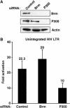
Brm is not involved in Tat activation of a nonintegrated LTR template. (A) HeLa cells were transfected twice with siRNA against Brm, P300 or control siRNA. The levels of endogenous Brm, P300 and actin were determined by Western blot analysis. (B) At 24 h after siRNA treatment, cells were transfected with an expression vector for Tat-Flag and the LTR-Luc reporter gene. The luciferase activity was determined 24 h after Tat transfection, and normalized to Renilla. The mean relative luciferase activities (plus standard errors) obtained from three independent transfection experiments are shown.
Figure 7
Figure 7
Tat recruits Brm and BAF155 to the HIV-1 LTR in vivo. HeLa LTR-Luc cells were treated with 2 μg/μl of recombinant GST or GST-Tat protein for 4 h. (A, B, upper panels) Tat activation of the HIV-1 promoter was quantified by RT–PCR. Total RNA was extracted from a sample of GST- or GST-Tat-treated cells and reverse transcribed. RT products were PCR-amplified using oligonucleotides within the luciferase reporter gene. RNA sample not reverse transcribed were used as control. An irrelevant RNA was used as a negative control for PCR. PCR products were resolved on 1% agarose/TAE gels containing ethidium bromide. After crosslinking, chromatin was extracted and submitted to immunoprecipitation using antibodies against: (A) GST, CyclinT1, Brm, BAF155 or SPT5. DNA was then extracted and subjected to PCR amplification using primers specific for the promoter/nuc-1 region of the HIV-1 LTR, or (B) GST, PCAF and Brm and the extracted DNA was subjected to real-time PCR analysis.
Figure 8
Figure 8
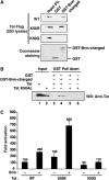
Interaction between Tat and Brm is regulated by acetylation. (A) Residue K50 of Tat is involved in Brm binding. Total lysates from 293 cells transfected with expression vector encoding Tat WT, Tat K50R or Tat K50Q were incubated with GST or GST-Brm-charged fusion proteins retained on glutathione-Sepharose beads. After intensive washes, beads were resuspended in Laemmli buffer and Tat level was assessed by Western blotting using an anti-Flag antibody. The bottom panel shows Coomassie blue staining of samples run in parallel. (B) Acetylation at Lys 50 of Tat inhibits its interaction with Brm. GST (lanes 3 and 5) or GST-Brm-charged (lanes 4 and 6) were incubated with 100 ng of chemically synthesized Tat 1–86 nonacetylated (Tat) or acetylated at Lys 50 (Tat K50Ac). Bead-bound material was analyzed after intensive washes using a mixture of anti-Tat antibodies. (C) Inhibition of Tat acetylation increased Brm synergy. Tat-Flag expression vectors (10 ng of Tat WT, Tat K50R or Tat K50Q) were cotransfected with a Brm expression vector in HeLa LTR-Luc. The luciferase activity was determined 24 h after Tat transfection, and normalized to Renilla. Fold activation was calculated relative to transfection in the absence of Tat expression plasmid. Values are the mean of three independent transfection experiments (plus standard errors).

Similar articles

Cited by

References

    1. Armstrong JA, Bieker JJ, Emerson BM (1998) A SWI/SNF-related chromatin remodeling complex, E-RC1, is required for tissue-specific transcriptional regulation by EKLF in vitro. Cell 95: 93–104 - PubMed
    1. Barker N, Hurlstone A, Musisi H, Miles A, Bienz M, Clevers H (2001) The chromatin remodelling factor Brg-1 interacts with beta-catenin to promote target gene activation. EMBO J 20: 4935–4943 - PMC - PubMed
    1. Belandia B, Orford RL, Hurst HC, Parker MG (2002) Targeting of SWI/SNF chromatin remodelling complexes to estrogen-responsive genes. EMBO J 21: 4094–4103 - PMC - PubMed
    1. Benkirane M, Chun RF, Xiao H, Ogryzko VV, Howard BH, Nakatani Y, Jeang KT (1998) Activation of integrated provirus requires histone acetyltransferase. p300 and P/CAF are coactivators for HIV-1 Tat. J Biol Chem 273: 24898–24905 - PubMed
    1. Bochar DA, Wang L, Beniya H, Kinev A, Xue Y, Lane WS, Wang W, Kashanchi F, Shiekhattar R (2000) BRCA1 is associated with a human SWI/SNF-related complex: linking chromatin remodeling to breast cancer. Cell 102: 257–265 - PubMed

Publication types