. 2006 Apr 19;25(8):1730-40.
doi: 10.1038/sj.emboj.7601073. Epub 2006 Apr 6.

Antiviral effect of the mammalian translation initiation factor 2alpha kinase GCN2 against RNA viruses

Affiliations

Antiviral effect of the mammalian translation initiation factor 2alpha kinase GCN2 against RNA viruses

Juan J Berlanga et al. EMBO J. .

Abstract

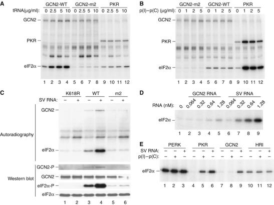
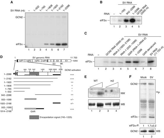
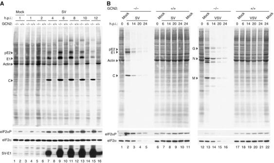
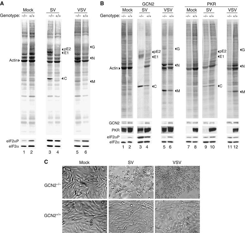
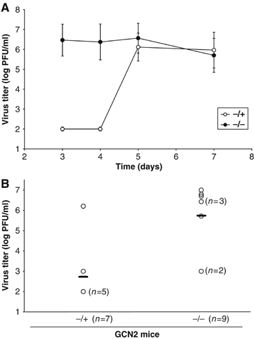
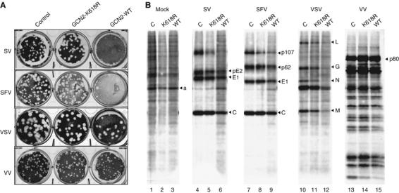
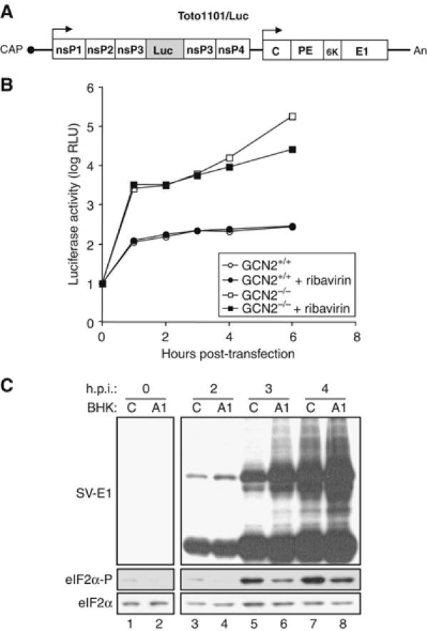
In mammals, four different protein kinases, heme-regulated inhibitor, double-stranded RNA-dependent protein kinase (PKR), general control non-derepressible-2 (GCN2) and PKR-like endoplasmic reticulum kinase, regulate protein synthesis in response to environmental stresses by phosphorylating the alpha-subunit of the initiation factor 2 (eIF2alpha). We now report that mammalian GCN2 is specifically activated in vitro upon binding of two nonadjacent regions of the Sindbis virus (SV) genomic RNA to its histidyl-tRNA synthetase-related domain. Moreover, endogenous GCN2 is activated in cells upon SV infection. Strikingly, fibroblasts derived from GCN2-/- mice possess an increased permissiveness to SV or vesicular stomatitis virus infection. We further show that mice lacking GCN2 are extremely susceptible to intranasal SV infection, demonstrating high virus titers in the brain compared to similarly infected control animals. The overexpression of wild-type GCN2, but not the catalytically inactive GCN2-K618R variant, in NIH 3T3 cells impaired the replication of a number of RNA viruses. We determined that GCN2 inhibits SV replication by blocking early viral translation of genomic SV RNA. These findings point to a hitherto unrecognized role of GCN2 as an early mediator in the cellular response to RNA viruses.


Figures

Figure 1
Figure 1
SV RNA stimulates GCN2-mediated phosphorylation of eIF2α. Purified wild-type GCN2 (GCN2-WT), the HisRS mutant (GCN2-m2) and PKR were assayed for their ability to phosphorylate eIF2α in the absence or the presence of increasing concentrations of uncharged bovine liver tRNA (Sigma) (A) or of poly(I)–poly(C) (Sigma) (B), as indicated. Phosphoproteins were analyzed by SDS–PAGE and autoradiography. Note an unknown phosphoprotein above the eIF2α band (A), which seems to be the consequence of the unspecific copurification of a kinase activity bound to TALON affinity resin. (C) In vitro eIF2α kinase assay of purified GCN2-WT, GCN2-K618R or GCN2-m2, in the absence or presence of SV RNA. Proteins were resolved into 10% SDS–PAGE and transferred to an immobilon-P membrane. The membrane was exposed to autoradiography (upper panels) and then probed with different antisera to detect eIF2α phosphorylated at serine 51 (eIF2α-P), total eIF2α and phosphorylated, and total GCN2 (lower panels) as indicated. (D) Kinase reactions were performed in the presence of the indicated concentrations of SV RNA or GCN2 RNA (as a negative control) and analyzed as described in (A). (E) Phosphorylation of eIF2α by Drosophila PERK, or mammalian PKR, GCN2 and HRI in the absence or presence of either poly(I)–poly(C) (1 μg/ml) or SV RNA (2.5 μg/ml, 0.64 nM). The analysis was carried out as described in (A).
Figure 2
Figure 2
Activation of GCN2 by SV RNA involves two nonadjacent regions at the 5′-end of the viral RNA and the HisRS domain of GCN2. (A) The 5′-end-containing RNAs encompassing the indicated nucleotides of SV RNA were assayed for their ability to activate GCN2 as described in Figure 1. (B) Activation of GCN2 by the indicated fragments of viral RNA. GCN2 RNA was assayed as a negative control. (C) Fragments 1920–2168 (RNA-2) and 502–1099 (RNA-1) were tested alone or in combination, upon joining them by DNA recombinant technology (GAR construct; GCN2 Activating RNA). The heat-denaturated GAR fragment was also assayed, as well as native SV RNA 1–2288 and a full-length RNA (1–11 703). All RNAs were used at the same concentration (0.64 nM). In (B) and (C), only eIF2α phosphorylation is shown. (D) Schematic diagram depicting regions of SV genomic RNA involved in activation of GCN2 according to data from (A) to (C). GCN2 activation by the different RNA fragments is expressed as % of activity with respect to those obtained with full-length SV RNA: (+++), 75–100% activity, (++), 50–75% activity, (+), 25–50% activity and (+/−) less than 25% activity. Data are expressed as the mean of at least three independent experiments. GAR (hatched and black boxes) and the encapsidation signal in the SV genome (shadow box) are also drawn. (E) GCN2-(WT) and the HisRS mutant GCN2-m2 were purified as described in Materials and methods. Two different amounts of proteins were resolved in a 7.5% SDS–PAGE, transferred to a nitrocellulose membrane and probed with a 32P-labeled GAR fragment in a Northwestern blot assay (NW, top panel). The amount of loaded protein was monitored by reprobing the membrane with anti-myc antibody (WB, bottom panel). (F) MEFs were mock-infected or infected with SV at an MOI=50, and cell lysates were prepared at 2 h.p.i. and subjected to immunoprecipitation with anti-GCN2 antibody. Immune complexes were assayed for their ability to phosphorylate eIF2α (top panel). The amount of GCN2 was monitored by immunoblot using anti-MGCN2 antibody (bottom panel). The amount of 32P incorporated in eIF2α was quantified in a Phosphorimager BAS-1500 (Fujifilm). A representative experiment out of three that yielded similar results is shown. Numbers under the top panel represent the increase in eIF2α phosphorylation in immune complexes from SV-infected cells compared to those from mock-infected ones. Values indicate the mean±s.d.
Figure 3
Figure 3
Cells were not infected (Mock) or infected with either SV at an MOI of 50 (A) or the indicated viruses at an MOI of 10 (SV) or 5 (VSV) (B) and metabolically labeled with [35S]Met-Cys for 30 min at the indicated h.p.i. Cells were lysed in a sample buffer and equivalent amounts of total protein were subjected to 12% SDS–PAGE, transferred to nitrocellulose membranes and subjected to autoradiography (upper panels). The membranes were then probed with specific antisera for the detection of SV-E1 protein, eIF2α-P and total eIF2α (bottom panels) as indicated. The position of the main viral protein bands of each virus as well as that of the actin band in mock-infected cells is also indicated.
Figure 4
Figure 4
Cells were not infected (Mock) or infected with the indicated viruses at an MOI of either 10 (SV) and 5 (VSV) (A) or 50 (SV) and 25 (VSV) (B). Viral protein synthesis was analyzed at 6 h.p.i. as described in Figure 3 (upper panels). The membranes were then probed with specific antisera for detection of GCN2, PKR, eIF2α-P and total eIF2α (bottom panels) as indicated. (C) Result of GCN2 depletion on the cytopathic effect produced by the indicated viruses. Cells were infected as in (A) and phase-contrast micrographs of mock- or virus-infected cell monolayers were taken at 18–20 h.p.i.
Figure 5
Figure 5
SV-infected GCN2−/− mice show high brain titers compared with control (GCN2−/+) mice. GCN2−/− and control mice were infected i.n. with 1 × 10⁶ PFU and killed on different days after inoculation. Brain homogenates (3 ml) were titered by plaque assay on cell monolayers, as described under Materials and methods. (A) Viral titers at different days after inoculation. Two or three animals were used for each data point. Individual titers did not vary by more than one log. Data are representative of three independent experiments, and error bars represent s.d. of the mean values. (B) Viral titers at day 3 after inoculation. Viral titers below 102 PFU are not detectable by this method. Open circles represent individual titers, and the horizontal line corresponds to the mean of all individual titers (−/+=2.74±1.57; −/−=5.73±1.59). The differences in viral titers found between the two groups of mice are statistically significant (P<0.01).
Figure 6
Figure 6
(A) NIH 3T3 cells bearing an empty vector (control), or expressing either the wild-type (GCN2-WT) or the K618R mutant form (GCN2-K618R) of GCN2 were infected with dilutions of the indicated viruses. After 3 days, lysis plaques were visualized. (B) NIH 3T3 cells were infected with viruses at an MOI of 10 PFU/cell and labeled with [35S]Met-Cys for 1 h at 5 h.p.i., except for vaccinia infections, where the labeling period started at 16 h.p.i. Cells were lysed in a sample buffer and equivalent amounts of total protein were analyzed by SDS–PAGE followed by autoradiography. C stands for cells bearing an empty vector.
Figure 7
Figure 7
GCN2 blocks translation of incoming SV RNA by phosphorylating eIF2α. (A) Schematic representation of luciferase-expressing SV variant Toto1101/Luc. Solid lines represent UTRs, and the location of the cap and poly(A) tail (An) are indicated. The approximate locations of the nsPs, nsP1, nsP2, nsP3 and nsP4, and structural protein capsid (C), glycoproteins (PE and E1) and the 6 kDa protein (6K) are shown by open boxes. The shaded box within nsP3 corresponds to the Firefly luciferase encoding region. (B) MEFs derived from GCN2−/− and control mice were transfected with Toto1101/Luc RNA and at the indicated times luciferase activity was measured. Where indicated, cells were treated with 0.5 mM of ribavirin. Data are representative of three independent experiments. RLU, relative light units. (C) Control (C) and A1-transduced (A1) BHK cells were infected with SV (MOI=1). At the indicated h.p.i., cell lysates were analyzed by immunoblot with specific antisera for the detection of SV-E1 protein, eIF2α-P and total eIF2α as indicated.

References

    1. Abraham N, Stojdl DF, Duncan PI, Methot N, Ishii T, Dube M, Vanderhyden BC, Atkins HL, Gray DA, McBurney MW, Koromilas AE, Brown EG, Sonenberg N, Bell JC (1999) Characterization of transgenic mice with targeted disruption of the catalytic domain of the double-stranded RNA-dependent protein kinase, PKR. J Biol Chem 274: 5953–5962 - PubMed
    1. Balachandran S, Roberts PC, Brown LE, Truong H, Pattnaik AK, Archer DR, Barber GN (2000) Essential role for the dsRNA-dependent protein kinase PKR in innate immunity to viral infection. Immunity 13: 129–141 - PubMed
    1. Berlanga JJ, Herrero S, de Haro C (1998) Characterization of the hemin-sensitive eukaryotic initiation factor 2α kinase from mouse nonerythroid cells. J Biol Chem 273: 32340–32346 - PubMed
    1. Berlanga JJ, Santoyo J, de Haro C (1999) Characterization of a mammalian homolog of the GCN2 eukaryotic initiation factor 2α kinase. Eur J Biochem 265: 754–762 - PubMed
    1. Bick MJ, Carroll JW, Gao G, Goff SP, Rice CM, MacDonald MR (2003) Expression of the zinc-finger antiviral protein inhibits alphavirus replication. J Virol 77: 11555–11562 - PMC - PubMed

Publication types

MeSH terms