. 2006 May 3;25(9):1915-23.
doi: 10.1038/sj.emboj.7601071. Epub 2006 Apr 6.

Cockayne syndrome B protein regulates the transcriptional program after UV irradiation

Affiliations

Cockayne syndrome B protein regulates the transcriptional program after UV irradiation

Luca Proietti-De-Santis et al. EMBO J. .

Abstract

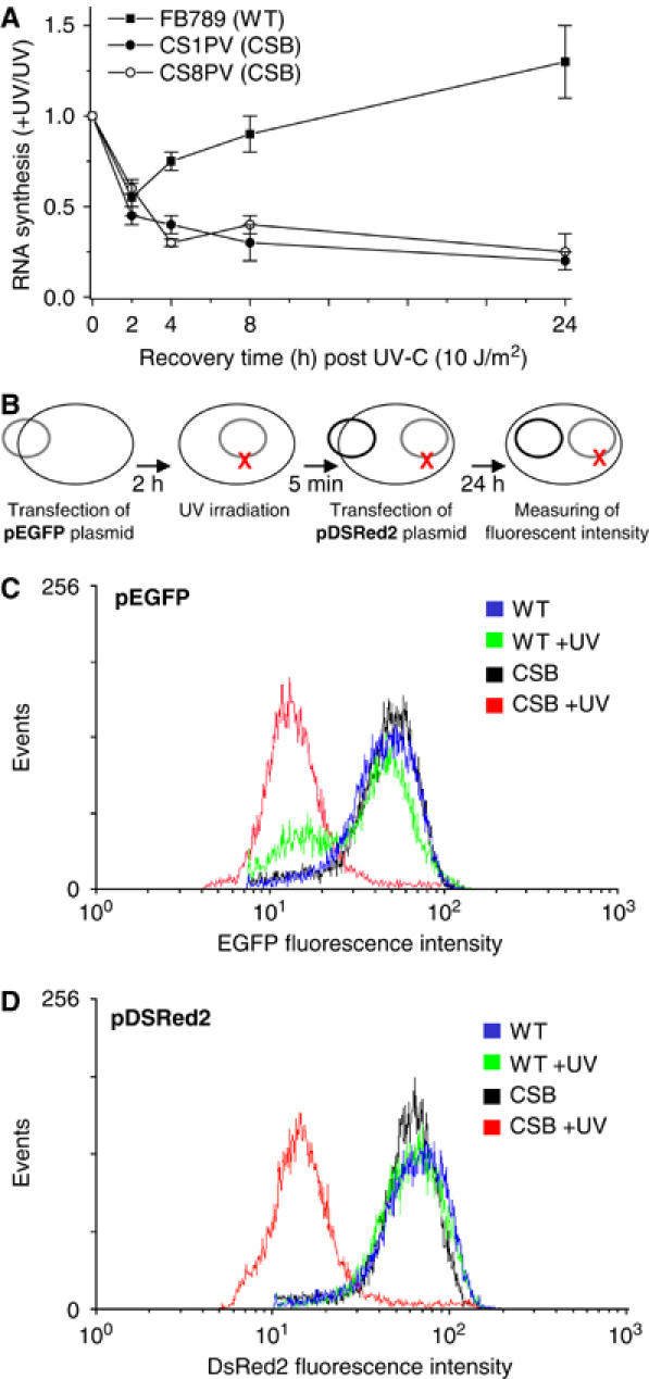
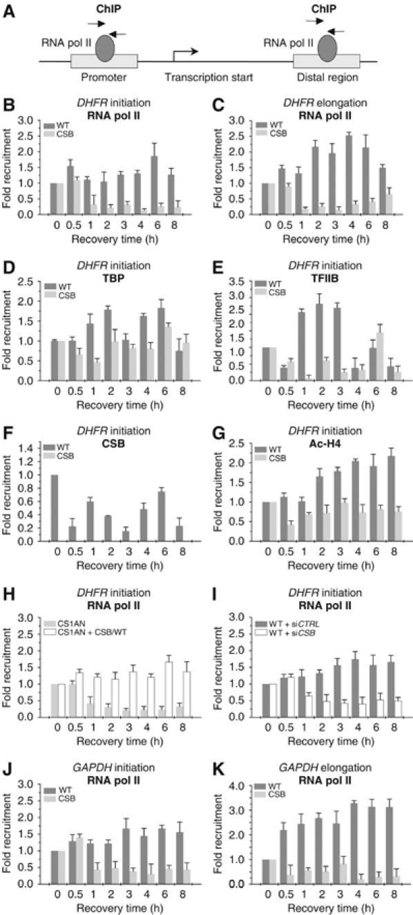
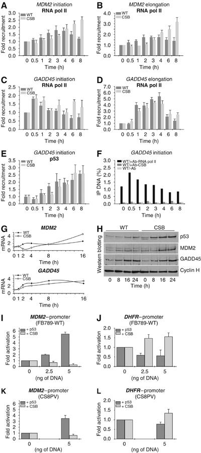
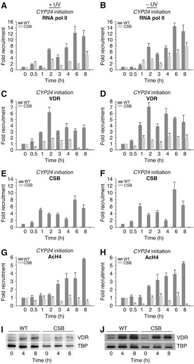
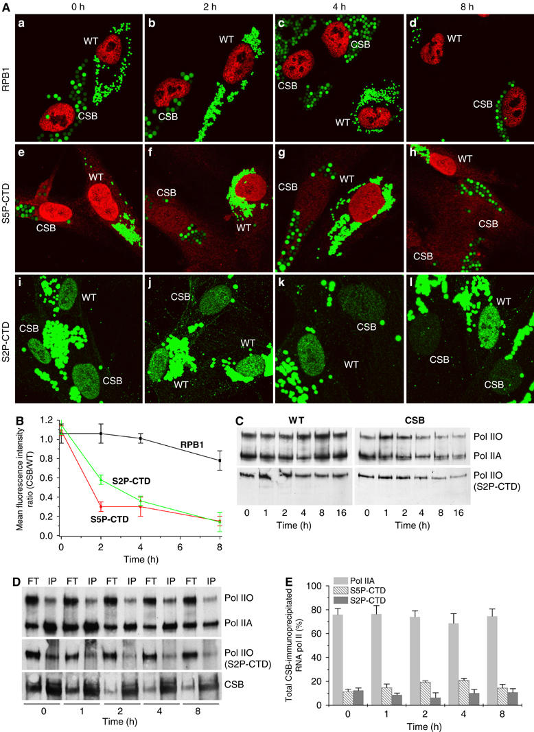
The phenotype of the human genetic disorder Cockayne syndrome (CS) is not only due to DNA repair defect but also (and perhaps essentially) to a severe transcription initiation defect. After UV irradiation, even undamaged genes are not transcribed in CSB cells. Indeed, neither RNA pol II nor the associated basal transcription factors are recruited to the promoters of the housekeeping genes, around of which histone H4 acetylation is also deficient. Transfection of CSB restores the recruitment process of RNA pol II. On the contrary, the p53-responsive genes do not require CSB and are transcribed in both wild-type and CSB cells upon DNA damage. Altogether, our data highlight the pivotal role of CSB in initiating the transcriptional program of certain genes after UV irradiation, and also may explain some of the complex traits of CS patients.


Figures

Figure 1
Figure 1
(A) RNA synthesis recovery of WT (FB789) and CSB (CS1PV and CS8PV) primary fibroblasts after UV irradiation. After prelabelling with [14C]thymidine (0.02 μCi/ml) for 2 days, unirradiated or UV (10 J/m2) irradiated cells were pulse labelled for 30 min with [3H]uridine at different incubation times after irradiation, and the acid-insoluble radioactivity was determined. Schematic representation (B) of the temporal order of transfection and irradiation. Histograms generated from flow cytometry analysis of WT and CS8PV cells 24 h after transfection with pEGFP (C) and pDSRed2 plasmid (D). Cells were either unirradiated or UV irradiated after the pEGFP transfection while pDSRed2 was transfected after UV irradiation. Relative EGFP and DSRed2 fluorescence are represented on the X-axis, and number of events (cells) is represented on the Y-axis. Similar patterns of fluorescence were observed in at least three independent experiments.
Figure 2
Figure 2
Kinetics of RNA pol II occupancy at the promoter or distal exon of DHFR and GAPDH genes (A) after UV irradiation (10 J/m2). Soluble chromatin was prepared from WT and CS8PV cells at indicated times after UV treatment and subjected to ChIP assay using the indicated antibodies. Real-time PCR using specific primers was performed to test the relative enrichment for either the proximal promoter (initiation) or the distal regions (elongation) of the gene as compared to the unirradiated samples. Kinetics of RNA pol II occupancy either at the promoter (B) or at a distal region (C) of DHFR gene after UV irradiation, in WT and CSB deficient cells. Kinetics of TBP (D), TFIIB (E), CSB (F) and acetylated H4 (G) at the promoter of DHFR gene. RNA pol II occupancy after UV irradiation, at the promoter of DHFR, either in CSB (CS1AN) or in CS1AN cells (CS1AN+CSB/WT) transfected with WT CSB gene (H). RNA pol II occupancy at the promoter of DHFR gene in WT cells previously transfected with a pool of oligonucleotides either of control (siCTRL) or silencing CSB gene (siCSB) (I). RNA pol II occupancy either at the promoter (J) or at a distal region (K) of GAPDH gene in WT and CSB cells. The results are expressed as fold enrichment relative to the untreated cells. Data are from three independent experiments. The values shown are means±s.d.
Figure 3
Figure 3
Kinetics of RNA pol II occupancy at the promoter and distal exon of MDM2 (A, B) and GADD45 (C, D) genes after UV irradiation (10 J/m2). Kinetics of p53 (E) and CSB (F) protein occupancy at the promoter of GADD45 gene after UV irradiation. (G) RT–PCR of MDM2 and GADD45 genes. RNA was extracted at the time indicated after UV irradiation. (H) Western blot analysis of p53, MDM2, GADD45 and cyclin H protein amount from nuclear extracts of either WT or CS8PV cells. Nuclear extracts were collected, after irradiation, at the time indicated. WT and CSB cells were cotransfected with either pMDM2-Luc (I, K) or pDHFR-Luc (J, L) in combination with either empty vectors or constructs expressing p53 or CSB proteins at the indicated concentrations. The results are expressed as fold activation relative to the cells transfected with the empty vector. In (F), results are expressed as the percentage of the immunoprecipitated DNA as compared to the input. Values obtained using CSB antibody do not show any enrichment as compared to the ones obtained without any antibody (negative control), which evidences the absence of CSB at the promoter.
Figure 4
Figure 4
ChIP analysis on the CYP24 promoter in WT and CS8PV cells. Soluble chromatin was prepared from either UV irradiated (left panels) or unirradiated (right panels) cells co-treated with vit-D (10−7 M) for the indicated time and immunoprecipitated with antibodies against RNA pol II (A, B), VDR (C, D), CSB (E, F) or the acetylated histone H4 (G, H). The genomic DNAs were analyzed by quantitative PCR. Western blot analyzes (I, J) of VDR protein from nuclear extracts of WT and CSB cells, either UV irradiated or unirradiated, at different time points after vit-D treatment.
Figure 5
Figure 5
(A) Confocal microscopy analysis of RNA pol II concentration in WT or CS8PV cells using antibodies that recognize either RPB1, the largest subunit of RNA pol II (panels a–d), or the hyperphosphorylated CTD domain, at either serine 5 (S5P-CTD, panels e–h) or serine 2 (S2P-CTD, panels i–l). Cells were prelabelled with green latex fluorescent beads of different size and spotted on the same slide to further analysis of the RNA pol II concentration at the time indicated after UV irradiation (10 J/m2). (B) Graph represents mean fluorescent intensity (protein concentration) ratios calculated by dividing the mean fluorescence, relative to the protein staining, of CSB cells by the mean fluorescence of the WT cells as obtained by confocal scanner fluorescence, measuring of up to 100 cells. (C) Western blot analysis of RNA pol II amount from chromatin-bound fraction of either WT or CSB cells using antibodies that recognize RPB1, regardless its phosphorylation state (upper panel), or the phosphorylated S2P-CTD (lower panel). Pol IIO and Pol IIA indicate the hyper- and hypophosphorylated form of the CTD-RNA pol II, respectively. (D) Nuclear extracts from either unirradiated or UV-irradiated cells were immunoprecipitated (IP) using an antibody against CSB. The IP and the flowthrough (FT) fractions were analyzed by Western blot using antibody thaat recognize RPB1 (upper panel), the phosphorylated S2P-CTD RNA pol II (middle panel) or CSB (lower panel). (E) Graph represents the percentage of RNA pol IIA, and the phosphorylated S5P-CTD and S2P-CTD RNA pol II, which were CSB-IP at different time after UV irradiation.

References

    1. Balajee AS, May A, Dianov GL, Friedberg EC, Bohr VA (1997) Reduced RNA polymerase II transcription in intact and permeabilized Cockayne syndrome group B cells. Proc Natl Acad Sci USA 94: 4306–4311 - PMC - PubMed
    1. Beerens N, Hoeijmakers JH, Kanaar R, Vermeulen W, Wyman C (2005) The CSB protein actively wraps DNA. J Biol Chem 280: 4722–4729 - PubMed
    1. Bird AW, Yu DY, Pray-Grant MG, Qiu Q, Harmon KE, Megee PC, Grant PA, Smith MM, Christman MF (2002) Acetylation of histone H4 by Esa1 is required for DNA double-strand break repair. Nature 419: 411–415 - PubMed
    1. Bradsher J, Auriol J, Proietti de Santis L, Iben S, Vonesch JL, Grummt I, Egly JM (2002) CSB is a component of RNA pol I transcription. Mol Cell 10: 819–829 - PubMed
    1. Bregman DB, Halaban R, van Gool AJ, Henning KA, Friedberg EC, Warren SL (1996) UV-induced ubiquitination of RNA polymerase II: a novel modification deficient in Cockayne syndrome cells. Proc Natl Acad Sci USA 93: 11586–11590 - PMC - PubMed

Publication types

MeSH terms