. 2006 Apr 19;25(8):1611-22.
doi: 10.1038/sj.emboj.7601053. Epub 2006 Apr 6.

SH3P7/mAbp1 deficiency leads to tissue and behavioral abnormalities and impaired vesicle transport

Affiliations

SH3P7/mAbp1 deficiency leads to tissue and behavioral abnormalities and impaired vesicle transport

Sabine Connert et al. EMBO J. .

Abstract

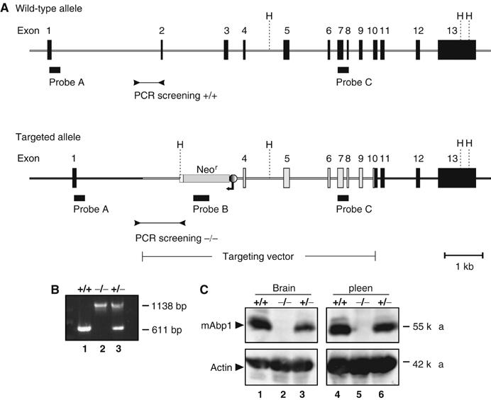
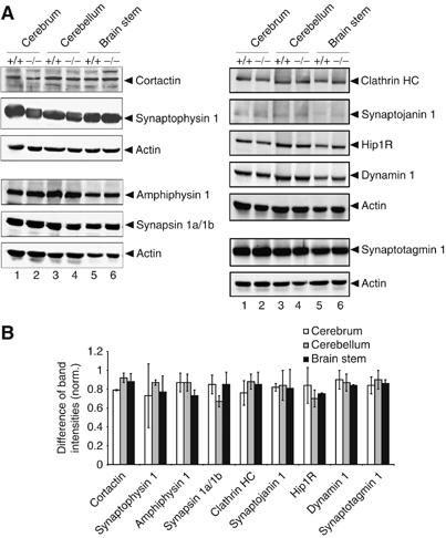
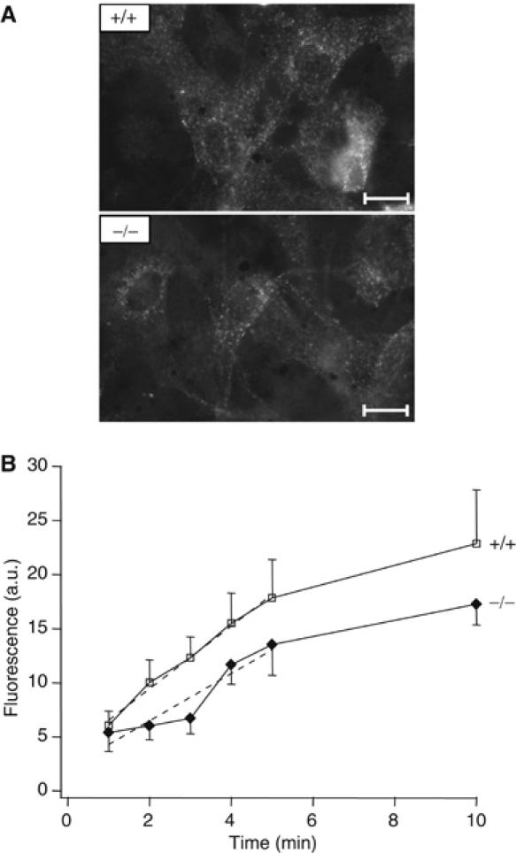
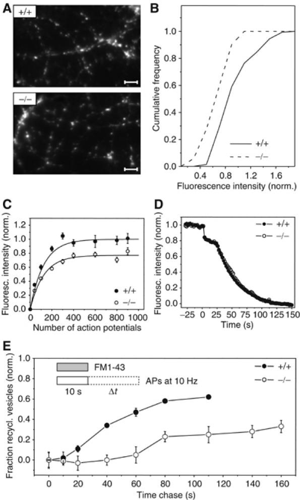
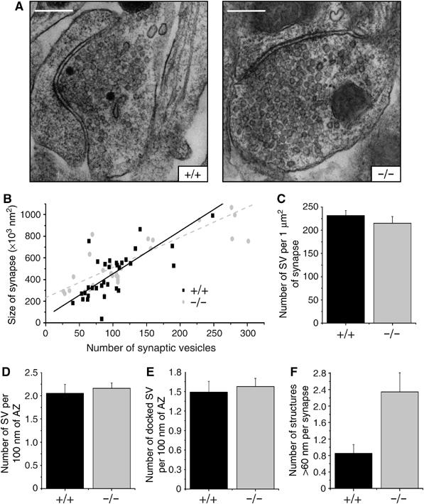
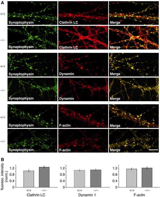
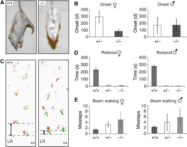
The intracellular adaptor protein SH3P7 is the mammalian ortholog of yeast actin-binding protein 1 and thus alternatively named as mAbp1 (or HIP55). Structural properties, biochemical analysis of its interaction partners and siRNA studies implicated mAbp1 as an accessory protein in clathrin-mediated endocytosis (CME). Here, we describe the generation and characterization of mice deficient for SH3P7/mAbp1 owing to targeted gene disruption in embryonic stem cells. Mutant animals are viable and fertile without obvious deficits during the first weeks of life. Abnormal structure and function of organs including the spleen, heart, and lung is observed at about 3 months of age in both heterozygous and homozygous mouse mutants. A moderate reduction of both receptor-mediated and synaptic endocytosis is observed in embryonic fibroblasts and in synapses of hippocampal neurons, respectively. Recycling of synaptic vesicles in hippocampal boutons is severely impaired and delayed four-fold. The presynaptic defect of SH3P7/mAbp1 mouse mutants is associated with their constricted physical capabilities and disturbed neuromotoric behaviour. Our data reveal a nonredundant role of SH3P7/mAbp1 in CME and places its function downstream of vesicle fission.


Figures

Figure 1
Figure 1
Targeted disruption of murine mabp1 and genotype analysis. (A) A diagram of the mabp1 locus before and after homologous recombination of the targeting construct is shown. Exons 2 and 3 of mabp1 were replaced by a neomycin resistance cassette in opposite transcriptional orientation. Black boxes indicate location of probes A, B, and C for Southern blot analysis (data not shown) and primers for genotyping the F2 generation by PCR analysis (B) performed on tail DNA of wt mice (lane 1), mabp1−/− mice (lane 2) and mabp1+/− mice (lane 3), respectively. H=HindIII, fragment sizes are indicated on the right in base pairs. (C) The expression of mAbp1 and actin as a control (upper and lower panel, respectively) is monitored in cleared cellular lysates from total brain (lanes 1–3) and spleen (lanes 4–6) of wt mice (lanes 1 and 4) and mutant mice harboring either one or two inactivated mabp1 alleles (lanes 2, 5 and 3, 6, respectively) by immunoblot analysis. Relative molecular mass of marker proteins is indicated on the right in kDa.
Figure 2
Figure 2
Loss of mAbp1 does not affect expression of functionally related proteins. (A) Expression levels of amphiphysin, synapsinIa/Ib, synaptotagmin1, clathrin HC, synaptojanin1, Hip1R, dynamin, cortactin, and synaptophysin were analyzed with specific antibodies in cerebrum (lanes 1 and 2), cerebellum (lanes 3 and 4) and brain stem (lanes 5 and 6). Actin levels were used as loading control. (B) Plotted is the difference between band intensities of the analyzed proteins in wt and mabp1−/− mice normalized to the corresponding band intensity of the wt level. Band intensities were quantified with Gel Pro-Analyzer software (INTAS, Göttingen, Germany) and normalized to actin. The diagram depicts relative differences between the expression levels of the analyzed proteins in mabp1−/− and wt mice. Error bars represent standard deviations calculated on the basis of two independent experiments.
Figure 3
Figure 3
Altered anatomy of the spleen, lung, and heart in mabp1-deficient mice. (A) Splenomegaly was identified in mabp1−/− mice (scale bar 0.25 cm). (B) Abnormal structure is observed in lobes of mabp1−/− lung compared to that of wt control mice (scale bars 0.25 cm). (C) mabp1−/− mice possess an enlarged heart with an enormously enlarged left atrium (encircled, scale bars 0.25 cm). (D) Four chamber dilatations and a left atrial thrombus is observed in mabp1−/− mice (hematoxilin/eosin (HE) staining, left panel), additionally all chambers of the mabp1−/− heart show extensive fibrosis (red) (Sirius red staining, right panel). For comparison, an age-matched wt heart is shown. RV=right ventricle; LV=left ventricle; and LA=left atrium (scale bars 2 mm).
Figure 4
Figure 4
Receptor-mediated endocytosis is slightly reduced in mabp1-deficient mice. (A) Representative fluorescence images of mabp1+/+ and mabp1−/− MEFs show the uptake and internalization of Cy2-labelled transferrin (10 min incubation; scale bars 1.45 μm). (B) Mean fluorescence intensities of single MEFs after incubation with Cy2-labelled mouse transferrin for 1–5 and 10 min at 37°C (errors in s.e.m.; n=69, 45, 49, 45, 62, 42 for t=1, 2, 3, 4, 5, 10 min for mabp1+/+ and n=43, 74, 57, 37, 26, 28 for mabp1−/−, respectively). Initial internalization rates were determined by regression analysis (dashed lines) over the first 5 time points (slopes of 2.9±0.16 for mabp1+/+ cells and 2.2±0.45 for mabp1−/− cells, respectively) revealing a moderate reduction of approximately 20% of receptor-mediated endocytosis in mabp1-deficient cells (errors of slopes are the confidence intervals at 95% level).
Figure 5
Figure 5
Synaptic vesicle recycling in hippocampal synapses of mabp1-deficient mice is severely impaired. (A) Exemplar images of FM1–43-stained axonal processes of cultured hippocampal neurons derived from wt and mabp1−/− mice. No obvious differences in arborization, bouton density, or distribution can be seen (scale bar 3.2 μm). (B) Cumulative distribution of vesicle turnover during a loading train of 40 APs, measured by FM1–43 at n(3)=144 and n(2)=51 individual synaptic boutons for mabp1+/+ and mabp1−/−, respectively. Fluorescence intensities are normalized to the average FM fluorescence of mabp1+/+ boutons. The mean value of the distribution for mabp1−/− neurons is 0.75±0.03 (s.e.m.). (C) Size of the functional synaptic vesicle pool in dependence of stimulus length for mabp1−/− (open circles) and mabp1+/+ boutons (filled circles). Boutons were loaded by different AP numbers and fully destained with 900 APs at 10 Hz. Error bars are confidence intervals of the mean at 95% level. Numbers of experiments were n=3, 4, 5, 2, 4, 2, 3, 3 for mabp1+/+ and n=2, 4, 6, 2, 4, 4, 2, 4 for mabp1−/− neurons, respectively, each comprising typically 50–100 individual boutons. The total recycling pool size estimated from a single exponential fit (solid lines) for mabp1−/− is 0.77±0.02 (s.d.) of that determined from a fit for mabp1+/+ (1±0.02 (s.d.)). (D) Average FM1–43 release kinetics of maximally loaded (900 APs at 10 Hz) synaptic boutons for two consecutive destaining stimuli (40 AP at 20 Hz and 900 AP at 10 Hz) are similar for mabp1+/+ and mabp1−/− neurons. Data are normalized to fluorescence signals before destaining after subtraction of nonreleasable background. (E) Repriming kinetics determined by FM1–43 pulse-chase experiments. Plotted is the fraction of dye released during a given chase time Δt normalized to the maximum upload ΔF0 for chase time Δt=0, that is, (ΔF0FΔt)/ΔF0. Under prolonged stimulation, reformation of fusion-competent vesicles is severely impaired and delayed. The minimum repriming time of 10–20 s for control mabp1+/+ is delayed to 60–80 s in mabp1−/− synapses. After 100 s, only 25 versus 60% of retrieved membrane is releasable again in mabp1−/− synapses. Numbers of experiments were n=4, 3, 3, 3, 2, 2, 2 for mabp1+/+ and n=5, 4, 4, 4, 3, 3, 2, 2, 2 for mabp1−/− neurons, respectively, each comprising typically 50–100 individual boutons. Error bars are confidence intervals of the mean at 95% level.
Figure 6
Figure 6
Ultrastructural analysis of synaptic boutons in hippocampal cultures. (A) Representative synaptic boutons from wt and mabp1-deficient hippocampal cultures (scale bars 0.3 μm). (B) The distributions of the number of vesicles per bouton area are similar in wt (n=34) and mabp1−/− synapses (n=30). Total numbers of vesicles are 3729 for wt and 3630 for mabp1−/− synapses. (C) Average number of synaptic vesicles (SV) per 1 μm2 bouton area are similar in wt (231.63±10.92) and mabp1−/− synapses (214.83±14.87). (D) Average numbers of morphologically docked vesicles per AZ length are similar in mAbp1-positive (n=20) and mAbp1-negative synapses (n=25). (E) Average numbers of vesicles close to the AZ (<20 nm) per AZ length are similar in wt (n=20) and mabp1−/− synapses (n=25). (F) The number of endosome-like intermediates per synapse is significantly higher in mabp1-deficient (2.497±0.464) than in wt (0.853±0.212) synapses.
Figure 7
Figure 7
Localization of F-actin, dynamin1 and CLC in mabp1-deficient and wt hippocampal neurons. (A) Hippocampal neurons at 12–14 days in vitro (DIV) were stained with anti-dynamin1 and anti-CLC antibodies as well as with phalloidin together with anti-synaptophysin1 and imaged by laser scanning confocal microscopy (scale bar 10 μm). (B) Quantification of CLC, dynamin1, and F-actin expression in synaptophysin-positive puncta. Diagrams showing mean values of fluorescence intensities with s.e.m. normalized to synaptophysin1 fluorescence intensities.
Figure 8
Figure 8
Characterization of behavioral abnormalities of mabp1−/− mice. (A) wt mice spread their limbs during a tail-suspension test while mabp1−/− mice react with hind- and forelimb clasping. (B) The onset of mAbp1 deficiency symptoms for female mabp1+/− (n=16) and mabp1−/− (n=29) mice as well as male mabp1+/− (n=21) and mabp1−/− (n=24) mice were investigated over a period of 1 year. No symptoms were identified for mabp1+/+ littermates used as controls (female n=12, male n=10). Error bars represent standard deviations. (C) Evaluation of foot placement was performed by recording footprints of mice with differently painted forelimb (red) and hindlimb (green). Analysis of path geometry revealed differences in left–right (LR) alteration between mabp1−/− (n=13) and mabp1+/+ mice (n=9) upon the onset of the disease symptoms (scale bar 1 cm). (D) In a Rotarod test, time to fall off (s) was determined in five test sessions performed every second day at a fixed daytime. A maximum score of 300 s was given to those mice not falling off within this period of time. The average time to fall off is shown with standard deviations. Female mabp1+/+ (n=2), mabp1+/− (n=3), and mabp1−/− (n=3) mice as well as male mabp1+/+ (n=2), mabp1+/− (n=3), and mabp1−/− (n=4) mice with no obvious mAbp1 deficiency symptoms were employed. (E) Beam walking experiments, performed three times at intervals of 1 week, revealed deficits in motor abilities in a gene dose-dependent manner. The average number of missteps was determined for female mabp1+/+ (n=4), mabp1+/− (n=6), and mabp1−/− (n=4) mice and male mabp1+/+ (n=4), mabp1+/− (n=4), and mabp1−/− (n=5) mice with no obvious symptoms. Error bars represent standard deviations.

References

    1. Benesch S, Polo S, Lai FP, Anderson KI, Stradal TE, Wehland J, Rottner K (2005) N-WASP deficiency impairs EGF internalization and actin assembly at clathrin-coated pits. J Cell Sci 118: 3103–3115 - PubMed
    1. Betz WJ, Bewick GS (1992) Optical analysis of synaptic vesicle recycling at the frog neuromuscular junction. Science 255: 200–203 - PubMed
    1. Danino D, Hinshaw JE (2001) Dynamin family of mechanoenzymes. Curr Opin Cell Biol 13: 454–460 - PubMed
    1. Di Paolo G, Sankaranarayanan S, Wenk MR, Daniell L, Perucco E, Caldarone BJ, Flavell R, Picciotto MR, Ryan TA, Cremona O, De Camilli P (2002) Decreased synaptic vesicle recycling efficiency and cognitive deficits in amphiphysin 1 knockout mice. Neuron 33: 789–804 - PubMed
    1. Ensenat D, Yao Z, Wang XS, Kori R, Zhou G, Lee SC, Tan TH (1999) A novel Src homology 3 domain-containing adaptor protein, HIP-55, that interacts with hematopoietic progenitor kinase 1. J Biol Chem 274: 33945–33950 - PubMed

Publication types

MeSH terms

LinkOut - more resources