

Chromatic pattern-reversal electroretinograms (ChPERGs) are spared in multiple system atrophy compared with Parkinson's disease
- PMID: 16601931
- PMCID: PMC2001254
- DOI: 10.1007/s10072-006-0522-1
Chromatic pattern-reversal electroretinograms (ChPERGs) are spared in multiple system atrophy compared with Parkinson's disease
Abstract
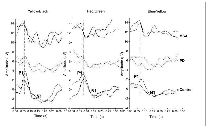
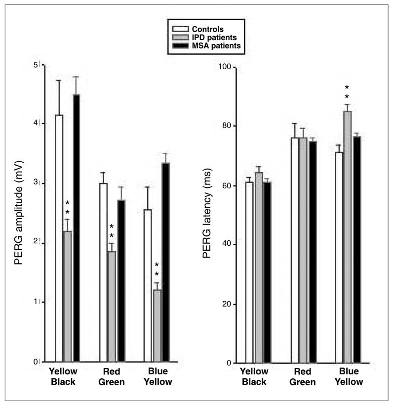
Idiopathic Parkinson's disease (IPD) patients have abnormal visual evoked potentials (VEPs) and pattern electroretinograms (PERGs), attributed to dopaminergic transmission deficiency in visual pathway, probably the retina. VEP abnormalities are not reported in multiple system atrophy (MSA). The aim of this study was to investigate and compare chromatic (Ch) red-green (R-G) and blue-yellow (B-Y), and luminance yellow-black (Y-Bk) PERGs in patients with MSA and IPD. We investigated 6 MSA patients (mean age: 62+/-7.4 years) not undergoing any pharmacological treatment, as well as 12 early IPD patients (mean age: 60.1+/-8.3 years) and 12 age-matched normal observers. ChPERGs were recorded monocularly in response to full-field equiluminant R-G, B-Y and Y-Bk horizontal gratings. In MSA only responses to R-G stimuli showed minimal insignificant changes (slight but not significant amplitude reduction without any significant latency delay); no significant abnormality was detected for B-Y and luminance Y-Bk stimuli. By contrast, in IPD all responses were reduced in amplitude and delayed in latency, above all for B-Y stimuli. Present data indicate that both chromatic and achromatic PERGs are virtually unaffected in MSA, whereas in early IPD they are clearly impaired, suggesting different pathogenic retinal mechanisms and a useful simple tool for distinguishing MSA from IPD.
Figures
References
-
- Bodis-Wollner I, Yahr M. Measurement of visual evoked potentials in Parkinson’s disease. Brain. 1978;101:661–671. - PubMed
-
- Bodis-Wollner I, Marx MS, Mitra S, et al. Visual dysfunction in Parkinson’s disease. Loss in spatiotemporal contrast sensitivity. Brain. 1987;110:1675–1698. - PubMed
-
- Stanzione P, Pierelli P, Peppe A. Pattern visual evoked potentials and electroretinogram abnormalities in Parkinson’s disease: effect of l-dopa therapy. Clin Vis Sci. 1989;4:115–127.
-
- Peppe A, Stanzione P, Pierelli F, et al. Visual alterations in “de novo” Parkinson’s disease: pattern electroretinogram latencies are more delayed and more reversible by levodopa than are visual evoked potentials. Neurology. 1995;45:1144–1148. - PubMed
-
- Stanzione P, Pierantozzi M, Semprini R, et al. Increasing doses of l-sulpiride reveal dose- and spatial frequency-dependent effects of D2 selective blockade in the human electroretinogram. Vision Res. 1995;35:2659–2664. - PubMed
Publication types
MeSH terms
Grants and funding
LinkOut - more resources
Full Text Sources
Medical
