. 2006 Apr;4(4):e99.
doi: 10.1371/journal.pbio.0040099. Epub 2006 Mar 28.

Testing electrostatic complementarity in enzyme catalysis: hydrogen bonding in the ketosteroid isomerase oxyanion hole

Affiliations

Testing electrostatic complementarity in enzyme catalysis: hydrogen bonding in the ketosteroid isomerase oxyanion hole

Daniel A Kraut et al. PLoS Biol. 2006 Apr.

Abstract

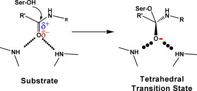
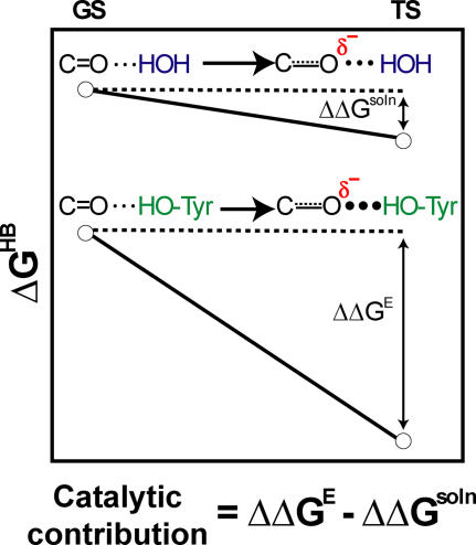
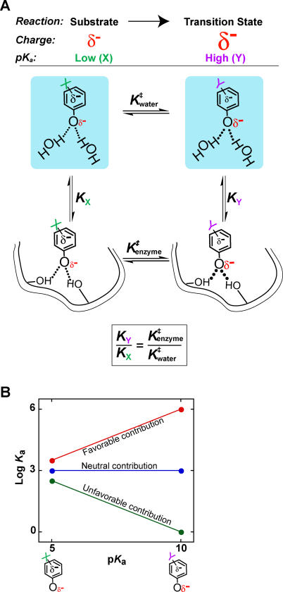
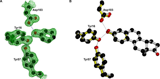
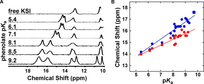
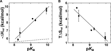
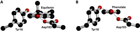
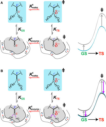
A longstanding proposal in enzymology is that enzymes are electrostatically and geometrically complementary to the transition states of the reactions they catalyze and that this complementarity contributes to catalysis. Experimental evaluation of this contribution, however, has been difficult. We have systematically dissected the potential contribution to catalysis from electrostatic complementarity in ketosteroid isomerase. Phenolates, analogs of the transition state and reaction intermediate, bind and accept two hydrogen bonds in an active site oxyanion hole. The binding of substituted phenolates of constant molecular shape but increasing pK(a) models the charge accumulation in the oxyanion hole during the enzymatic reaction. As charge localization increases, the NMR chemical shifts of protons involved in oxyanion hole hydrogen bonds increase by 0.50-0.76 ppm/pK(a) unit, suggesting a bond shortening of 0.02 A/pK(a) unit. Nevertheless, there is little change in binding affinity across a series of substituted phenolates (DeltaDeltaG = -0.2 kcal/mol/pK(a) unit). The small effect of increased charge localization on affinity occurs despite the shortening of the hydrogen bonds and a large favorable change in binding enthalpy (DeltaDeltaH = -2.0 kcal/mol/pK(a) unit). This shallow dependence of binding affinity suggests that electrostatic complementarity in the oxyanion hole makes at most a modest contribution to catalysis of 300-fold. We propose that geometrical complementarity between the oxyanion hole hydrogen-bond donors and the transition state oxyanion provides a significant catalytic contribution, and suggest that KSI, like other enzymes, achieves its catalytic prowess through a combination of modest contributions from several mechanisms rather than from a single dominant contribution.


Figures

Figure 1
Figure 1. Catalysis Is Preferential Transition State Stabilization
Figure 2
Figure 2. Geometric or Electrostatic Complementarity in the Lysozyme Reaction
(A) Simplified mechanism of lysozyme. Asp52 attacks the substrate, and the general acid Glu35 protonates the leaving group oxygen. The reaction proceeds through a loose transition state, in which the bond to the leaving group is nearly broken with only a small amount of bond formation to the incoming aspartate. Instead, positive charge accumulates on the C1 carbon of the sugar and the ring oxygen atom (δ +). This transition state has a half-chair or sofa-like conformation, distinct from the ground state chair conformation [ 31, 36]. (B) Simplified structure of a transition-state analog that binds tightly to lysozyme [ 32]. This analog has the same half-chair conformation as the transition state. It also has a similar charge distribution, with positive charge localized on the carbon and oxygen atoms in the ring and negative charge on the carbonyl oxygen.
Figure 3
Figure 3. The Serine Protease Reaction Showing Interactions in the Oxyanion Hole
Figure 4
Figure 4. Geometric and Electrostatic Changes in the KSI Reaction
(A) Mechanism of KSI catalyzed isomerization of 5-androstene-3,17-dione (substrate) to 4-androstene-3,17-dione (product). In the first step a general base, Asp40, removes a proton from the steroid to form a dienolate intermediate (via a dienolate-like transition state), which receives hydrogen bonds from an oxyanion hole consisting of Tyr16 and protonated Asp103. In the second step of the reaction the steroid is reprotonated at a different position to give the product. (B) Geometric changes accompanying the first half of the KSI reaction. Oxygen is shown in red, carbon in grey, hydrogen in blue. The ring geometry changes in the transition state and in the intermediate, becoming more planar in the intermediate. The sp 2-hybridized carbonyl oxygen in the substrate becomes a predominantly sp 3-hybridized oxyanion. Structures were generated using CaCHE 4.93 (Fujitsu, Tokyo, Japan) MOPAC PM5 geometry optimization [ 150] and rendered using CS Chem3D Pro 5.0 (CambridgeSoft, Cambridge, Massachusetts, United States). (C) Electrostatic changes at the carbonyl group accompanying the first half of the KSI reaction. The larger “δ−” refers to increased negative charge on the oxygen atom as the reaction proceeds. The dienolate-like transition state is expected to be between the substrate and dienolate intermediate in charge arrangement, but closer to the high-energy intermediate. (D) Schematic depiction of the steroid equilenin bound at the KSI active site. Equilenin geometrically and electrostatically resembles the dienolate reaction intermediate and transition state. (E) Schematic depiction of a single-ringed phenolate bound at the active site of KSI D40N, the mutant enzyme used for this work. The Asp40Asn mutation mimics the protonated aspartate found in the intermediate and equilenin complexes, see (A) and (D), and leads to tighter binding of phenolate and other intermediate analogs [ 69, 71].
Figure 5
Figure 5. Potential Catalytic Contribution from a Greater Strengthening of Hydrogen Bonds Accompanying Charge Redistribution in an Enzymatic Environment than in Aqueous Solution
As charge increases on the carbonyl oxygen going from the ground state to the transition state, hydrogen bonds from either water or an enzyme tyrosine will strengthen. Results from small molecule studies indicate that in a nonaqueous environment such as an enzyme active site, this strengthening (ΔΔG E) can be greater than in aqueous solution (ΔΔG soln), where little strengthening is observed [ 68, 151, 152]. This potential differential strengthening is indicated by the different sizes of hydrogen-bonded dots and the larger change in ΔG between the ground state and the transition state for the enzymatic reaction compared with the solution reaction. Adapted from [ 68].
Figure 6
Figure 6. Schematic Depiction of an Experimental Method to Isolate and Probe the Role of Electrostatic Complementarity to the Transition State
(A) In the reaction to be probed there is an accumulation of negative charge in the transition state. Two phenolates with different substituents (X and Y) and therefore different p K a values and different charge densities on the oxygen atom are used to mimic the increase in charge localization going from the ground state to the transition state. As charge localization on the oxygen increases, hydrogen bonds may strengthen in both water and in the enzyme active site (larger dots for hydrogen bonds formed by phenolate Y). The equilibria and represent the exchange of one phenolate for the other in water and on the enzyme, respectively, mimicking the change in charge localization along the reaction coordinate. The equilibria K X and K Y are the affinities of the two phenolates, i.e., the equilibrium for their transfer from water to the enzyme. (B) The log of the affinities of the substituted phenolates from (A) are plotted versus p K a. If the phenolate with substituent Y binds more strongly than that with substituent X (red; K Y > K X), then the enzyme is better than water at stabilizing increased charge localization and there is a favorable contribution to catalysis from electrostatic complementarity. The sign and steepness of the slope, as established using a series of substituted phenolates, can determine the sign and magnitude of the contribution to catalysis.
Figure 7
Figure 7. Crystal Structure of Phenolate Bound at the Active Site of pKSI D40N
(A) Electron density map (1.5 σ) shows that phenolate is bound at the oxyanion hole, receiving short hydrogen bonds from Tyr16 and Asp103. (B) Overlay of the phenolate•KSI structure (yellow) and the intermediate analog equilenin•KSI structure (grey; PDB code 1OGX [ 72]) at the active site.
Figure 8
Figure 8. 1H NMR Downfield Chemical Shifts for Substituted Phenolates Bound to tKSI D40N
(A) Representative spectra, with phenol p K a shown on the left. From top to bottom: free enzyme, 3,4-dinitrophenol, 3-fluoro-4-nitrophenol, 4-nitrophenol, 3-fluoro-5-trifluoromethylphenol, 3,4-dichlorophenol, and 3-iodophenol. (B) Correlation between increasing phenolate p K a and increasing chemical shift of observed downfield peaks. Circles are the two downfield peaks observed for phenolate binding. A linear fit gives slopes of 0.76 ± 0.06 and 0.50 ± 0.06 ppm/p K a unit for the most downfield (blue) and the second-most downfield (red) peak, respectively. The square is the main downfield peak observed with the intermediate analog equilenin (p K a = 9.7), for comparison.
Figure 9
Figure 9. Binding Assay for Phenolates via Competition with a Fluorescently Labeled Equilenin (EqA488–1)
(A) Structure of EqA488. Synthesis resulted in two isomers, and the isomer referred to as EqA488-1 was used in further experiments, as described in Materials and Methods. (B) Addition of pKSI D40N (E) to 0.1 nM EqA488–1 (Eq) (pH 6.9) leads to quenching of fluorescence at 515 nm (excitation at 480 nm). Each point is the average of two replicates (with errors smaller than the points). Data were fit to Equation 1 and gave = 0.7 ± 0.1 nM for this determination. Additional replicates gave = 1.0 ± 0.3 nM. The accuracy of this determination does not impact the comparison of the affinities of the substituted phenolates relative to one another. (C) Addition of 4-nitrophenol (P) to a solution of 0.1 nM EqA488 and 5 nM pKSI D40N pH 6.9 leads to recovery of fluorescence. Data were fit to Equation 2 and gave = 11.0 ± 0.9 μM. This observed affinity was first converted to an apparent affinity (= 1.8 ± 0.2 μM; Equation 3) of the phenol for the enzyme at (pH 6.9). This apparent affinity was then converted into the pH-independent affinity ( = 26 ± 2 nM; Equation 4) of the phenolate form of the ligand (PO ) for the protonated form of the enzyme (EOH) using the known phenol and enzyme ionization constants ( = 7.1 and = 5.5, respectively; see Materials and Methods). (D) Binding schemes from which Equations 1–4 were derived.
Figure 10
Figure 10. Dependence of the Affinity of a Series of meta- and para-Substituted Fluorophenolates for KSI D40N on p K a
Data for pKSI D40N is shown in (A) and data for tKSI D40N is shown in (B), with slopes of 0.11 ± 0.03 and 0.10 ± 0.03, respectively.
Figure 11
Figure 11. Dependence of Changes in Enthalpy and Entropy of Binding of a Series of meta- and para-Substituted Fluorophenolates to pKSI D40N on p K a
(A) The relative value of ΔH binding as a function of p K a (uncorrected for the enzyme ionization enthalpy, which is constant across the series of phenolates) has a slope (solid line) of −2.0 ± 0.2 kcal/mol/p K a unit. The dotted line is the relative dependence of −ΔG on p K a from Figure 6 for comparison (−0.2 kcal/mol/p K a unit). (B) The relative TΔS binding as a function of p K a has a slope of 2.0 ± 0.3 kcal/mol.
Figure 12
Figure 12. Structure of Equilenin (1OGX) and Phenolate Bound to pKSID40N, Showing the Hydrogen Bonds from Tyr16 and Asp103 That Come from Above and Below the Plane of the Steroid
(A) For equilenin (PDB Code 1OGX [72]), average angles out of the plane of the steroid ring are 28° and 46°. (B) For phenolate, the average angles are 15° and 47°.
Figure 13
Figure 13. Model for the KSI Active Site Environment and Energetic Consequences
Charge accumulates on the oxyanion in the transition state. Transfer from water to a mock active site (or mutant) lacking hydrogen bonds (A) results in a desolvation penalty that is larger in the transition state than in solution as the KSI active site provides only a limited number of dipoles (colored bars) partially pre-oriented to stabilize the transition state charge distribution. Hydrogen bonds, when added back to the active site (B), shorten and strengthen (larger dots and purple arrow) in the transition state on the enzyme, as suggested by the large value of −ΔΔH. Enzyme dipoles rearrange and become more conformationally restricted in the transition state, shown in both (A) and (B), as suggested by the large value of Δ(TΔS). The energetic sum of these interactions results in a free energy reaction profile similar to that in solution.
Figure 14
Figure 14. Determination of Thermodynamic Values for Phenolate Binding to KSI

Comment in

Similar articles

Cited by

References

    1. Page MI, Jencks WP. Entropic contributions to rate accelerations in enzymic and intramolecular reactions and the chelate effect. Proc Natl Acad Sci U S A. 1971;68:1678–1683. - PMC - PubMed
    1. Fersht AR. Catalysis, binding and enzyme-substrate complementarity. Proc R Soc Lond B Biol Sci. 1974;187:397–407. - PubMed
    1. Jencks WP. Binding energy, specificity, and enzymic catalysis: The Circe effect. Adv Enzymol Relat Areas Mol Biol. 1975;43:219–410. - PubMed
    1. Bruice TC. A view at the millennium: The efficiency of enzymatic catalysis. Acc Chem Res. 2002;35:139–148. - PubMed
    1. Bruice TC, Benkovic SJ. Bioorganic mechanisms. New York: W.A. Benjamin; 1966.

Publication types

MeSH terms

Associated data