

Offline persistence of memory-related cerebral activity during active wakefulness
- PMID: 16602824
- PMCID: PMC1413571
- DOI: 10.1371/journal.pbio.0040100
Offline persistence of memory-related cerebral activity during active wakefulness
Abstract
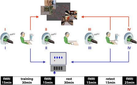
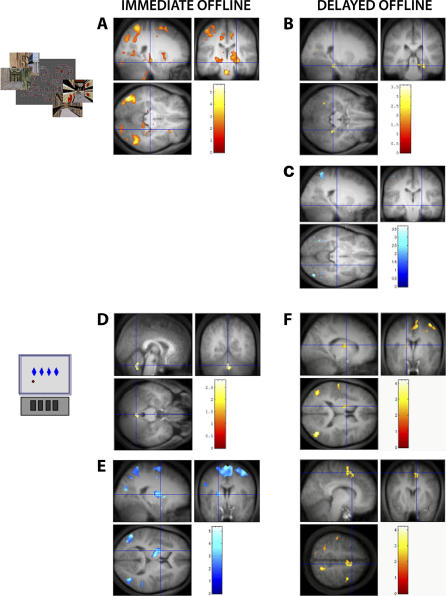
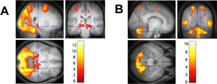
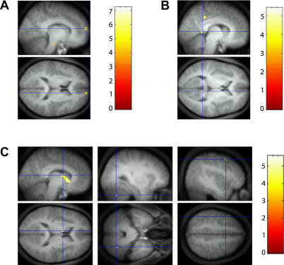
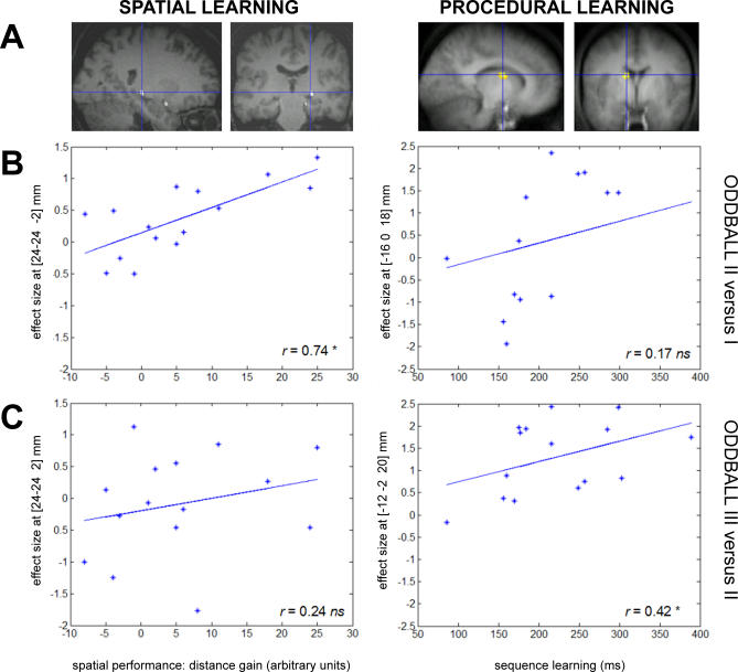
Much remains to be discovered about the fate of recent memories in the human brain. Several studies have reported the reactivation of learning-related cerebral activity during post-training sleep, suggesting that sleep plays a role in the offline processing and consolidation of memory. However, little is known about how new information is maintained and processed during post-training wakefulness before sleep, while the brain is actively engaged in other cognitive activities. We show, using functional magnetic resonance imaging, that brain activity elicited during a new learning episode modulates brain responses to an unrelated cognitive task, during the waking period following the end of training. This post-training activity evolves in learning-related cerebral structures, in which functional connections with other brain regions are gradually established or reinforced. It also correlates with behavioral performance. These processes follow a different time course for hippocampus-dependent and hippocampus-independent memories. Our experimental approach allowed the characterization of the offline evolution of the cerebral correlates of recent memories, without the confounding effect of concurrent practice of the learned material. Results indicate that the human brain has already extensively processed recent memories during the first hours of post-training wakefulness, even when simultaneously coping with unrelated cognitive demands.
Figures
Comment in
-
The neural persistence of memory: Retention begins while you're still awake.PLoS Biol. 2006 Apr;4(4):e116. doi: 10.1371/journal.pbio.0040116. Epub 2006 Mar 28. PLoS Biol. 2006. PMID: 20076554 Free PMC article. No abstract available.
Similar articles
-
Labile or stable: opposing consequences for memory when reactivated during waking and sleep.Nat Neurosci. 2011 Mar;14(3):381-6. doi: 10.1038/nn.2744. Epub 2011 Jan 23. Nat Neurosci. 2011. PMID: 21258327
-
Nap and melatonin-induced changes in hippocampal activation and their role in verbal memory consolidation.J Pineal Res. 2007 Nov;43(4):336-42. doi: 10.1111/j.1600-079X.2007.00482.x. J Pineal Res. 2007. PMID: 17910601 Clinical Trial.
-
Odor cues during slow-wave sleep prompt declarative memory consolidation.Science. 2007 Mar 9;315(5817):1426-9. doi: 10.1126/science.1138581. Science. 2007. PMID: 17347444
-
Sharp-wave ripples as a signature of hippocampal-prefrontal reactivation for memory during sleep and waking states.Neurobiol Learn Mem. 2019 Apr;160:11-20. doi: 10.1016/j.nlm.2018.01.002. Epub 2018 Jan 10. Neurobiol Learn Mem. 2019. PMID: 29331447 Free PMC article. Review.
-
Hippocampal ripples as a mode of communication with cortical and subcortical areas.Hippocampus. 2020 Jan;30(1):39-49. doi: 10.1002/hipo.22997. Epub 2018 Nov 13. Hippocampus. 2020. PMID: 30069976 Review.
Cited by
-
Baseline brain activity fluctuations predict somatosensory perception in humans.Proc Natl Acad Sci U S A. 2007 Jul 17;104(29):12187-92. doi: 10.1073/pnas.0611404104. Epub 2007 Jul 6. Proc Natl Acad Sci U S A. 2007. PMID: 17616583 Free PMC article. Clinical Trial.
-
Post-learning molecular reactivation underlies taste memory consolidation.Front Syst Neurosci. 2011 Sep 26;5:79. doi: 10.3389/fnsys.2011.00079. eCollection 2011. Front Syst Neurosci. 2011. PMID: 21991247 Free PMC article.
-
Visual-procedural memory consolidation during sleep blocked by glutamatergic receptor antagonists.J Neurosci. 2008 May 21;28(21):5513-8. doi: 10.1523/JNEUROSCI.5374-07.2008. J Neurosci. 2008. PMID: 18495885 Free PMC article. Clinical Trial.
-
Sleep modulates the neural substrates of both spatial and contextual memory consolidation.PLoS One. 2008 Aug 13;3(8):e2949. doi: 10.1371/journal.pone.0002949. PLoS One. 2008. PMID: 18698363 Free PMC article.
-
Posterior cingulate cortex-related co-activation patterns: a resting state FMRI study in propofol-induced loss of consciousness.PLoS One. 2014 Jun 30;9(6):e100012. doi: 10.1371/journal.pone.0100012. eCollection 2014. PLoS One. 2014. PMID: 24979748 Free PMC article.
References
-
- Peigneux P, Laureys S, Fuchs S, Destrebecqz A, Collette F, et al. Learned material content and acquisition level modulate cerebral reactivation during post-training rapid-eye-movements sleep. NeuroImage. 2003;20:125–134. - PubMed
-
- Peigneux P, Laureys S, Fuchs S, Collette F, Perrin F, et al. Are spatial memories strengthened in the human hippocampus during slow wave sleep? Neuron. 2004;44:535–545. - PubMed
-
- Maquet P, Laureys S, Peigneux P, Fuchs S, Petiau C, et al. Experience-dependent changes in cerebral activation during human REM sleep. Nature Neurosci. 2000;3:831–836. - PubMed
-
- Huber R, Ghilardi MF, Massimini M, Tononi G. Local sleep and learning. Nature. 2004;430:78–81. - PubMed
Publication types
MeSH terms
LinkOut - more resources
Full Text Sources
Other Literature Sources
Medical
