

Review
doi: 10.1016/j.yfrne.2006.02.001.
Epub 2006 Apr 17.
Hippocampal neuroplasticity induced by early-life stress: functional and molecular aspects
Affiliations
- PMID: 16603235
- PMCID: PMC2937188
- DOI: 10.1016/j.yfrne.2006.02.001
Item in Clipboard
Review
Hippocampal neuroplasticity induced by early-life stress: functional and molecular aspects
Front Neuroendocrinol.
2006 Jul.
Abstract
Whereas genetic factors contribute crucially to brain function, early-life events, including stress, exert long-lasting influence on neuronal function. Here, we focus on the hippocampus as the target of these early-life events because of its crucial role in learning and memory. Using a novel immature-rodent model, we describe the deleterious consequences of chronic early-life 'psychological' stress on hippocampus-dependent cognitive tasks. We review the cellular mechanisms involved and discuss the roles of stress-mediating molecules, including corticotropin releasing hormone, in the process by which stress impacts the structure and function of hippocampal neurons.
Figures
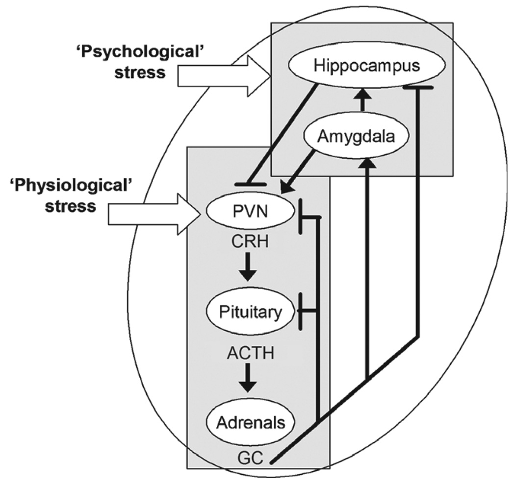
A simplified diagram of the circuits involved in ‘physiological’ and ‘psychological’ stress responses. Physiological stress signals activate the neuroendocrine hypothalamic–pituitary–adrenal axis (outlined by the rectangular box). Physiological stress signals converge on the hypothalamic paraventricular nucleus (PVN) causing the release of corticotropin-releasing hormone (CRH) into the hypothalamo–pituitary portal system. CRH elicits secretion of adrenocorticotropic hormone (ACTH) from the pituitary and ACTH induces the release of glucocorticoids (GC) from the adrenal gland. GCs cross the blood–brain barrier and interact with type-2 corticoid receptors (GR) in the hippocampus, PVN and pituitary to ‘shut-off’ the neuroendocrine response to stress (indicated by the blunt-ended lines). In contrast, activation of GR in the central nucleus of the amygdala increases CRH expression in this region [30,113] and is generally considered to facilitate stress responses. Psychological stress engages additional brain regions and circuits, involving, at a minimum, the hippocampal formation. Arrows denote facilitatory projections.
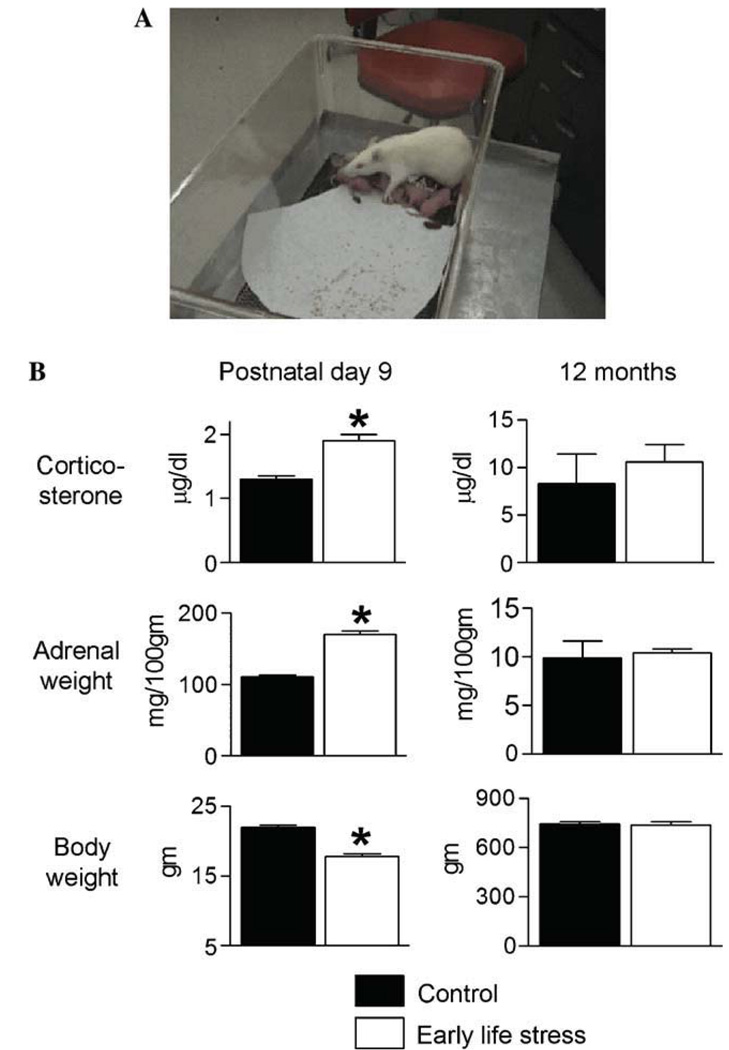
The design and consequences of a chronic, ‘psychological’ early-life stress paradigm. (A) Photograph of a cage at the moment of onset of the stress paradigm (postnatal day 2). A single paper towel is provided to the dam for creating a ‘nest’. The dam will rapidly shred it for this purpose. (B) Parameters indicative of stress immediately after the early-life stress period (postnatal day 9; left column) and in adult male rats (12 months of age; right column). Elevated basal corticosterone levels, higher adrenal gland weights, and modestly lower body weight are found in chronically stressed 9-day old rats. These changes are no longer apparent in adult rats. Black bar, control group; white bar, early-life stress group. *P< 0.05. (Modified from 31 with permission.)
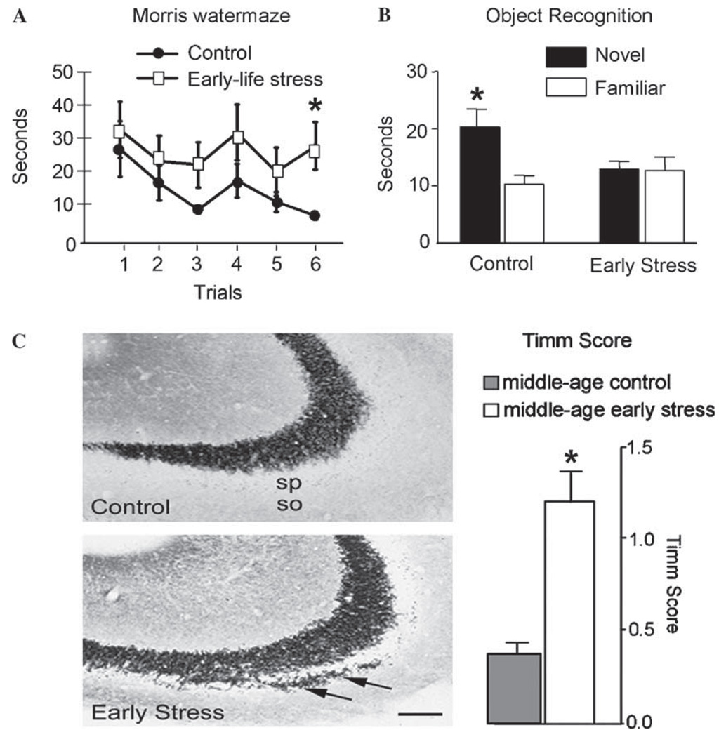
Functional and structural consequences of early-life psychological stress between P2 and P9 on the hippocampus of adult male rats. (A) Escape latencies (time to reach the hidden platform) on the testing day (day 3) are shown after 2 training days. Early-life stressed rats (white squares) require significantly longer time to locate the hidden platform in the Morris watermaze test when compared to age-matched controls (black circles; paired t test, P < 0.05). T test analyses of each trial demonstrate significantly different escape latencies at specific trials (marked by *). (B) Using the object recognition paradigm, control rats are able to discriminate between familiar and novel objects (remembering the familiar object from the day before and exploring it for significantly shorter time). In contrast, early-life stress rats spend the same amount of time exploring the familiar and novel objects, indicating impairment of recognition memory. (Modified from [31] with permission.) (C) Sections of CA3 pyramidal cell Welds from control and early-life stress rats subjected to Timm’s stain for visualizing the high zinc content of mossy fiber terminals (axons of the CA3 innervating granule cells). In early-life stressed rats, these terminals are abnormally abundant within CA3 stratum oriens (so; arrows). Quantification of the Timm’s stained sections confirmed that mossy fiber sprouting is significantly increased in early-life stress animals. sp, stratum pyramidale. Scale bar, 50 µm. *P < 0.05. (Modified from [31], with permission.)
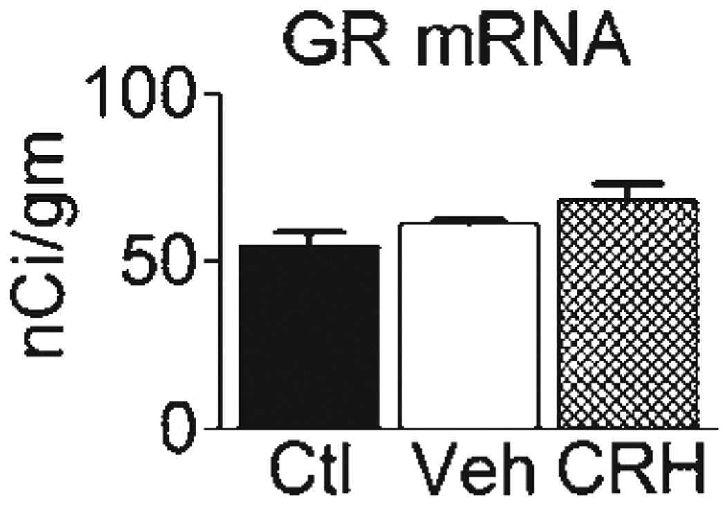
Administration of CRH early in life does not significantly influence GR-mRNA expression in hippocampal CA1 long-term. Adult male rats that were treated with CRH early in life express similar levels of hippocampal GR mRNA when compared to vehicle-treated controls. Note that a trend towards increased GR mRNA levels is found in vehicle-treated versus naive controls. Ctl, adult group, not infused at all on P10; Veh, adult rats infused with vehicle (water) on P10; CRH, adult rats treated with CRH early in life (see [28] for infusion procedures and details).
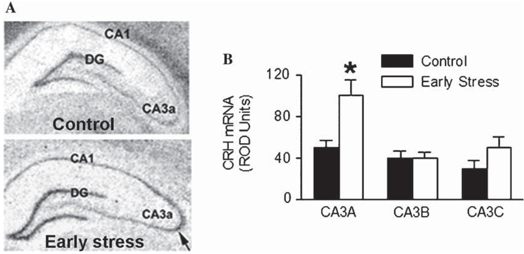
Elevated CRH mRNA expression in adult (12-month old) rats that had experienced early-life stress. (A) Photomicrographs of coronal brain sections from control and early-life stress animals after in situ hybridization for CRH mRNA. (B) Relative optical density (ROD) of the CRH signal was analyzed in hippocampal subfields. CRH mRNA expression is significantly enhanced in hippocampal subfield CA3a of early-life stressed animals when compared to that of controls (arrow). DG, dentate gyrus. *P< 0.05.
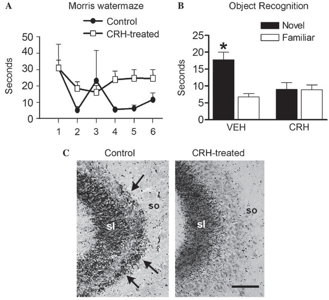
Administration of synthetic CRH into the brain recapitulates the structural and functional effects of early-life stress. (A) Adult rats that were treated with CRH early in life suffer from hippocampus-dependent memory dysfunction in the Morris watermaze. CRH-treated rats (white squares) take significantly longer to locate the hidden platform in the watermaze when compared to controls (black circles) [F(2,132) = 5.53, P< 0.01]. (B) Impaired memory is further evident by the performance of CRH-treated rats on the non-aversive, relatively stress-free object recognition test. Here, CRH-treated rats fail to distinguish the familiar object from the novel object, indicating impairment of recognition memory. (C) Sections of CA3A pyramidal cell regions from control and CRH-treated adult (12-month old), subjected to Timm’s stain for visualizing zinc-rich mossy fiber terminals. In CRH-treated rats, these terminals are abnormally abundant within the CA3 stratum oriens (so; arrows). sl, stratum lucidum. Scale bar = 50 µm. *P < 0.05. (Modified from [28] with permission.)
References
-
- Aldenhoff JB, Gruol DL, Rivier J, Vale W, Siggins GR. Corticotropin releasing factor decreases postburst hyperpolarizations and excites hippocampal neurons. Science. 1983;221:875–877. - PubMed
-
- Alfarez DN, Joels M, Krugers HJ. Chronic unpredictable stress impairs long-term potentiation in rat hippocampal CA1 area and dentate gyrus in vitro. Eur. J. Neurosci. 2003;17:1928–1934. - PubMed
-
- Altman J, Bayer SA. Prolonged sojourn of developing pyramidal cells in the intermediate zone of the hippocampus and their settling in the stratum pyramidale. J. Comp. Neurol. 1990;301:343–364. - PubMed
-
- Amaral DG, Dent JA. Development of the mossy fibers of the dentate gyrus: I. A light and electron microscopic study of the mossy fibers and their expansions. J. Comp. Neurol. 1981;195:51–86. - PubMed
Publication types
MeSH terms
Substances
Grants and funding
LinkOut - more resources
Full Text Sources
Other Literature Sources
Medical
