. 2006 May;20(7):959-61.
doi: 10.1096/fj.05-5134fje. Epub 2006 Apr 7.

Receptor-mediated oral delivery of a bioencapsulated green fluorescent protein expressed in transgenic chloroplasts into the mouse circulatory system

Affiliations

Receptor-mediated oral delivery of a bioencapsulated green fluorescent protein expressed in transgenic chloroplasts into the mouse circulatory system

Arati Limaye et al. FASEB J. 2006 May.

Abstract

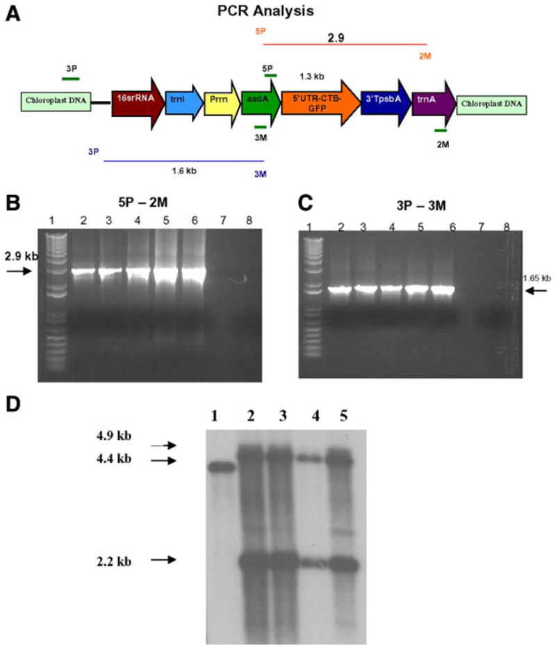
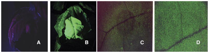
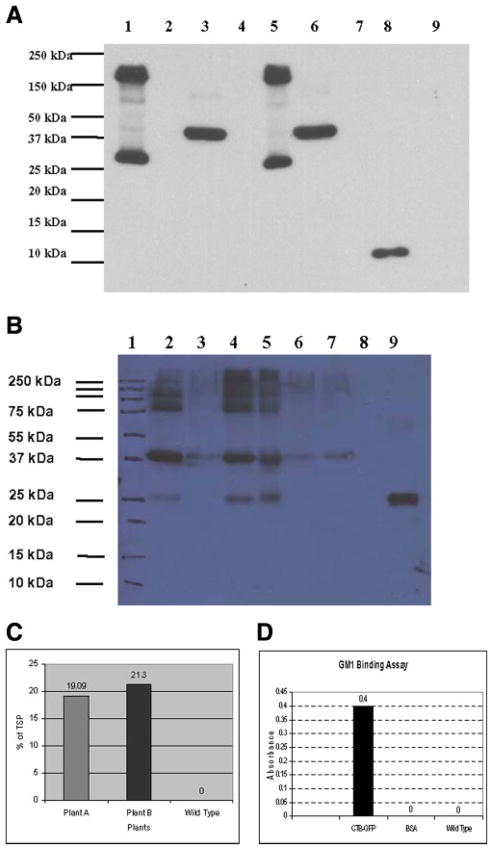
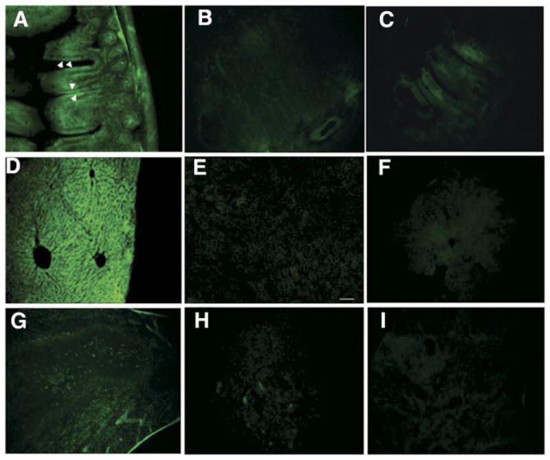
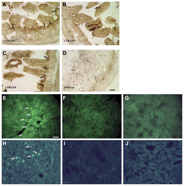
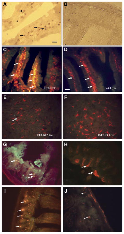
Oral delivery of biopharmaceutical proteins expressed in plant cells should reduce their cost of production, purification, processing, cold storage, transportation, and delivery. However, poor intestinal absorption of intact proteins is a major challenge. To overcome this limitation, we investigate here the concept of receptor-mediated oral delivery of chloroplast-expressed foreign proteins. Therefore, the transmucosal carrier cholera toxin B-subunit and green fluorescent protein (CTB-GFP), separated by a furin cleavage site, was expressed via the tobacco chloroplast genome. Polymerase chain reaction (PCR) and Southern blot analyses confirmed site-specific transgene integration and homoplasmy. Immunoblot analysis and ELISA confirmed expression of monomeric and pentameric forms of CTB-GFP, up to 21.3% of total soluble proteins. An in vitro furin cleavage assay confirmed integrity of the engineered furin cleavage site, and a GM1 binding assay confirmed the functionality of CTB-GFP pentamers. Following oral administration of CTB-GFP expressing leaf material to mice, GFP was observed in the mice intestinal mucosa, liver, and spleen in fluorescence and immunohistochemical studies, while CTB remained in the intestinal cell. This report of receptor-mediated oral delivery of a foreign protein into the circulatory system opens the door for low-cost production and delivery of human therapeutic proteins.


Figures

Figure 1
Figure 1
PCR analysis for the confirmation of transgene integration. A) Schematic representation of the transgene cassette. B) 5P/2M—These primers land on the aadA and trnA regions (flanking the CTB-GFP). A 2.9 kb PCR product was obtained from the PCR analysis of trasgenic plants. C) 3P/3M—The 3P primer lands on the native chloroplast genome and the 3M primer lands on the aadA gene. A 1.6 kb PCR product was obtained from the PCR anlysis of the transgenic plants. Lane 1: 1 kb plus ladder. Lanes 2–5: Transgenic lines of CTB-GFP. Lane 6: Positive Control. Lane 7: Empty. Lane 8: Wild-type Plant. D) Southern blot analysis of the plants. Lane 1: WT showing 4.4 kb fragment. Lanes 2–5: Transgenic plants showing 4.9 and 2.2 kb hybridizing fragments. Flanking sequence shown in Figure 1A was used as the probe.
Figure 2
Figure 2
Visualization of GFP fluorescence in transgenic plants under UV light. A) Wild-type (untransformed) plant seen under UV light. B) CTB- GFP expressing leaf showing fluorescence observed under UV light. C) Wild-type leaf under a low-magnification microscope. D) CTB-GFP expressing leaf showing fluorescence under a low-magnification microscope.
Figure 3
Figure 3
Immunoblot analysis, furin cleavage assay and quantification of CTB-GFP expressed in chloroplasts of transgenic lines. A) Immunoblot demonstrating the expression of CTB-GFP in transgenic plant crude extracts: Lane 1: Un-boiled crude extract of transgenic line A. Lane 3: boiled crude extract of transgenic line A. Lane 5: Unboiled crude extract of transgenic line B. Lane 6: Boiled crude extract of transgenic line A. Lane 8: Purified CTB standard 200 ng. Lane 9: Wild-type plant crude extract. Lanes 2, 4, 7: empty. B) Furin cleavage assay of the plant extract: Lane 1: Marker. Lane 2: CTB-GFP, pH 6.0, with furin, no PMSF. Lane 3: CTB-GFP no incubation, no furin. Lane 4: CTB-GFP pH 6.0 with furin and PMSF. Lane 5: CTB-GFP, pH 7.0, with furin and PMSF. Lane 6: CTB-GFP, pH 6.0, with PMSF, no furin. Lane 7: CTB-GFP, pH 6.0, no PMSF, no furin. Lane 8: Blank. Lane 9: Purified recombinant GFP standard. C) Expression levels in % of CTB-GFP in total soluble protein (TSP) of the CTB-GFP expressing plants. D) GM1 ganglioside binding assayshowing the presence of CTB-GFP functional pentamers.
Figure 4
Figure 4
Cryosections of the intestine and liver of the mice fed with CTB-GFP or wild-type plant leaves material. A) GFP in the ileum of a mouse following oral delivery of the CTB-GFP expressing plant leaf material. Arrows show numerous columnar cells of the intestinal mucous membrane, which have up-taken the CTB-GFP. Various cells in the connective tissue beneath the epithelium also show the presence of GFP. B) Section of the ileum of a mouse fed by the wild-type (untransformed) plant leaf material. C) Section of the ileum of a mouse fed by the IFN-GFP leaf material. D) GFP in hepatocytes of a mouse liver following oral delivery of CTB-GFP expressing plant. E) Section of the liver of a mouse fed by the wild-type plant material. F) Section of the liver of a mouse fed by IFN- GFP expressing plant material. G) GFP in the spleen of a mouse following oral delivery of CTB-GFP expressing plant. Arrows show various splenic cells with GFP. H) Section of the spleen of a mouse fed by the wild-type plant material. I) Section of the spleen of a mouse fed by IFN-GFP expressing plant material. Scale bar: 50 μm.
Figure 5
Figure 5
Immunohistochemical localization of the GFP in mouse ileum, liver, and spleen. A–C) are sections of the ileum of the mice fed with CTB-GFP expressing plant leaf. Arrows indicate presence of GFP in the intestinal epithelium as well as cells of the crypts. D) Shows a section of the ileum of a mouse fed with wild-type (untransformed) plant leaf materials. E) GFP- immunoreactivity in hepatocytes (arrows) in a mouse fed orally by CTB-GFP expressing plant. F) Section of the liver from a mouse fed by wild-type (untransformed) plant. G) Section of the liver from a mouse fed by IFN-GFP expressing plant. H) GFP-immunoreactivity in the spleen of mouse fed orally by CTB-GFP expressing plant. Arrows indicate various cells with a higher GFP content. I) Section of the spleen from a mouse fed by wild-type (untransformed) plant. J) Section of the spleen from a mouse fed by wild-type (untransformed) plant. Scale bar for A–D = 50 μm, Scale bar for E–J = 25 μm.
Figure 6
Figure 6
Immunohistochemistry of ileum, liver, and spleen tissues of mice fed with CTB-GFP expressing leaves or IFN-GFP expressing leaves or wild-type leaves. A) Shows a section of the intestine of a CTB-GFP treated mouse. The arrows indicate CTB in the submucosa of the intestinal villi. B) Shows a section of mouse ileum fed with wild-type plant, immunostained for CTB. C–F) Double staining for macrophage (red) and CTB (green) in mouse intestine and liver. C) Arrows show macrophages in the sub-mucosa of the intestine containing CTB, in a mouse fed with CTB-GFP expressing plant leaf material. The merged color is yellow. D) Arrows indicate F4/80-positive cells (macrophages, in red) in a merged picture in the intestine of a mouse fed with WT leaf material. E) A merged picture showing double staining for macrophage (Kupffer cells) and CTB in mouse liver. Arrows show macrophages (red) in the liver. No sign of CTB (green) was found in the liver of CTB-GFP fed mouse. F) Liver section of an IFN-GFP fed mouse used as a negative control for CTB. Macrophages are seen in red. G) F4/80 Ab was used as a marker of macrophages in the intestine. Arrows indicate macrophages, which have entrapped GFP (yellow after merging the red and the green). Many of the macrophages are not associated with GFP. H) Many macrophages are seen in the intestine of mouse fed with IFN-GFP expressing plant leaf material, which do not show GFP immunoreactivity. I, J) CD11c (red) and GFP (green) immunoreactivities in the mouse intestine. I) Arrows indicate CD11c (red, presumably dendritic cells, due to having a star shape morphology) with internalized GFP (green), which can be seen in yellow color when the red and green channels were merged. J) Arrows indicate CD11c-positive cells in intestine of mice fed with IFN-GFP expressing plant leaf material. Scale bar for A and B = 25 μm. Scale bar for C–J = 50 μm.

Similar articles

Cited by

References

    1. Daniell H, Carmona-Sanchez O, Burns BE. Chloroplast derived antibodies, biopharmaceuticals and edible vaccines. In: Fischer R, Schillberg S, editors. Molecular Farming. WILEY-VCH Verlag Publishers; Weinheim, Germany: 2004. pp. 113–133.
    1. Petridis D, Sapidou E, Calandranis J. Computer aided process analysis and economic evaluation for biosynthetic human insulin production–a case study. Biotechnol Bioeng. 1995;48:529–541. - PubMed
    1. Giddings G, Allison G, Brooks D, Carter A. Transgenic plants as factories for biopharmaceuticals. Nat Biotechnol. 2000;18:1151–1155. - PubMed
    1. Arntzen C, Plotkin S, Dodet B. Plant-derived vaccines and antibodies: potential and limitations. Vaccine. 2005;23:1753–1756. - PubMed
    1. Mason HS, Warzecha H, Mor T, Arntzen C. Trends Mol Med. 2002;8:324. - PubMed

Publication types

MeSH terms

LinkOut - more resources