. 2006 Apr 18;103(16):6196-201.
doi: 10.1073/pnas.0506926103. Epub 2006 Apr 7.

Spatiotemporal control of spindle midzone formation by PRC1 in human cells

Affiliations

Spatiotemporal control of spindle midzone formation by PRC1 in human cells

Changjun Zhu et al. Proc Natl Acad Sci U S A. .

Abstract

We have examined the role of PRC1, a midzone-associated, microtubule bundling, Cdk substrate protein, in regulating the spatiotemporal formation of the midzone in HeLa cells. Cdk-mediated phosphorylation of PRC1 in early mitosis holds PRC1 in an inactive monomeric state. During the metaphase-to-anaphase transition, PRC1 is dephosphorylated, promoting PRC1 oligomerization. Using time-lapse video microscopy, RNA interference, 3D immunofluorescence reconstruction imaging, and rescue experiments, we demonstrate that the dephosphorylated form of PRC1 is essential for bundling antiparallel, nonkinetochore, interdigitating microtubules to establish the midzone that is necessary for cytokinesis. Our results thus indicate that PRC1 is an essential factor in controlling the spatiotemporal formation of the midzone in human cells.


Conflict of interest statement

Conflict of interest statement: No conflicts declared.

Figures

Fig. 1.
Fig. 1.
Effects of Cdk phosphorylation on PRC1 oligomerization and spindle localization. (A) Purified, bacterially expressed, His-tagged PRC1 was incubated with or without baculovirus-expressed, purified Cdc2/cyclin B1 in the presence of ATP. The reactions were subjected to immunoblotting analysis by using α-PRC1 or α-PRC1P. (B) Mitotic HeLa cell lysates were incubated with α-PRC1 antibody, and the immunoprecipitates then were incubated with or without phosphatases (GST-Cdc14A or CIAP). The reactions were subjected to immunoblotting analysis by using α-PRC1 or α-PRC1P. (C) HeLa cells or HeLa cells expressing EYFP-PRC1ΔN184 were lysed in 1% Nonidet P-40 lysis buffer, and cell lysates were subjected to sucrose gradient sedimentation (5% to ≈30%) by ultracentrifugation. The sediments were collected and subjected to immunoblotting analysis by using α-PRC1, α-PRC1P, or anti-GFP antibody. (D) Asynchronous HeLa cells grown on coverslips were fixed with 3% formaldehyde and then stained with α-PRC1P (red), α-PRC1 (blue), anti-α-tubulin antibody (green), and DAPI (DNA, white). (Scale bars: 5 μm.) (E and F) HeLa cells grown on 35-mm glass-bottom dishes were transfected with 0.5 μg of pEYFP-PRC1 and 0.5 μg of pECFP-H2B together with (F) or without (E) 0.1 μg of pEGFP-cyclin B1Δ90. Thirty-six hours after transfection, cells were placed on a 37°C heated stage, and time-lapse images were collected under a Zeiss Axiovert 100M inverted fluorescence microscope at 2-min intervals by using an automatic digital charge-coupled device camera for 9–10 h. The movies were edited by using slidebook 3.0 and director mx (Adobe Systems, San Jose, CA). Representative images of the movies are shown (EYFP-PRC1, pseudocolored red; ECFP-H2B, pseudocolored green). Red arrows indicate spindle midzone/midbody localization of PRC1 in control cells and abnormal mitotic spindle localization of PRC1 in pEGFP-cyclin B1Δ90-transfected cells.
Fig. 2.
Fig. 2.
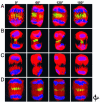
Three-dimensional reconstruction images of anaphase spindle morphology and structure in a control cell, PRC1-depleted cells, and a PRC1-depleted cell expressing exogenous EYFP-PRC1ΔC. HeLa cells were transfected with control siRNA (A), PRC1 siRNA (B and C), or PRC1 siRNA together with pEYFP-PRC1ΔC* plasmid (D). Forty-eight hours after transfection, cells were fixed and stained with α-PRC1 (green in AC), anti-α-tubulin antibody (red in AD), and DAPI (blue in AD). EYFP-PRC1ΔC is shown in green in D. Three-dimensional images were reconstructed from a Z-series (0.3 μm stepwise) of immunostained cells by using volocity. quicktime movies of 3D images were exported according to the manufacturer’s instructions. Representative images of the movies are shown.
Fig. 3.
Fig. 3.
The ability of ectopically expressed PRC1 oligomerization/Cdk phosphorylation mutants to rescue mitotic and cytokinetic defects, including midzone formation, in PRC1-depleted cells. HeLa cells grown on coverslips were transfected with 0.2 μg of pEYFPC1-PRC1ΔC* (A), pEYFPC1-PRC1ΔN184ΔC (B), pEYFPC1-PRC1ΔC*AA (C), or pEYFPC1-PRC1ΔC*EE (D). Twenty-four hours after transfection, cells were transfected with 100 nM PRC1 siRNA. Forty-eight hours after the second transfection, cells were fixed and stained for α-PRC1 (blue), anti-tubulin antibody (red), and DNA (white). Expression of EYFP-PRC1ΔC mutant proteins is shown in green. (Scale bars: 5 μm.)
Fig. 4.
Fig. 4.
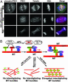
Interdigitating MT bundles in Kif4 siRNA-treated cells. (A) HeLa cells grown on coverslips were transfected with 100 nM Kif4 siRNA as described in ref. . Two days after transfection, cells were fixed and stained with α-PRC1P (green), α-PRC1 (red), anti-α-tubulin antibody (blue), and DAPI (DNA, white). (Scale bars: 5 μm.) (B) The proposed model for temporal and spatial control of midzone formation by PRC1 in mammalian cells (see text for details).

References

    1. D’Avino P. P., Savoian M. S., Glover D. M. J. Cell Sci. 2005;118:1549–1558. - PubMed
    1. McCollum D. Curr. Biol. 2004;14:R953–R955. - PubMed
    1. Mishima M., Kaitna S., Glotzer M. Dev. Cell. 2002;2:41–54. - PubMed
    1. Mishima M., Pavicic V., Gruneberg U., Nigg E. A., Glotzer M. Nature. 2004;430:908–913. - PubMed
    1. Bowerman B. Nature. 2004;430:840–842. - PubMed

Publication types