. 2006 May;18(5):1199-212.
doi: 10.1105/tpc.105.038364. Epub 2006 Apr 7.

Gamma-tubulin is essential for acentrosomal microtubule nucleation and coordination of late mitotic events in Arabidopsis

Affiliations

Gamma-tubulin is essential for acentrosomal microtubule nucleation and coordination of late mitotic events in Arabidopsis

Pavla Binarová et al. Plant Cell. 2006 May.

Abstract

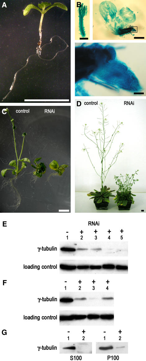
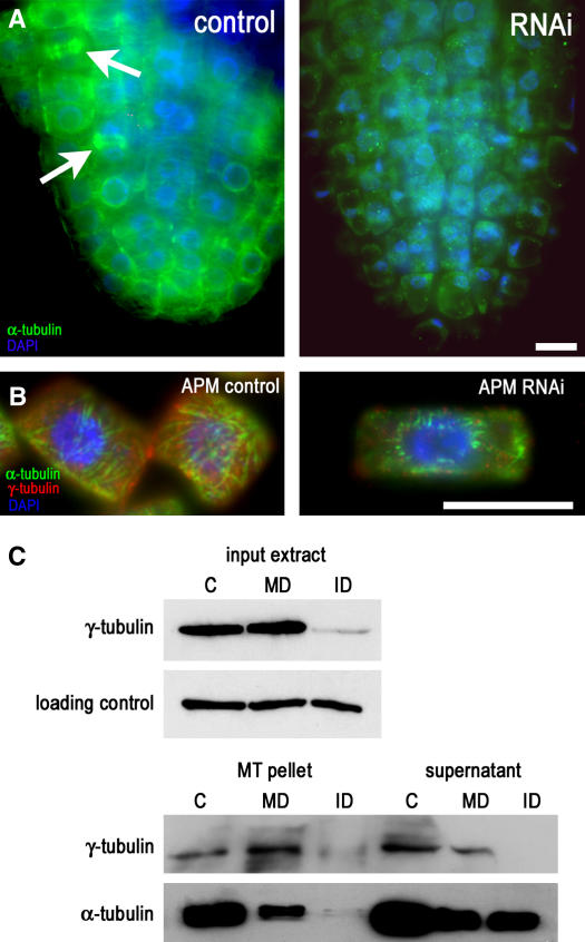
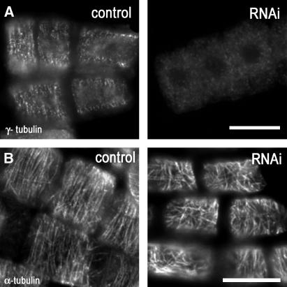
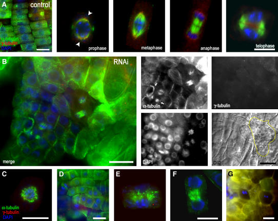
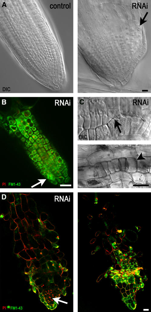
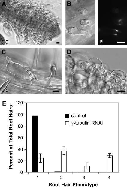
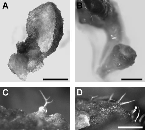
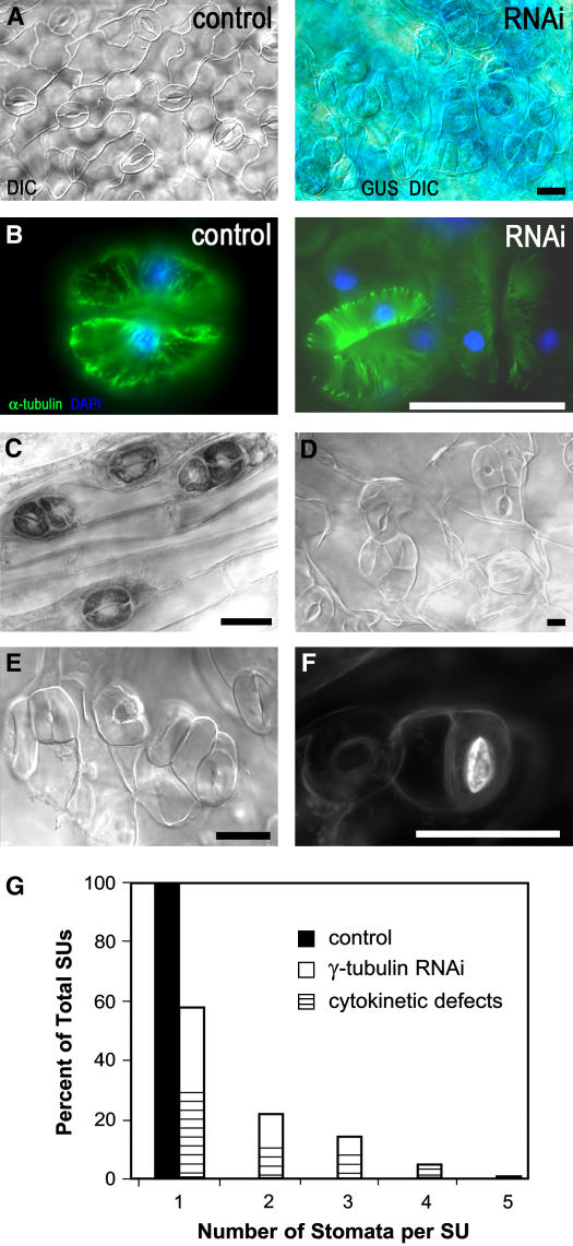
Gamma-tubulin is required for microtubule (MT) nucleation at MT organizing centers such as centrosomes or spindle pole bodies, but little is known about its noncentrosomal functions. We conditionally downregulated gamma-tubulin by inducible expression of RNA interference (RNAi) constructs in Arabidopsis thaliana. Almost complete RNAi depletion of gamma-tubulin led to the absence of MTs and was lethal at the cotyledon stage. After induction of RNAi expression, gamma-tubulin was gradually depleted from both cytoplasmic and microsomal fractions. In RNAi plants with partial loss of gamma-tubulin, MT recovery after drug-induced depolymerization was impaired. Similarly, immunodepletion of gamma-tubulin from Arabidopsis extracts severely compromised in vitro polymerization of MTs. Reduction of gamma-tubulin protein levels led to randomization and bundling of cortical MTs. This finding indicates that MT-bound gamma-tubulin is part of a cortical template guiding the microtubular network and is essential for MT nucleation. Furthermore, we found that cells with decreased levels of gamma-tubulin could progress through mitosis, but cytokinesis was strongly affected. Stepwise diminution of gamma-tubulin allowed us to reveal roles for MT nucleation in plant development, such as organization of cell files, anisotropic and polar tip growth, and stomatal patterning. Some of these functions of gamma-tubulin might be independent of MT nucleation.


Figures

Figure 1.
Figure 1.
Developmental Abnormalities in Arabidopsis Lines with RNAi-Induced Downregulation of γ-Tubulin. (A) Control uninduced Arabidopsis seedling transformed with dexamethasone-inducible RNAi construct 10 d after germination. Bar = 1 cm. (B) Strong phenotype in a representative dexamethasone-induced RNAi γ-tubulin seedling, 10 d after germination on medium with dexamethasone, showing growth arrest and lethality at the cotyledon stage. Histochemical GUS staining revealed strong inducible expression of the coregulated marker. As shown at left and in the enlarged boxed area, the main root is arrested and new auxiliary meristems are activated. Bars = 1 mm. (C) Mild RNAi phenotype in two representative dexamethasone-induced γ-tubulin RNAi seedlings. Shown are a control uninduced plant and two RNAi plants 20 d after induction with arrested development. Bar = 1 cm. (D) Weak γ-tubulin RNAi phenotype with reduced elongation growth. Control and dexamethasone-induced γ-tubulin RNAi plants were grown in soil for 50 d under continuous dexamethasone treatment. Bar = 1 cm. (E) Immunoblot analysis of γ-tubulin in total protein extracts from dexamethasone-inducible γ-tubulin RNAi plants with the strong phenotype as shown in (B) arrested at the cotyledon stage. Lane 1, uninduced control; lanes 2 and 3, pooled seedlings 5 d after induction; lanes 4 and 5, pooled seedlings 10 d after induction. (F) γ-Tubulin in seedlings with the mild phenotype as shown in (C). γ-Tubulin was analyzed by immunoblotting in total protein extracts from individual γ-tubulin RNAi plants. Lane 1, uninduced control; lane 2, plants with dexamethasone-induced RNAi for 15 d; lane 3, dexamethasone induction for 20 d; lane 4, plants with ethanol-induced RNAi for 15 d. Samples were loaded at the same protein content (40 μg/lane). Shown are typical examples of protein gel blot analysis selected from ∼100 analyzed RNAi plants, all showing a corresponding reduction in γ-tubulin levels with the severity of the phenotypes. (G) Distribution of γ-tubulin in cytoplasmic and membrane fractions from RNAi plants 15 d after induction. S100, high-speed supernatant; P100, microsomal pellet. Lane 1, uninduced control; lane 2, induced RNAi plant. To compare the relative distribution of γ-tubulin, pelleted material was resuspended in a volume equal to that of the corresponding supernatant.
Figure 2.
Figure 2.
RNAi Depletion and Immunodepletion of γ-Tubulin Impair Plant MT Nucleation. (A) α-Tubulin labeling in an RNAi plant (strong phenotype as shown in Figure 1B) grown for 10 d on dexamethasone. Mitotic microtubular arrays were immunolabeled in control root tip cells (arrows), whereas in RNAi seedlings, MTs were largely absent and cells had extreme vacuolization and small nuclei close to the cell walls. DAPI, 4′,6-diamidino-2-phenylindole. Bar = 10 μm. (B) Recovery of MTs after depolymerization by the drug APM. In control uninduced plants, MT regrowth was observed at 15 min after removal of APM. In plants (mild phenotype as shown in Figure 1C) with dexamethasone RNAi expression induced for 10 d, the level of γ-tubulin was partially reduced, and a few short, thick bundles of MTs were recovered in the vicinity of nuclei 1 h after APM removal. Bar = 10 μm. (C) In vitro polymerization of MTs in extracts immunodepleted for γ-tubulin. Taxol-driven polymerization from input extracts is shown in the top panel, followed by immunoblot analysis for α- and γ-tubulin of pelleted MTs and supernatants in the bottom panel. No MTs were pelleted from extracts immunodepleted for γ-tubulin. C, control; ID, immunodepleted; MD, mock-depleted.
Figure 3.
Figure 3.
Organization of Cortical MTs Is Sensitive to Decreased γ-Tubulin Levels. (A) γ-Tubulin and DNA labeling in roots of control and dexamethasone-inducible RNAi plants (mild phenotype). γ-Tubulin was distributed on the cortex and along cortical MTs in a patchy pattern in control plants, and the signal for γ-tubulin rapidly disappeared from cortical MTs in RNAi-expressing plants 5 d after induction with dexamethasone. Bar = 10 μm. (B) Parallel arrangements of cortical MTs in control plants became randomized in RNAi plants 5 d after induction with dexamethasone. Bar = 10 μm.
Figure 4.
Figure 4.
γ-Tubulin RNAi in Dividing Cells of Arabidopsis Roots Affect Cytokinesis and the Organization of Cell Files. (A) Whole-mount α-tubulin (green) and γ-tubulin (red) immunolabeling and DAPI staining of DNA (blue) of control uninduced seedlings having regularly arranged cell files. γ-Tubulin in the root cells of the control exhibits bipolar localization from prophase (arrowheads) to telophase when it accumulates in the phragmoplast and the forming cell plate area. (B) Whole-mount α-tubulin (green) and γ-tubulin (red) immunolabeling and DAPI staining of DNA (blue) of RNAi-expressing seedlings with mild phenotypes and reduced γ-tubulin levels 10 d after dexamethasone induction. Shown are a merged image (left panel), α-tubulin, γ-tubulin, DAPI staining, and DIC image (four small right panels). MTs are still present in mitotic cells, with spindles focused to acentrosomal poles, but cytokinesis became defective. The phragmoplasts are misaligned (arrows), and the organization of cell files is disrupted. The yellow line indicates the shape of a binuclear cell in the DIC image. Dividing cells are surrounded by enlarged cells with callose deposits. (C) Collapsed spindle with MTs randomly arranged in the vicinity of chromosomes in a cell with severely depleted γ-tubulin 15 d after induction with dexamethasone. (D) Misorientation of cell division planes in roots expressing ethanol-induced RNAi for 10 d. (E) Phragmoplast with bundled and disorganized MTs in roots expressing dexamethasone-induced RNAi for 10 d. (F) Early solid phragmoplast persists between separated nuclei with already decondensed chromatin, as revealed by DAPI staining. The expanded ring-like phragmoplast is present in this late stage of cytokinesis in the control cells (A). (G) Cells are often binuclear in seedlings 20 d after induction, with severe depletion of γ-tubulin. Bars = 10 μm.
Figure 5.
Figure 5.
The Organization of Root Cell Files Is Distorted with RNAi-Induced Diminution of γ-Tubulin. (A) DIC images of a root from an uninduced control plant (left) with cells organized as files and the root of an RNAi plant (right) 5 d after dexamethasone induction with radial expansion and with binuclear cells (arrow). (B) Confocal laser scanning microscopy single optical section of a root tip of an RNAi plant 10 d after dexamethasone induction. FM1-43 membrane staining and propidium iodide (PI) nuclear staining showed that cells in the meristematic zone became vacuolated, with nuclei shifted from a central position, whereas cell division sites were often misaligned (arrow). (C) Patches of epidermal cells with misaligned cell walls (top, arrow) or clusters of isodiametric epidermal cells (bottom, arrowhead) in the elongation zone of RNAi plants 10 d after induction of RNAi with ethanol shown in DIC images. (D) Confocal laser scanning microscopy single optical sections through the central root (left) and close to the surface layer (right) show that all cell files are distorted in roots grown for 15 d with dexamethasone-induced RNAi expression. FM1-43 and propidium iodide staining are as in (B). Bars = 10 μm.
Figure 6.
Figure 6.
Ectopic Root Hair Formation and Loss of Polar Root Hair Growth in γ-Tubulin RNAi Plants. (A) Ectopic root hairs formed along the entire root length in a γ-tubulin RNAi plant 8 d after dexamethasone induction. Bar = 10 μm. (B) Two nuclei (right) stacked beneath an extremely large vacuole (left) in a short and wide root hair 10 d after dexamethasone induction. Bar = 10 μm. (C) Two or more growth axes emerged from one root hair bulge 10 d after dexamethasone induction. Bar = 10 μm. (D) Root hair bulges with failure of tip growth 12 d after dexamethasone induction. Bar = 10 μm. (E) Percentage of root hair phenotypes in the control plants transformed with the empty cassette grown on dexamethasone and in dexamethasone-inducible RNAi plants. Bars 1, root hairs of wild-type phenotype; bars 2, short wide root hairs, as shown in (B); bars 3, root hairs with two or more growth axes from one root hair bulge, as shown in (C); bars 4, root hair bulges with failure of the transition to tip growth, as shown in (D). Values shown represent means of the percentage counted for 10 plants; n = 1000. Error bars represent sd. Similar results were obtained with the ethanol-inducible RNAi lines.
Figure 7.
Figure 7.
Leaf Phenotypes of RNAi Plants. (A) Thick, curled leaves of plants with the mild phenotype, as shown in Figure 1C, grown for 15 d with RNAi induction. Leaves have roughened surfaces. (B) The youngest leaves developed with reduced γ-tubulin levels have club-shaped blades. (C) Trichomes with extremely swollen bases that appear with high frequency. (D) Unbranched trichomes with bulged tips developed on leaves later upon RNAi induction, and the youngest malformed leaves were hairless (B). Bars = 250 μm.
Figure 8.
Figure 8.
Stomatal Patterning and Differentiation Are Altered in RNAi Plants. (A) Leaf epidermis of a control plant (left) and an RNAi plant of the weak phenotype grown in soil with dexamethasone induction for 21 d (right). Stomata are in clusters of two to four. Bar = 25 μm. (B) Immunolocalization analysis of α-tubulin showing the presence of MTs in the control stomata (left) and in the stomata cluster from the γ-tubulin RNAi plant shown in (A) (right). Bar = 25 μm. (C) Clustered stomata also appeared on inflorescence stems that developed with induced RNAi. Bar = 25 μm. (D) and (E) Stomata were clustered and cytokinetic defects appeared in mild phenotype plants with more severe depletion of γ-tubulin (as shown in Figure 1C) upon dexamethasone (D) or ethanol (E) RNAi induction for 15 d. Bar = 25 μm. (F) Callose deposits in a stomata with aberrant division after RNAi induction for 15 d. Bar = 25 μm. (G) Frequencies of stomata units (SUs) with clustered stomata and with or without the cytokinetic defect in ethanol- and dexamethasone-induced RNAi-expressing plants with the defects shown in (D) to (F). n = 500 for the control and 500 for RNAi plants.

References

    1. Baskin, T.I., Beemster, G.T., Judy-March, J.E., and Marga, F. (2004). Disorganization of cortical microtubules stimulates tangential expansion and reduces the uniformity of cellulose microfibril alignment among cells in the root of Arabidopsis. Plant Physiol. 135 2279–2290. - PMC - PubMed
    1. Bergmann, D.C., Lukowitz, W., and Somerville, C.R. (2004). Stomata development and pattern controlled by a MAPKK kinase. Science 304 1461–1462. - PubMed
    1. Bibikova, T.N., Blancaflor, E.B., and Gilroy, S. (1999). Microtubules regulate tip growth and orientation in root hairs of Arabidopsis thaliana. Plant J. 17 657–665. - PubMed
    1. Binarova, P., Cenklova, V., Hause, B., Kubatova, E., Lysak, M., Dolezel, J., Bogre, L., and Draber, P. (2000). Nuclear gamma-tubulin during acentriolar plant mitosis. Plant Cell 12 433–442. - PMC - PubMed
    1. Binarova, P., Cihalikova, J., and Dolezel, J. (1993). Localization of MPM-2 recognized phosphoproteins and tubulin during cell cycle progression in synchronized Vicia faba root meristem cells. Cell Biol. Int. 17 847–856. - PubMed

Publication types

MeSH terms

Associated data

LinkOut - more resources