. 2006 May;12(5):807-18.
doi: 10.1261/rna.2338706. Epub 2006 Apr 7.

RDE-4 preferentially binds long dsRNA and its dimerization is necessary for cleavage of dsRNA to siRNA

Affiliations

RDE-4 preferentially binds long dsRNA and its dimerization is necessary for cleavage of dsRNA to siRNA

Greg S Parker et al. RNA. 2006 May.

Abstract

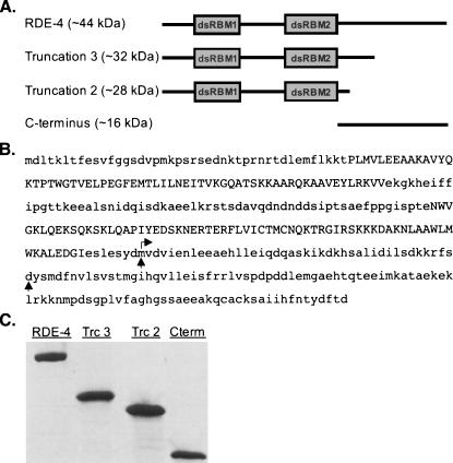
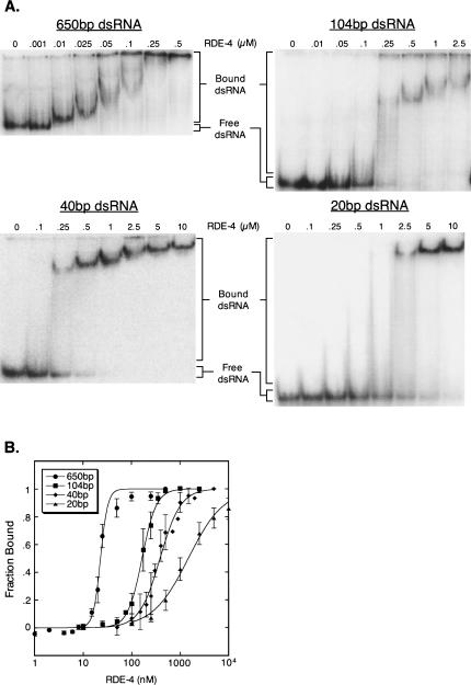
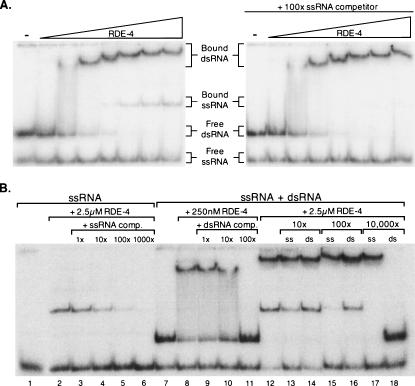
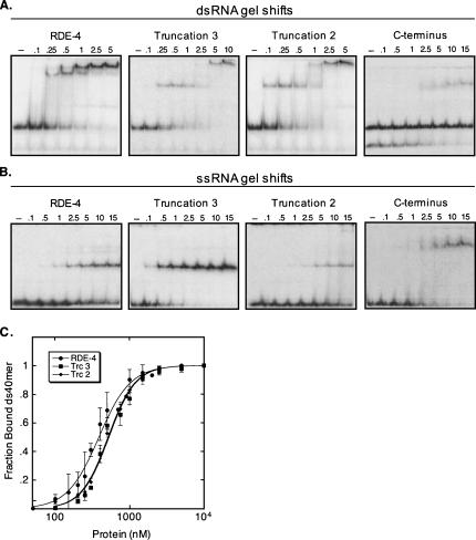
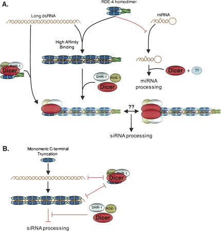
In organisms ranging from Arabidopsis to humans, Dicer requires dsRNA-binding proteins (dsRBPs) to carry out its roles in RNA interference (RNAi) and micro-RNA (miRNA) processing. In Caenorhabditis elegans, the dsRBP RDE-4 acts with Dicer during the initiation of RNAi, when long dsRNA is cleaved to small interfering RNAs (siRNAs). RDE-4 is not required in subsequent steps, and how RDE-4 distinguishes between long dsRNA and short siRNA is unclear. We report the first detailed analysis of RDE-4 binding, using purified recombinant RDE-4 and various truncated proteins. We find that, similar to other dsRBPs, RDE-4 is not sequence-specific. However, consistent with its in vivo roles, RDE-4 binds with higher affinity to long dsRNA. We also observe that RDE-4 is a homodimer in solution, and that the C-terminal domain of the protein is required for dimerization. Using extracts from wild-type and rde-4 mutant C. elegans, we show that the C-terminal dimerization domain is required for the production of siRNA. Our findings suggest a model for RDE-4 function during the initiation of RNAi.


Figures

FIGURE 1.
FIGURE 1.
RDE-4 protein constructs. (A) Schematic diagram of RDE-4 protein constructs with double-stranded RNA binding motifs (dsRBMs) shown as boxes and the molecular weights indicated. (B) Amino acid sequence of RDE-4. Vertical arrows indicate the last amino acid before the engineered stop codon of truncations 2 and 3. Horizontal arrow indicates start position for the C terminus protein. Truncation 2 contains eight additional nonnative amino acids (GMPPNNCS) at its C terminus (see Materials and Methods). (C) SYPRO Red-stained SDS-PAGE gel of purified RDE-4, Truncations 2 and 3 (Trc2 & Trc3) and C terminus (Cterm).
FIGURE 2.
FIGURE 2.
Gel mobility shift analyses of RDE-4 and dsRNA substrates of different lengths. (A) Increasing concentrations of RDE-4 were added to 32P-labeled 650-bp dsRNA (top left), 104-bp dsRNA (top right), 40-bp dsRNA (bottom left), and 20-bp dsRNA (bottom right). Complex formation was analyzed by native gel electrophoresis. Bands corresponding to bound and free dsRNA are labeled. Previous studies note that cooperative ligands bind to nucleic acid lattices in clusters, and as the binding density increases, the average size of the bound clusters increases (Kowalczykowski et al. 1986). The gradual decrease in mobility for the 650-bp complex is likely due to additional protein binding events, possibly in “clusters,” but future studies will be required to confirm this. (B) RNA binding isotherms for RDE-4. Radioactivity in gels as in A were quantified to determine fraction bound = [dsRNA]bound/[dsRNA]total (see Materials and Methods). All RNA of slower mobility than dsRNAfree was considered as bound. Data points with error bars (standard deviation) represent average values (2 ≤ n ≤ 4) and were fit using the Hill formalism, where fraction bound = 1/(1 + (Kdn/[P]n)). Resulting Hill constants for the 650-bp, 104-bp, 40-bp, and 20-bp RNAs were 4.98 ± 1.0, 3.53 ± 0.1, 2.04 ± 0.2, and 1.18 ± 0.1, respectively, but it is important to note that these values may derive from statistical considerations rather than intrinsic cooperativity (see Discussion).
FIGURE 3.
FIGURE 3.
Competition assays. (A) Mobility shift assay showing that RDE-4 binds to dsRNA and ssRNA. Increasing concentrations of RDE-4, as specified in Figure 2A, were added to the preparation of 32P-labeled 40-bp dsRNA, in the absence (left panel) or presence (right panel) of competitor ssRNA (sequence matching one strand of the 40-bp dsRNA). The panel on the right verifies the second complex is RDE-4 binding to ssRNA, since it was competed by 100× unlabeled ssRNA without affecting dsRNA binding (compare complexes in left and right panels). (B) Competition assays at differing protein and competitor concentrations. RNAs are either purified 32P-labeled 40-nt ssRNA (lanes 1–6) or the mix of 32P-labeled dsRNA and 32P-labeled ssRNA used in part A (lanes 7–18). Competitor RNAs are 104 nt in length, either single stranded or double stranded, and are different in sequence to that of the labeled RNAs.
FIGURE 5.
FIGURE 5.
Gel mobility shift analyses comparing binding affinities of RDE-4, Truncation 3, Truncation 2, and the C terminus. (A) 32P-labeled 40-bp dsRNA was incubated with increasing amounts of protein (μM), as indicated above each gel. The 40-bp dsRNA preparations contained minimal amounts of contaminating ssRNA, except for that used with the C terminus protein, and in this case the observed complex is with ssRNA. (B) 32P-labeled 40-nt ssRNA was incubated with increasing amounts of protein (μM), as indicated above each gel. (C) Forty base-pair dsRNA binding isotherms for RDE-4, Truncation 3 and Truncation 2. The C terminus protein did not bind dsRNA and was not plotted. Data points with error bars (standard deviation) represent average values (2 ≤ n ≤ 3) and were fit using the Hill formalism as in Figure 2B. Resulting Hill coefficients for RDE-4, Truncation 2 and Truncation 3 were 2.04 ± 0.2, 2.39 ± 0.1 and 2.39 ± 0.2, respectively.
FIGURE 4.
FIGURE 4.
Sedimentation equilibrium data for RDE-4 and Truncation 3. The lower panels show data for three concentrations of RDE-4 (+ = 15 μM, ⋄ = 10 μM, ◯ = 5 μM) and Truncation 3 (+ = 20 μM, ⋄ = 10 μM, ◯ = 5 μM). RDE-4 data fit to a single-species of MW 86,280 Da ± 3400. The MWobs/MWcalc = 1.99, indicating RDE-4 formed a stable dimer in solution. Truncation 3 data fit to a single-species of MW 35,863 Da ± 3500 and Truncation 2 data fit to a single-species of MW 29,572 Da ± 1750 (data not shown) indicating both truncations are monomeric. The upper panels show the corresponding residuals for each fit.
FIGURE 6.
FIGURE 6.
Reconstitution of siRNA production. Reactions were 30-min preincubations of a 650-bp 32P-internally labeled dsRNA ± recombinant RDE-4 proteins, followed by 1-h incubations with either wild-type (N2) or rde-4(ne337) embryo extracts (40-μg total protein). dsRNA cleavage products were analyzed by denaturing gel electrophoresis. (A) The autoradiogram shows products of reactions incubated without (-extract) or with extracts prepared from N2 and rde-4(ne337) embryos. The production of siRNA was reconstituted in the mutant extracts by adding recombinant RDE-4 at the concentrations indicated. Reconstituted Dicer activity is maximal at +100 nM RDE-4 and is inhibited by higher concentrations of RDE-4. The far left lane shows markers prepared from a 32P-end-labeled 25-bp DNA ladder. (B) As in A but with the addition of recombinant RDE-4 to N2 extracts. (C) As in A but showing reconstitution assays using full-length and truncated RDE-4; protein was added to a final concentration of 100 nM.
FIGURE 7.
FIGURE 7.
Model of RDE-4 function during the initiation of RNAi. The C-terminal dimerization domain of RDE-4 is represented by a green oval and the dsRBMs by dark blue circles. (A) RDE-4 binds long dsRNA with high affinity. The left branch illustrates the siRNA production pathway, with RDE-4 shown in two complexes, one with dsRNA and one without. It is unknown if the Dicer/RDE-1/DRH-1 complex is preformed in the absence of RDE-4, so these proteins are shown separately in the center of the figure, before recruitment to the RDE-4-bound dsRNA. According to our model, RDE-4 dimerization is important for the assembly of active RDE-4/Dicer complexes via one of two proposed scenarios, proper Dicer recruitment to dsRNA, or facilitating Dicer dimerization. For simplicity, only one Dicer complex is illustrated for each scenario. The right branch depicts the miRNA production pathway. RDE-4 is potentially excluded from miRNA maturation due to its low affinity for short RNA duplexes. A dsRBP partner of Dicer during miRNA processing has yet to be reported for C. elegans, and its potential existence is represented by the blue oval with question marks. (B) C-terminal truncation proteins that cannot dimerize are able to bind dsRNA but are unable to form an active Dicer complex. See Discussion for details.

References

    1. Andrews D., Butler J.S., Al-Bassam J., Joss L., Winn-Stapley D.A., Casjens S., Cingolani G. Bacteriophage P22 tail accessory factor GP26 is a long triple-stranded coiled-coil. J. Biol. Chem. 2005;280:5929–5933. - PubMed
    1. Bagga S., Bracht J., Hunter S., Massirer K., Holtz J., Eachus R., Pasquinelli A.E. Regulation by let-7 and lin-4 miRNAs results in target mRNA degradation. Cell. 2005;122:553–563. - PubMed
    1. Bass B.L., Weintraub H. A developmentally regulated activity that unwinds RNA duplexes. Cell. 1987;48:607–613. - PubMed
    1. Bass B.L., Hurst S.R., Singer J.D. Binding properties of newly identified Xenopus proteins containing dsRNA-binding motifs. Curr. Biol. 1994;4:301–314. - PubMed
    1. Brenner S. The genetics of Caenorhabditis elegans . Genetics. 1974;77:71–94. - PMC - PubMed

Publication types

MeSH terms

LinkOut - more resources