. 2006 May;116(5):1410-24.
doi: 10.1172/JCI27607. Epub 2006 Apr 6.

Cystamine and cysteamine increase brain levels of BDNF in Huntington disease via HSJ1b and transglutaminase

Affiliations

Cystamine and cysteamine increase brain levels of BDNF in Huntington disease via HSJ1b and transglutaminase

Maria Borrell-Pagès et al. J Clin Invest. 2006 May.

Abstract

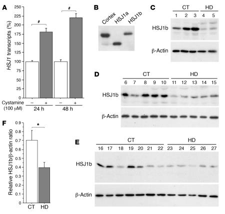
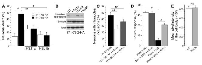
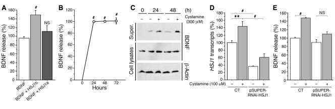
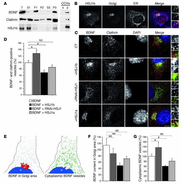
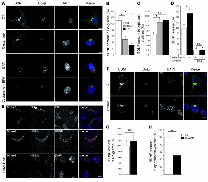
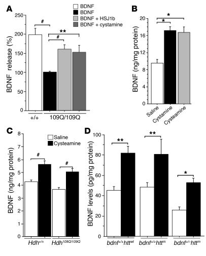
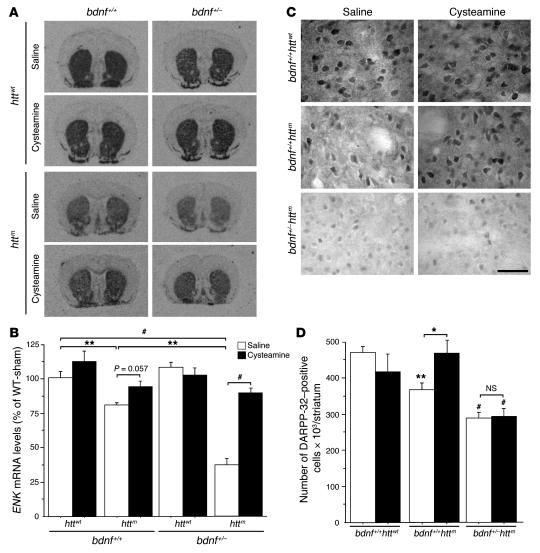
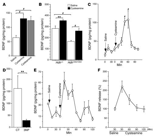
There is no treatment for the neurodegenerative disorder Huntington disease (HD). Cystamine is a candidate drug; however, the mechanisms by which it operates remain unclear. We show here that cystamine increases levels of the heat shock DnaJ-containing protein 1b (HSJ1b) that are low in HD patients. HSJ1b inhibits polyQ-huntingtin-induced death of striatal neurons and neuronal dysfunction in Caenorhabditis elegans. This neuroprotective effect involves stimulation of the secretory pathway through formation of clathrin-coated vesicles containing brain-derived neurotrophic factor (BDNF). Cystamine increases BDNF secretion from the Golgi region that is blocked by reducing HSJ1b levels or by overexpressing transglutaminase. We demonstrate that cysteamine, the FDA-approved reduced form of cystamine, is neuroprotective in HD mice by increasing BDNF levels in brain. Finally, cysteamine increases serum levels of BDNF in mouse and primate models of HD. Therefore, cysteamine is a potential treatment for HD, and serum BDNF levels can be used as a biomarker for drug efficacy.


Figures

Figure 1
Figure 1. Cystamine increases HSJ1 transcript levels in neuronal cells, while HSJ1b is decreased in postmortem brain extracts from HD patients.
(A) Data revealed a statistically significant increase in HSJ1 transcripts induced by cystamine treatment in comparison to control at 24 hours (Student’s t test, t[21] = 5.77; P < 0.0001) and at 48 hours (Student’s t test, t[10] = 9.88; P < 0.0001). (B) Protein extracts prepared from 1 control human cortical postmortem sample and from HEK 293T cells transfected with HSJ1a or HSJ1b were immunoblotted with an anti-HSJ1 antibody. The major brain isoform of HSJ1 proteins was the HSJ1b isoform. (CE) Protein extracts were prepared from whole striatum (C), putamen (D), and caudate nucleus (E) of control (CT) and HD individuals and analyzed as in B. Immunoblotting with an anti–β-actin antibody was used as a control. (F) Quantification of the Western blots presented in CE showed a statistically significant decrease in the protein level of HSJ1b in HD samples (n = 12) compared with control samples (n = 15) (Student’s t test, t[25] = 2.33; P = 0.028). *P < 0.05, #P < 0.0001.
Figure 2
Figure 2. HSJ1 proteins and polyQ-huntingtin–induced toxicity and dysfunction.
(A) Striatal neurons were transfected with 171-17Q-HA or 171-73Q-HA and HSJ1a, HSJ1b, or the corresponding empty vectors. Data (ANOVA, F5,40 = 6.89; P < 0.0001) demonstrated that cell death was significantly increased by 171-73Q-HA construct (post-hoc Fisher’s test, P < 0.01) and blocked by cotransfection with HSJ1a (post-hoc Fisher’s test, P = 0.0043) or HSJ1b (post-hoc Fisher’s test, P < 0.0001). (B) Cell extracts prepared from 171-73Q-HA–transfected HEK 293T cells were analyzed by immunoblotting using an anti-HA antibody. (C) Striatal neurons were transfected with 171-17Q-HA or 171-73Q-HA together with HSJ1a, HSJ1b, or the corresponding empty vectors. Data (ANOVA, F2,15 = 4.43; P = 0.031) revealed a statistically significant decrease in the percentage of neurons with intranuclear inclusions in the presence of HSJ1a (post-hoc Fisher’s test, P = 0.0094) but not of HSJ1b (NS). (D) Data (ANOVA, F10,87 = 23.44; P < 0.0001) revealed a statistically significant decrease in mechanosensation of touch receptor neurons in the tail of animals expressing the exon 1–128Q-GFP construct compared with neurons expressing exon 1–19Q-GFP (Student’s t test, t[16] = 16.12; P < 0.0001). Loss of touch response mediated by exon 1–128Q-GFP was inhibited by expression of HSJ1b (Student’s t test, t[16] = 9.01; P < 0.0001). (E) Morphometric analysis revealed no change in the aggregation of fusion proteins in the cell bodies of neurons from HSJ1b-expressing animals (Student’s t test, t[198] = 1.22; NS). **P < 0.01, #P < 0.001.
Figure 3
Figure 3. HSJ1b and cystamine increase BDNF release.
(A) Neuronal cells were transfected with BDNF, HSJ1a, HSJ1b, or the corresponding empty vectors. Forty-eight hours after transfection, cells were washed with PBS and incubated for 30 minutes with DMEM, and supernatants (super.) were collected. Data (ANOVA, F 2,31 = 9.17; P = 0.0007) revealed that HSJ1b (post-hoc Fisher’s test, P = 0.0002), but not HSJ1a (NS), induced a statistically significant increase in BDNF release. (B) Data (ANOVA, F 3,39 = 323.66; P < 0.0001) revealed a statistically significant increase in BDNF content in the supernatant of 100 μM cystamine-treated cells at 24, 48, and 72 hours (post-hoc Fisher’s test, P < 0.0001). (C) Cells transfected with BDNF and treated with cystamine were analyzed by immunoblotting with anti-BDNF and anti–β-actin antibodies. (D) Data (ANOVA, F 3,29 = 26.01; P < 0.0001) revealed a statistically significant decrease in HSJ1 transcripts in cells transfected with siRNA-HSJ1 compared with control cells with or without cystamine treatment (post-hoc Fisher’s test, P < 0.0001). Cystamine increased HSJ1 transcripts in control conditions (post-hoc Fisher’s test, P = 0.002) but not in the presence of siRNA-HSJ1 (NS). (E) Cystamine did not increase BDNF release when HSJ1b levels were lowered by RNAi-HSJ1 (ANOVA, F 3,19 = 7.59; P = 0.0016). Cells were cotransfected with BDNF and pSUPER-RNAi-HSJ1 and treated with cystamine 48 hours after transfection. There was a significant increase in BDNF release in cystamine-treated cells compared with control cells (post-hoc Fisher’s test, P = 0.0005). **P < 0.01, #P < 0.001.
Figure 4
Figure 4. HSJ1b increases BDNF processing from the Golgi to the cytoplasm.
(A) BDNF, clathrin, and HSJ1b were present in the same cellular compartments: small vesicle fraction (P3) and CCV fraction (p). (B) HSJ1b partially colocalized with the Golgi apparatus. (C) HSJ1b enhanced colocalization between BDNF and clathrin in the Golgi region, whereas RNAi-HSJ1 disrupted it. Scale bars: 10 μm. (D) Quantification (ANOVA, F 3,64 = 7.71; P = 0.0002) revealed that expression of HSJ1b significantly increased the amount of BDNF vesicles that were clathrin positive compared with control cells (post-hoc Fisher’s test, P = 0.032), while pSUPER-RNAi-HSJ1 significantly decreased it (post-hoc Fisher’s test, P = 0.019). Overexpression of HSJ1a had no effect (NS). (E) Scheme showing the measurement of BDNF in the Golgi area (left) and in the cytoplasm (sorted vesicles, right). (F) Quantification (ANOVA, F 3,67 = 2,92; P = 0.04) revealed that lowering HSJ1b by interference significantly decreased BDNF content in the Golgi area (post-hoc Fisher’s test, P = 0.005). There was no difference between cells expressing HSJ1b or HSJ1a compared with control conditions (NS). (G) Quantification (ANOVA, F 3,47 = 5,84; P = 0.0018) revealed that HSJ1b increased the amount of BDNF vesicles in the cytoplasm in comparison to control cells (post-hoc Fisher’s test, P = 0.048), whereas pSUPER-RNAi-HSJ1 decreased it (post-hoc Fisher’s test, P = 0.038). The difference was not significant in HSJ1a-overexpressing cells (NS). *P < 0.05, **P < 0.01.
Figure 5
Figure 5. Cystamine and TGase 2 regulate BDNF secretion.
(A) Cystamine treatment (100 μM, 30 minutes) of BDNF-GFP–transfected cells decreased colocalization between BDNF and GM130. BFA dispersed BDNF vesicles and GM130 with or without cystamine. (B) Quantification (ANOVA, F 2,46 = 7.10; P = 0.0021) revealed that cystamine significantly decreased BDNF in the Golgi area compared with control cells at 30 minutes (post-hoc Fisher’s test, P = 0.011) and 2 hours (post-hoc Fisher’s test, P = 0.0007). (C) Data (ANOVA, F 2,46 = 4.83; P = 0.0125) revealed that cystamine treatment significantly increased BDNF content in cytoplasmic vesicles compared with control cells at 30 minutes (post-hoc Fisher’s test, P = 0.021) and 2 hours (post-hoc Fisher’s test; P = 0.0049). (D) Data (ANOVA, F 3,32 = 45.6; P < 0.0001) revealed that BFA significantly reduced BDNF release in control and cystamine-treated cells (post-hoc Fisher’s test, P < 0.0001). Cystamine had an effect on control (post-hoc Fisher’s test, P < 0.006) but not on BFA-treated cells (NS). (E) TGase 2 colocalization with HSJ1b at the Golgi (GMAP-210) was disrupted in pSUPER-RNAi-HSJ1–transfected cells. (F) TGase overexpression induced a decrease in cytoplasmic BDNF-containing vesicles. Scale bars: 10 μm. (G) TGase 2 did not modify BDNF content in the Golgi area compared with control cells (Student’s t test, t[13] = 0.4; NS). (H) TGase 2 induced a statistically significant decrease in cytoplasmic BDNF vesicles compared with control cells (Student’s t test, t[13] = 3.4; P = 0.0051). *P < 0.05, **P < 0.01, and #P < 0.001.
Figure 6
Figure 6. HSJ1b, cystamine, and cysteamine regulate BDNF levels in HD.
(A) BDNF release was decreased (ANOVA, F 3,33 = 17.45; P < 0.0001) in 109Q/109Q cells compared with control cells (post-hoc Fisher’s test, P < 0.0001). This decrease was rescued when cells were transfected with HSJ1b (post-hoc Fisher’s test, P < 0.0001) or treated with 100 μM cystamine for 30 minutes (post-hoc Fisher’s test, P = 0.001). (B) Data (ANOVA, F 2,29 = 3.63; P = 0.0392) revealed a statistically significant increase in the amount of BDNF in the brains of mice treated with cystamine (post-hoc Fisher’s test, P = 0.015) or with cysteamine (post-hoc Fisher’s test, P = 0.04) compared with controls. (C) Data (ANOVA, F 3,49 =11.66; P < 0.0001) revealed a statistically significant increase in the amount of BDNF in the brains of mice treated with cysteamine (post-hoc Fisher’s test, P < 0.001). (D) Cysteamine increased BDNF levels in the striatum of the different genotypes (ANOVA, F 5,14 = 9.20; P < 0.0004): wild-type animals (post-hoc Fisher’s test, P = 0.002); bdnf+/+httm mice (R6/1mice; post-hoc Fisher’s test, P = 0.009); and bdnf+/–httm mice (post-hoc Fisher’s test, P = 0.024). *P < 0.05, **P < 0.01, and #P < 0.001.
Figure 7
Figure 7. Cysteamine is neuroprotective in HD mice through a BDNF-dependent mechanism.
(A) In situ hybridization revealed that ENK mRNA were reduced in R6/1 (bdnf+/+httm) and dramatically reduced in bdnf+/–httm mice. At early stages, cysteamine treatment increased the levels of ENK mRNA in bdnf+/+httm and in bdnf+/–httm mice. (B) Quantification of ENK mRNA levels. WT-sham, wild-type saline-treated mice. (C) Cysteamine treatment of wild-type animals did not modify DARPP-32 immunostaining. In R6/1 animals, there was a decrease in the number of DARPP-32–positive cells that was recovered by cysteamine treatment. The loss of DARPP-32–positive neurons could not be prevented by cysteamine treatment in bdnf+/–httm mice. Scale bar: 50 μm. (D) Quantitative analysis of the changes in DARPP-32 immunostaining. *P < 0.05, **P < 0.01, and #P < 0.001; see statistical analysis in Methods.
Figure 8
Figure 8. Cysteamine increases blood levels of BDNF in rodents and in a primate HD model.
(A) Data (ANOVA, F 2,27 =17.13; P < 0.0001) revealed that the amount of BDNF in serum of mice treated with cystamine (post-hoc Fisher’s test, P < 0.0001) or with cysteamine (post-hoc Fisher’s test, P < 0.0001) was significantly increased. (B) The blood level of BDNF (ANOVA, F 3,50 =17.22; P < 0.0001) was reduced in Hdh109Q/109Q mice compared with wild-type littermates (post-hoc Fisher’s test, P = 0.0001) and rescued by cysteamine. The amount of BDNF in the serum of wild-type mice (post-hoc Fisher’s test, P = 0.0099) and Hdh109Q/109Q mice (post-hoc Fisher’s test, P = 0.0010) was increased upon cysteamine treatment. (C) There was an increase of BDNF in serum of 4 cysteamine-treated rats (ANOVA, F 10,64 = 9.29; P < 0.0001) after 45 minutes (post-hoc Fisher’s test, P < 0.0001) and 60 minutes (post-hoc Fisher’s test, P < 0.0001). (D) BDNF levels were reduced in the blood of 3NP-treated monkeys (Student’s t test, t[27] = 3.05; P = 0.005). **P < 0.01 and #P < 0.001. Data are from 4 controls and three 3NP-treated monkeys. (E and F) Blood BDNF levels were increased in HD monkeys injected with cysteamine. (E) Representative results for 1 monkey are shown. (F) Graph represents the results for the 2 injected monkeys. Error bars represent SEM.

Similar articles

Cited by

References

    1. Young A.B. Huntingtin in health and disease. J. Clin. Invest. 2003;111:299–302. doi: 10.1172/JCI200317742. - DOI - PMC - PubMed
    1. MacDonald M.E., Gines S., Gusella J.F., Wheeler V.C. Huntington’s disease. Neuromolecular Med. 2003;4:7–20. - PubMed
    1. Melino G., Piacentini M. ‘Tissue’ transglutaminase in cell death: a downstream or a multifunctional upstream effector? FEBS Lett. 1998;430:59–63. - PubMed
    1. Lesort M., Tucholski J., Miller M.L., Johnson G.V. Tissue transglutaminase: a possible role in neurodegenerative diseases. Prog. Neurobiol. 2000;61:439–463. - PubMed
    1. Green H. Human genetic diseases due to codon reiteration: relationship to an evolutionary mechanism. Cell. 1993;74:955–956. - PubMed

Publication types

MeSH terms