. 2006 May;116(5):1254-63.
doi: 10.1172/JCI27372. Epub 2006 Apr 6.

"Viral déjà vu" elicits organ-specific immune disease independent of reactivity to self

Affiliations

"Viral déjà vu" elicits organ-specific immune disease independent of reactivity to self

Doron Merkler et al. J Clin Invest. 2006 May.

Abstract

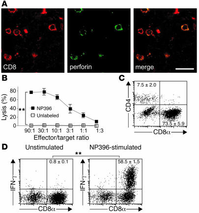
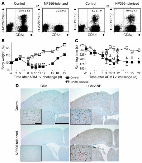
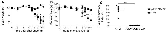
Autoimmune diseases are often precipitated by viral infections. Yet our current understanding fails to explain how viruses trigger organ-specific autoimmunity despite thymic tolerance extending to many non-lymphohematopoietic self antigens. Additionally, a key epidemiological finding needs to be explained: In genetically susceptible individuals, early childhood infections seem to predispose them to multiple sclerosis (MS) or type 1 diabetes years or even decades before clinical onset. In the present work, we show that the innate immune system of neonatal mice was sufficient to eliminate an attenuated lymphocytic choriomeningitis virus (LCMV) from most tissues except for the CNS, where the virus persisted in neurons (predisposing virus). Virus-specific cytotoxic T cells (CTLs) were neither deleted nor sufficiently primed to cause disease, but they were efficiently triggered in adulthood upon WT LCMV infection (precipitating virus). This defined sequence of viral infections caused severe CNS inflammation that was histomorphologically reminiscent of rasmussen encephalitis, a fatal human autoimmune disease. Yet disease in mice was mediated by antiviral CTLs targeting an epitope shared by the precipitating virus and the predisposing virus persisting in neurons (déjà vu). Thus the concept of "viral déjà vu" demonstrates how 2 related but independently encountered viral infections can cause organ-specific immune disease without molecular mimicry of self and without breaking self tolerance.


Figures

Figure 1
Figure 1. Inefficient induction of virus-specific CD8+ T cells in rLCMV/INDG carriers.
(A) Neonatal and adult C57BL/6 mice were infected with rLCMV/INDG i.c. or were left uninfected. On days 7 and 50, NP396-specific CD8+ T cells in the spleen (day 7) and blood (day 50) were enumerated by flow cytometry using MHC class I tetramers (H-2DbNP396). Numbers indicate percentages of NP396-specific CD8+ cells within the CD8+ T cell compartment (mean ± SD of 3–4 mice). (B) Neonatal (rectangles) or adult (triangles) C57BL/6 mice were infected with rLCMV/INDG i.c. or were left uninfected (circles). Mice were sacrificed 7 and 50 days later, and NP396-specific CTL activity was determined after in vitro restimulation of splenocytes in the presence (filled symbols) or absence (open symbols) of rIL-2. Shown are mean ± SEM of 3–4 mice per group. Cultures with rIL-2 were not significantly different from cultures without rIL-2 (not shown). (C) Mice were challenged with ARM i.v. 50 days after infection as described above. The frequency of NP396-specific CD8+ T cells in blood was measured using MHC class I tetramers. Values differing significantly from those of ARM-challenged mice without prior rLCMV/INDG infection (circles) are indicated. (D) rLCMV/INDG carrier mice (50 days old) and naive adult control mice were challenged with ARM i.v. Seven days later mice were sacrificed, and the NP396-specific primary ex vivo CTL activity of splenocytes was measured. Shown are mean ± SEM of 3 mice. Specific lytic activity of naive control splenocytes was < 3% (not shown). Most panels are representative of 2 similar experiments. *P < 0.05; **P < 0.01.
Figure 2
Figure 2. Persistence of rLCMV/INDG is restricted to CNS neurons.
(A) C57BL/6, RAG–/–, and A–/–RAG–/– mice were infected at birth with ARM or rLCMV/INDG i.c. or were left uninfected. At 35–50 days of age, the indicated organs were tested for viral S segment and NP mRNA by Northern hybridization (ethidium bromide staining of 28S rRNA is shown as loading control). An analogous pattern of rLCMV/INDG distribution in the body was observed in various experiments with neonatally infected C57BL/6, RAG–/–, and A–/–RAG–/– mice (8–10 mice per genotype). (B) Brain sections of neonatally rLCMV/INDG infected 5- to 7-week-old C57BL/6 mice were costained for LCMV-NP (red), nuclei (blue), and cell type–specific markers (green) as indicated. Scale bar: 50 μm. Insets show 2.5-fold-higher-magnification views of boxed regions. Representative images show neuron-restricted virus distribution as found in the entire CNS (n = 9 animals from 3 independent experiments with identical results; for overview images see Supplemental Figure 5).
Figure 3
Figure 3. Weight loss and impaired motor coordination in ARM-challenged rLCMV/INDG carriers.
On day –50, neonatal (rectangles) and adult (triangles) C57BL/6 mice were infected with rLCMV/INDG i.c., resulting in rLCMV/INDG carriers and rLCMV/INDG–immune mice, respectively. A control group of adult mice without rLCMV/INDG infection (circles) was also included. All mice were challenged with ARM i.v. on day 0. (A) Body weight was monitored at the indicated time points. (B) Motor coordination was tested on a rotating rod, and the running time was recorded. Shown are the mean ± SEM of 10–11 mice initially; due to terminal disease, individual rLCMV/INDG carriers had to be euthanized on days 8, 11 (3 mice), 17, and 26 after ARM challenge (broken lines). *P < 0.05, **P < 0.01 compared with the respective group’s highest value prior to disease onset.
Figure 4
Figure 4. T cell infiltration in the brains of ARM-challenged rLCMV/INDG carriers.
(A) Neonatal and adult C57BL/6 mice were infected at day –50 with rLCMV/INDG i.c., resulting in rLCMV/INDG carriers and rLCMV/INDG–immune mice, respectively. On day 0, these animals and a control group of adult mice without rLCMV/INDG infection were challenged with ARM i.v. Brain (top and center panels) and spinal cord sections (bottom panel) were prepared 8 days after ARM challenge and stained for LCMV-NP and CD3+ T cells. Representative images of 7–10 mice per group are shown. Scale bars: 500 μm (top); 100 μm (center); 20 μm (bottom). (B) Brain biopsy of a human patient with RE. A representative neuron surrounded by CD8+ T cells (brown) is shown. Scale bar: 20 μm. (C) C57BL/6 mice were infected with rLCMV/INDG as in A and sacrificed either immediately preceding ARM challenge 50 days later (groups I–III) or 8–12 days after ARM challenge (the peak of disease in rLCMV/INDG carriers; groups IV–VI) to determine the brain inflammatory index. Symbols represent individual mice from a total of 3 independent experiments; horizontal lines indicate means. Neonatally rLCMV/INDG-infected mice after ARM i.v. challenge (Group IV) exhibited dense T cell infiltration in brain, whereas the remaining groups were statistically indistinguishable from each other. Analogous results were also obtained for spinal cord (not shown). **P < 0.001 versus all other groups.
Figure 5
Figure 5. Virus-specific effector function of brain-infiltrating CD8+ T cells.
rLCMV/INDG carrier mice (50 days old) were challenged with ARM i.v. At the peak of disease 9 days later, brain tissue was processed for extraction of lymphocytes or for histological analysis of the infiltrate. (A) Brain-infiltrating T cells were stained for CD8 (red) and perforin (green) and were analyzed by confocal microscopy. Images are representative of multiple sections from at least 3 different animals. (B) Primary ex vivo CTL activity of brain-extracted lymphocytes on NP396-pulsed or unlabeled target cells. (C) Brain-infiltrating lymphocytes were analyzed by flow cytometry for surface expression of CD4 and CD8α. The percentage of single-positive cells is indicated (mean ± SD of 3 mice). (D) Brain-extracted lymphocytes were restimulated with NP396 peptide or with medium only (unstimulated), and IFN-γ–producing cells were enumerated by flow cytometry. Shown are percentages of IFN-γ–producing cells among CD8+ T cells (mean ± SD of 3 mice per group). One representative of 2 similar experiments is shown. Of note, the absence of infiltrates in naive mice and in rLCMV/INDG–immune mice challenged with ARM (see Figure 4A) rendered analogous tests for these groups impossible. **P < 0.01.
Figure 6
Figure 6. CNS disease is mediated by a single dominant specificity of antiviral CTL.
rLCMV/INDG carriers (50 days old) were either tolerized to NP396 by peptide treatment (open rectangles) or given diluent only (filled rectangles). On day 0, both groups of mice were challenged with ARM i.v. (A) The percentage of NP396- and GP33-specific CD8+ T cells in blood was measured on day 8 and is shown in each representative flow cytometry plot (mean ± SD of ≥3 mice per group). **P < 0.01. (B and C) The body weight (B) and motor coordination (C) of the same mice as in A were monitored. Shown are the mean ± SEM of initially 10 mice per group; due to severe illness, 2 nontolerized mice had to be euthanized on days 9 and 11 after ARM challenge (broken lines). *P < 0.05, **P < 0.01 compared with highest value prior to disease onset. (D) Separate groups of mice treated as in AC were sacrificed on day 8 after ARM challenge. Brain-infiltrating T cells (CD3) and persistent rLCMV/INDG (LCMV-NP) were assessed by histological analysis. Images are representative of histological sections of 3 mice per group. One representative of 2 similar experiments is shown. Scale bars: 1 mm; 100 μm (insets).
Figure 7
Figure 7. Absence of inflammatory CNS disease in rLCMV/INDG carriers challenged with rVSV/LCMV-GP instead of ARM.
rLCMV/INDG carriers (50 days old) were challenged either with ARM or with rVSV/LCMV-GP i.v. (A and B) Body weight (A) and running time on the rotating rod (B) were monitored at the indicated time points after ARM challenge. Shown are the mean ± SEM of 5 mice per group. *P < 0.05 compared with highest value prior to disease onset. (C) At the peak of disease in ARM-challenged animals (day 11), all mice were sacrificed, and the brain inflammatory index was determined. Symbols represent individual mice; horizontal lines indicate means. **P < 0.01.
Figure 8
Figure 8. Schematic interpretation of the viral déjà vu immunopathogenetic mechanism.
(A) Summary of the infection protocol eliciting CNS disease; viral distribution is denoted by color. (B) Mechanistic interpretation. In neonates and in adults, a substantial fraction of the rLCMV/INDG i.c. inoculum reaches the systemic circulation (38). Viral infection of secondary lymphoid organs is abortive due to efficient type I IFN–dependent control, whereas CNS infection is productive. In neonates but not in adults, viral gene expression in secondary lymphoid organs coincides with reduced T cell responsiveness. Hence adult mice mount a protective virus-specific (NP396 and other epitopes) CD8+ T cell response that clears rLCMV/INDG from the CNS, while virus persists in the neonate’s neurons. The antiviral CD8+ T cell response in neonates is inefficient, but ARM infection 50 days later triggers a vigorous response of NP396-specific CTLs (epitope shared by rLCMV/INDG and ARM) that causes CNS disease when reaching persistently rLCMV/INDG-infected neurons. Adult primary rLCMV/INDG infection has been cleared from the CNS, and hence only neonatally rLCMV/INDG-infected mice develop disease.

Similar articles

Cited by

References

    1. Marie P. Sclerose en plaque et maladie infecteuses. Prog. Med. 1884;12:287–289.
    1. Kurtzke J.F. Epidemiologic evidence for multiple sclerosis as an infection. Clin. Microbiol. Rev. 1993;6:382–427. - PMC - PubMed
    1. Ebers G.C., et al. A population-based study of multiple sclerosis in twins. N. Engl. J. Med. 1986;315:1638–1642. - PubMed
    1. Dyment D.A., Ebers G.C., Sadovnick A.D. Genetics of multiple sclerosis. Lancet Neurol. 2004;3:104–110. - PubMed
    1. Sibley W.A., Bamford C.R., Clark K. Clinical viral infections and multiple sclerosis. Lancet. 1985;1:1313–1315. - PMC - PubMed

Publication types