

Control of electrical alternans in canine cardiac purkinje fibers
- PMID: 16605736
- PMCID: PMC1566349
- DOI: 10.1103/PhysRevLett.96.104101
Control of electrical alternans in canine cardiac purkinje fibers
Abstract
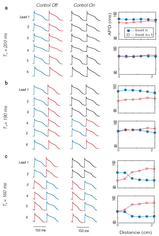
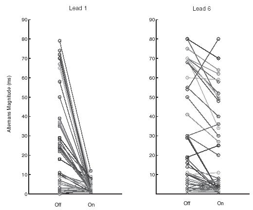
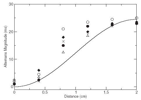
Alternation in the duration of consecutive cardiac action potentials (electrical alternans) may precipitate conduction block and the onset of arrhythmias. Consequently, suppression of alternans using properly timed premature stimuli may be antiarrhythmic. To determine the extent to which alternans control can be achieved in cardiac tissue, isolated canine Purkinje fibers were paced from one end using a feedback control method. Spatially uniform control of alternans was possible when alternans amplitude was small. However, control became attenuated spatially as alternans amplitude increased. The amplitude variation along the cable was well described by a theoretically expected standing wave profile that corresponds to the first quantized mode of the one-dimensional Helmholtz equation. These results confirm the wavelike nature of alternans and may have important implications for their control using electrical stimuli.
Figures
Similar articles
-
Spatiotemporal transition to conduction block in canine ventricle.Circ Res. 2002 Feb 22;90(3):289-96. doi: 10.1161/hh0302.104723. Circ Res. 2002. PMID: 11861417
-
Control of action potential duration alternans in canine ventricular tissue.Annu Int Conf IEEE Eng Med Biol Soc. 2010;2010:1997-2000. doi: 10.1109/IEMBS.2010.5627828. Annu Int Conf IEEE Eng Med Biol Soc. 2010. PMID: 21097010
-
Off-site control of repolarization alternans in cardiac fibers.Phys Rev E Stat Nonlin Soft Matter Phys. 2010 Jan;81(1 Pt 1):011915. doi: 10.1103/PhysRevE.81.011915. Epub 2010 Jan 25. Phys Rev E Stat Nonlin Soft Matter Phys. 2010. PMID: 20365407 Free PMC article.
-
Clinical utility of T-wave alternans.Card Electrophysiol Rev. 1997;1(3):390-4. doi: 10.1023/a:1009902030340. Card Electrophysiol Rev. 1997. PMID: 11541510 Review.
-
Cardiac alternans: diverse mechanisms and clinical manifestations.J Am Coll Cardiol. 1992 Aug;20(2):483-99. doi: 10.1016/0735-1097(92)90122-4. J Am Coll Cardiol. 1992. PMID: 1634690 Review.
Cited by
-
Continuous-time control of alternans in long Purkinje fibers.Chaos. 2014 Sep;24(3):033124. doi: 10.1063/1.4893295. Chaos. 2014. PMID: 25273204 Free PMC article.
-
Key aspects for effective mathematical modelling of fractional-diffusion in cardiac electrophysiology: a quantitative study.Commun Nonlinear Sci Numer Simul. 2020 May;84:105152. doi: 10.1016/j.cnsns.2019.105152. Epub 2019 Dec 25. Commun Nonlinear Sci Numer Simul. 2020. PMID: 32863678 Free PMC article.
-
A model for multi-site pacing of fibrillation using nonlinear dynamics feedback.J Biol Phys. 2007 Apr;33(2):145-53. doi: 10.1007/s10867-007-9049-9. Epub 2007 Dec 7. J Biol Phys. 2007. PMID: 19669546 Free PMC article.
-
Ephaptic Coupling as a Resolution to the Paradox of Action Potential Wave Speed and Discordant Alternans Spatial Scales in the Heart.Phys Rev Lett. 2023 May 26;130(21):218401. doi: 10.1103/PhysRevLett.130.218401. Phys Rev Lett. 2023. PMID: 37295103 Free PMC article.
-
Mechanisms of ventricular arrhythmias: a dynamical systems-based perspective.Am J Physiol Heart Circ Physiol. 2012 Jun 15;302(12):H2451-63. doi: 10.1152/ajpheart.00770.2011. Epub 2012 Mar 30. Am J Physiol Heart Circ Physiol. 2012. PMID: 22467299 Free PMC article. Review.
References
-
- Nolasco JB, Dahlen RW. Journal of Applied Physiology. 1968;25:191. - PubMed
-
- Tachibana H, Kubota I, Yamaki M, Watanabe T, Tomoike H. Am J Physiol. 1998;275:H116. - PubMed
-
- Pastore JM, Girouard SD, Laurita KR, Akar FG, Rosenbaum DS. Circulation. 1999;99:1385. - PubMed
-
- Pastore JM, Rosenbaum DS. Circ Res. 2000;87:1157. - PubMed
-
- Fox JJ, Riccio ML, Hua F, Bodenschatz E, Gilmour RF., Jr Circ Res. 2002;90:289. - PubMed
Publication types
MeSH terms
Grants and funding
LinkOut - more resources
Full Text Sources
Medical