. 2006 Aug 1;397(3):417-25.
doi: 10.1042/BJ20060125.

Role of pyruvate dehydrogenase kinase isoenzyme 4 (PDHK4) in glucose homoeostasis during starvation

Affiliations

Role of pyruvate dehydrogenase kinase isoenzyme 4 (PDHK4) in glucose homoeostasis during starvation

Nam Ho Jeoung et al. Biochem J. .

Abstract

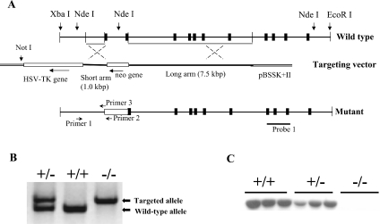
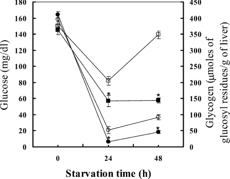
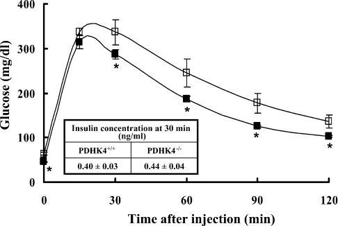
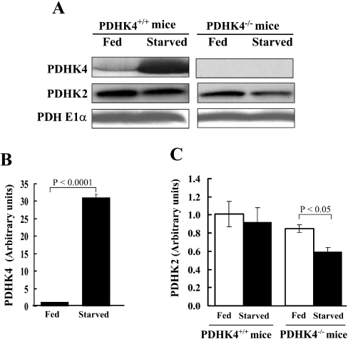
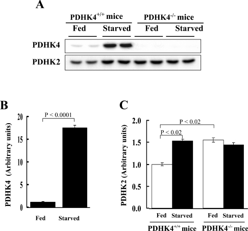
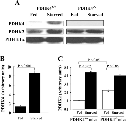
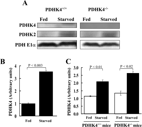
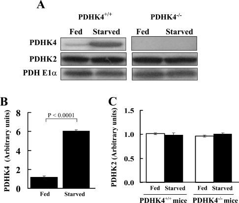
The PDC (pyruvate dehydrogenase complex) is strongly inhibited by phosphorylation during starvation to conserve substrates for gluconeogenesis. The role of PDHK4 (pyruvate dehydrogenase kinase isoenzyme 4) in regulation of PDC by this mechanism was investigated with PDHK4-/- mice (homozygous PDHK4 knockout mice). Starvation lowers blood glucose more in mice lacking PDHK4 than in wild-type mice. The activity state of PDC (percentage dephosphorylated and active) is greater in kidney, gastrocnemius muscle, diaphragm and heart but not in the liver of starved PDHK4-/- mice. Intermediates of the gluconeogenic pathway are lower in concentration in the liver of starved PDHK4-/- mice, consistent with a lower rate of gluconeogenesis due to a substrate supply limitation. The concentration of gluconeogenic substrates is lower in the blood of starved PDHK4-/- mice, consistent with reduced formation in peripheral tissues. Isolated diaphragms from starved PDHK4-/- mice accumulate less lactate and pyruvate because of a faster rate of pyruvate oxidation and a reduced rate of glycolysis. BCAAs (branched chain amino acids) are higher in the blood in starved PDHK4-/- mice, consistent with lower blood alanine levels and the importance of BCAAs as a source of amino groups for alanine formation. Non-esterified fatty acids are also elevated more in the blood of starved PDHK4-/- mice, consistent with lower rates of fatty acid oxidation due to increased rates of glucose and pyruvate oxidation due to greater PDC activity. Up-regulation of PDHK4 in tissues other than the liver is clearly important during starvation for regulation of PDC activity and glucose homoeostasis.


Figures

Figure 1
Figure 1. Targeting strategy for disruption of the mouse PDHK4 gene
(A) A 1.0 kb DNA fragment of the mouse PDHK4 gene promoter was used as the short arm; a 7.5 kb DNA fragment extending from exon 2 to intron 7 as the long arm. The targeting vector was linearized at a NotI site for electroporation into embryonic stem cells. After homologous recombination, the endogenous PDHK4 gene was disrupted by the neomycin gene of the targeting vector (labelled ‘Mutant’ in the diagram). Probe 1 was used to screen targeted embryonic stem cells by Southern-blot analysis. (B) Mice were genotyped for disruption of the PDHK4 gene by PCR with primers P1, P2 and P3. Template DNA was purified from toe clips. (C) Western-blot analysis was carried out with muscle extracts obtained from 24 h fasted mice [wild-type (+/+), heterozygous (+/–) and homozygous (–/–)]. Protein (50 μg) was separated by SDS/PAGE (12.5% gel) and then transferred on to a nitrocellulose membrane. Western-blot analysis was conducted with a rabbit antiserum for PDHK4.
Figure 2
Figure 2. Effect of starvation on blood glucose and hepatic glycogen in wild-type and PDHK4−/− mice
Blood glucose for wild-type (□) and PDHK4−/− mice (■), with n=14 and 12 in the groups respectively. Liver glycogen for wild-type (○) and PDHK4−/− (●) mice, with n=6 for each group. All data points are means±S.E.M. *Significantly different between wild-type and PDHK4−/− mice (P<0.01).
Figure 3
Figure 3. Glucose tolerance test in wild-type (□) and PDHK4−/− mice (■)
Glucose (2 g/kg of body mass) was administrated to overnight-fasted mice by intraperitoneal injection. All data points are means±S.E.M. with n=12 in each group. Insulin values, measured 30 min after glucose injection, are means±S.E.M. with n=6 in each group. *Significantly different between wild-type (PDHK4+/+) and PDHK4−/− mice (P<0.01).
Figure 4
Figure 4. Effect of starvation on PDHK protein expression in skeletal muscle of wild-type and PDHK4−/− mice
(A) Representative Western blots of PDHK4 and PDHK2 protein in gastrocnemius muscles of fed and 48 h starved wild-type (PDHK4+/+) and PDHK4−/− mice. (B) Histograms constructed from data obtained by Western-blot analysis, showing relative amounts of PDHK4 protein expressed in gastrocnemius muscle of fed and 48 h starved wild-type (PDHK4+/+) mice. n=4 in each group. (C) Histograms constructed from data obtained by Western-blot analysis, showing relative amounts of PDHK2 protein expressed in gastrocnemius muscle of fed and starved wild-type (PDHK4+/+) and PDHK4−/− mice. n=4 in each group.
Figure 5
Figure 5. Effect of starvation on PDHK protein expression in heart of wild-type and PDHK4−/− mice
(A) Representative Western blots of PDHK4 and PDHK2 protein in heart of fed and 48 h starved wild-type (PDHK4+/+) and PDHK4−/− mice. (B) Histograms constructed from data obtained by Western-blot analysis, showing relative amounts of PDHK4 protein expressed in heart of fed and 48 h starved wild-type (PDHK4+/+) mice. n=4 in each group. (C) Histograms constructed from data obtained by Western-blot analysis showing relative amounts of PDHK2 protein expressed in heart of fed and starved wild-type (PDHK4+/+) and PDHK4−/− mice. n=4 in each group.
Figure 6
Figure 6. Effect of starvation on PDHK protein expression in kidney of wild-type mice and PDHK4−/− mice
(A) Representative Western blots of PDHK4 and PDHK2 protein in kidney of fed and 48 h starved wild-type (PDHK4+/+) and PDHK4−/− mice. (B) Histograms constructed from data obtained by Western-blot analysis, showing relative amounts of PDHK4 protein expressed in kidney of fed and 48 h starved wild-type (PDHK4+/+) mice. n=4 in each group. (C) Histograms constructed from data obtained by Western-blot analysis, showing relative amounts of PDHK2 protein expressed in kidney of fed and starved wild-type (PDHK4+/+) and PDHK4−/− mice. n=4 in each group.
Figure 7
Figure 7. Effect of starvation on PDHK protein expression in liver of wild-type and PDHK4−/− mice
(A) Representative Western blots of PDHK4 and PDHK2 protein in liver of fed and 48 h starved wild-type (PDHK4+/+) and PDHK4−/− mice. (B) Histograms constructed from data obtained by Western-blot analysis, showing relative amounts of PDHK4 protein expressed in liver of fed and 48 h starved wild-type (PDHK4+/+) mice. n=4 in each group. (C) Histograms constructed from data obtained by Western-blot analysis, showing relative amounts of PDHK2 protein expressed in liver of fed and starved wild-type (PDHK4+/+) and PDHK4−/− mice. n=4 in each group.
Figure 8
Figure 8. Effect of starvation on PDHK protein expression in diaphragm of wild-type and PDHK4−/− mice
(A) Representative Western blots of PDHK4 and PDHK2 protein in diaphragm of fed and 48 h starved wild-type (PDHK4+/+) and PDHK4−/− mice. (B) Histograms constructed from data obtained by Western-blot analysis, showing relative amounts of PDHK4 protein expressed in diaphragm of fed and 48 h starved wild-type (PDHK4+/+) mice. n=4 in each group. (C) Histograms constructed from data obtained by Western-blot analysis, showing relative amounts of PDHK2 protein expressed in diaphragm of fed and starved wild-type (PDHK4+/+) and PDHK4−/− mice. n=4 in each group.

References

    1. Linn T. C., Pettit F. H., Reed L. J. α-Keto acid dehydrogenase complex, X: regulation of the activity of the pyruvate dehydrogenase complex from beef kidney mitochondria by phosphorylation and dephosphorylation. Proc. Natl. Acad. Sci. U.S.A. 1969;62:234–241. - PMC - PubMed
    1. Yeaman S. J. The 2-oxo acid dehydrogenase complexes: recent advances. Biochem. J. 1989;257:625–632. - PMC - PubMed
    1. Holness M. J., Sugden M. C. Regulation of pyruvate dehydrogenase complex activity by reversible phosphorylation. Biochem. Soc. Trans. 2003;31:1143–1151. - PubMed
    1. Roche T. E., Baker J. C., Yan X., Hiromasa Y., Gong X., Peng T., Dong J., Turkan A., Kasten S. A. Distinct regulatory properties of pyruvate dehydrogenase kinase and phosphatase isoforms. Prog. Nucleic Acid Res. Mol. Biol. 2001;70:33–75. - PubMed
    1. Behal R. H., Buxton D. B., Robertson J. G., Olson M. S. Regulation of the pyruvate dehydrogenase multienzyme complex. Annu. Rev. Nutr. 1993;13:497–520. - PubMed

Publication types

MeSH terms