. 2006;7(4):R28.
doi: 10.1186/gb-2006-7-4-r28. Epub 2006 Apr 7.

Genes regulated by estrogen in breast tumor cells in vitro are similarly regulated in vivo in tumor xenografts and human breast tumors

Affiliations

Genes regulated by estrogen in breast tumor cells in vitro are similarly regulated in vivo in tumor xenografts and human breast tumors

Chad J Creighton et al. Genome Biol. 2006.

Abstract

Background: Estrogen plays a central role in breast cancer pathogenesis. Although many studies have characterized the estrogen regulation of genes using in vitro cell culture models by global mRNA expression profiling, it is not clear whether these genes are similarly regulated in vivo or how they might be coordinately expressed in primary human tumors.

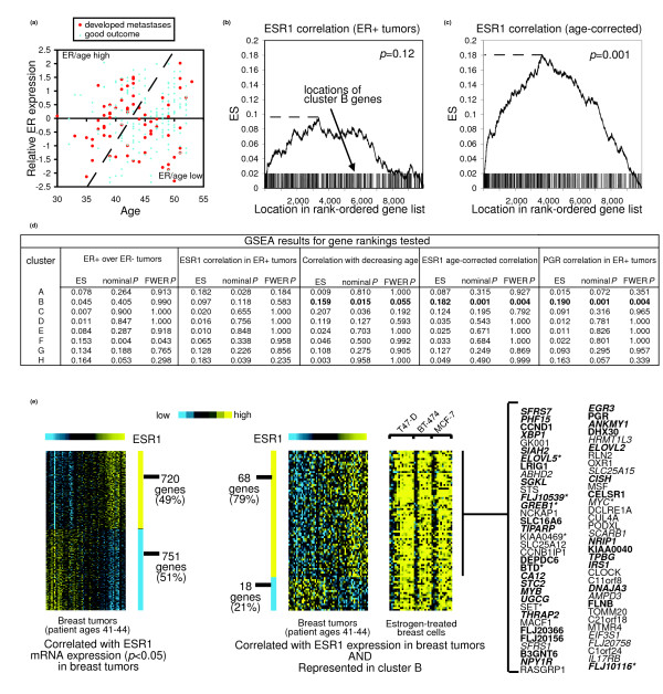
Results: We generated DNA microarray-based gene expression profiles from three estrogen receptor alpha (ERalpha)-positive breast cancer cell lines stimulated by 17beta-estradiol (E2) in vitro over a time course, as well as from MCF-7 cells grown as xenografts in ovariectomized athymic nude mice with E2 supplementation and after its withdrawal. When the patterns of genes regulated by E2 in vitro were compared to those obtained from xenografts, we found a remarkable overlap (over 40%) of genes regulated by E2 in both contexts. These patterns were compared to those obtained from published clinical data sets. We show that, as a group, E2-regulated genes from our preclinical models were co-expressed with ERalpha in a panel of ERalpha+ breast tumor mRNA profiles, when corrections were made for patient age, as well as with progesterone receptor. Furthermore, the E2-regulated genes were significantly enriched for transcriptional targets of the myc oncogene and were found to be coordinately expressed with Myc in human tumors.

Conclusion: Our results provide significant validation of a widely used in vitro model of estrogen signaling as being pathologically relevant to breast cancers in vivo.


Figures

Figure 1
Figure 1
Gene expression signatures of estrogen regulation in vitro and in vivo. (a) Expression data matrix of genes showing either induction or repression by E2 in vitro (p < 0.01 at 4, 8, 12, or 24 hour time points). Each row represents a gene; each column represents a sample. The level of expression of each gene in each sample is represented using a yellow-blue color scale (yellow, high expression); gray indicates missing data. Shown alongside our in vitro time course dataset are the expression values for the corresponding genes in two independent mRNA profile datasets of E2-treated breast cells ('Rae' dataset from [5] of cell lines MCF-7, BT-474, and T47-D; 'Finlin' dataset from [11] of MCF-7). (b) Alongside the in vitro datasets are the corresponding values for MCF-7 xenografts with or without E2 supplementation (E2 withdrawn for 24 hours and 48 hours in the -E2 group). Genes in cluster 'B' (a) that show significant up-regulation by E2 in each of the four datasets are listed (an asterisk indicates positively correlated (p < 0.05) with age-correlation ERα expression (Figure 2); bold type indicates having higher expression in ERα+ compared to ERα- breast tumors (p < 0.01) according to the 'van't Veer' dataset from [15]); italics indicates having higher expression in ERα- compared to ERα+ breast tumors). (c) Genes MYB (c-myb) and MYBL1 (A-MYB) are regulated by E2 in vivo. Expression patterns for genes from (b) were validated by RT-PCR. Shown are the mean and standard deviation of individual samples assayed in triplicate. Tumor volumes (expressed in mm3) are shown above each bar.
Figure 2
Figure 2
A significant number of genes induced by E2 in vitro are correlated with age-corrected mRNA expression of ERα in human breast tumors. (a) Scatter plot of ERα expression versus age in ERα+ breast tumor samples from the profile data from [14]. The dotted line is used to stratify the samples into ER/age high (above the line) and ER/age low (below the line) groups. Figure adapted from [18]. (b) GSEA of the cluster B genes (induced early by E2 and sustained over time; Figure 1a) against the overall ranking of genes according to similarity with ERα mRNA (ESR1) expression in the cohort of ERα+ breast tumor profiles from [14]. The ES statistic (the maximum of the ES running sum) is high if many genes in the set of interest appear near the top of the ranked list. Vertical bars along the ES plot denote occurrences of a cluster B gene. (c) GSEA of the cluster B genes against gene ranking by similarity with ESR1 expression as corrected for patient age (using dotted line in (a)). (d) GSEA results for estrogen-regulated gene clusters A to H against four different gene rankings tested. The FWER p-value corrects for multiple gene set testing. (e) Enrichment of cluster B genes within the set of genes showing positive correlation (p < 0.05) with ESR1 expression in a set of tumor profiles from patients within a narrow age range of 41 to 44 years. In the gene list: an asterisk indicates negatively correlated (p < 0.05) with age in ERα+ tumors; bold type indicates having higher expression in ERα+ compared to ERα- breast tumors (p < 0.01); and italics indicate induced by E2 in breast tumor xenografts (p < 0.05; Figure 1b).
Figure 3
Figure 3
E2-inducible genes are enriched in ER+/PR+ breast tumors compared to ER+/PR- breast tumors. Expression patterns for the cluster B genes (induced by E2 in vitro within 4 hours; Figure 1a) are shown alongside the corresponding patterns in MCF-7 xenografts (Figure 1b) and in two independent breast tumor datasets from van de Vijver et al. [14] and Wang et al. [20]. Genes showing significant correlation with PGR in both van de Vijver and Wang datasets (p < 0.05 in each) are highlighted and listed: italics indicate more highly expressed in ER+ compared to ER- tumors (p < 0.01); bold type indicates significantly correlated with age-corrected ER expression (p < 0.05; see Figure 2). Enrichment p value by one-sided Fisher's exact test.
Figure 4
Figure 4
Genes induced by E2 in vitro are enriched for transcriptional targets of Myc. A conditional Myc-estrogen receptor (Myc-ER) fusion protein was used to induce Myc transcriptional activity. Conditional activation of Myc occurs upon stimulation with the anti-estrogen 4-hydroxytamoxifen (OHT). Gene expression profiles were taken of primary human fibroblasts with Myc-ER to identify transcriptional targets of Myc [22] (CHX, cycloheximide). (a) Enrichment of Myc targets (p < 0.01) within each of the distinct clusters of estrogen-regulated genes (Figure 1). Values indicating significance of overlap (p < 0.05) between two given genes sets are in bold. (b) Expression data matrix for cluster B and C genes that were also represented in the Myc-ER dataset, alongside the corresponding values in both the Myc-ER dataset and in a profile dataset from [26] of human fibroblasts grown in high and low serum conditions, in which Myc is up-regulated in the high serum group. Myc targets (p < 0.01) are listed (asterisk indicates has predicted Myc TF site in promoter region; italics indicate cell cycle gene from [13]). (c) GSEA of the cluster B genes against the overall ranking of genes according to similarity with myc mRNA expression in a compendium of tumor profiles from breast and 10 other tissues types from [24]. (d) GSEA results for estrogen-regulated gene clusters A to H, genes induced by Myc-ER+OHT in fibroblasts (p < 0.01), and cell cycle genes [13], against gene rankings by Myc correlation in four different datasets (clusters A to H, FWER p, Myc and cell cycle, nominal p).

References

    1. Hall JM, Couse JF, Korach KS. The multifaceted mechanisms of estradiol and estrogen receptor signaling. J Biol Chem. 2001;276:36869–36872. doi: 10.1074/jbc.R100029200. - DOI - PubMed
    1. Elledge RM, Fuqua SA. Estrogen and progesterone receptors. In: Harris J, Lippman ME, Morrow M, Osborne C, editor. Diseases of the Breast. Philadelphia: Lippincott, Williams and Wilkins; 2000. pp. 471–488.
    1. Clemons M, Goss P. Estrogen and the risk of breast cancer. N Engl J Med. 2001;344:276–285. doi: 10.1056/NEJM200101253440407. - DOI - PubMed
    1. Jordan VC. Tamoxifen: a most unlikely pioneering medicine. Nat Rev Drug Discov. 2003;2:205–213. doi: 10.1038/nrd1031. - DOI - PubMed
    1. Rae JM, Johnson MD, Scheys JO, Cordero KE, Larios JM, Lippman ME. GREB 1 is a critical regulator of hormone dependent breast cancer growth. Breast Cancer Res Treat. 2005;92:141–149. doi: 10.1007/s10549-005-1483-4. - DOI - PubMed

Publication types

MeSH terms