Comparative Study
. 2006 Apr 17;203(4):883-95.
doi: 10.1084/jem.20051776. Epub 2006 Apr 10.

Tissue expression of PD-L1 mediates peripheral T cell tolerance

Affiliations
Comparative Study

Tissue expression of PD-L1 mediates peripheral T cell tolerance

Mary E Keir et al. J Exp Med. .

Abstract

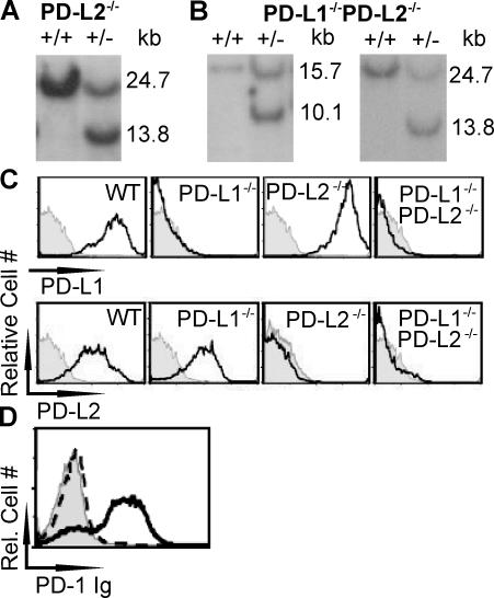
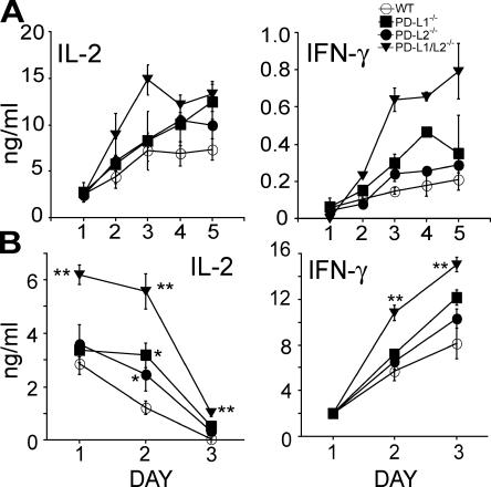
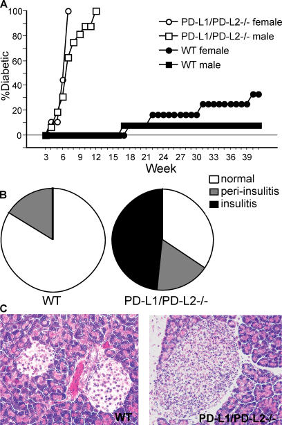
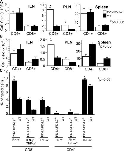
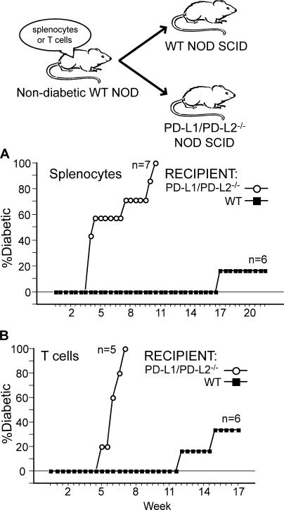
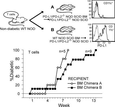
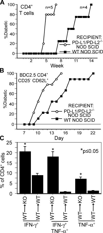
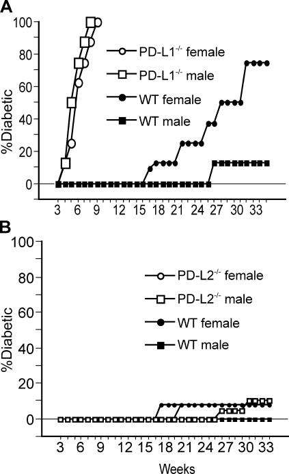
Programmed death 1 (PD-1), an inhibitory receptor expressed on activated lymphocytes, regulates tolerance and autoimmunity. PD-1 has two ligands: PD-1 ligand 1 (PD-L1), which is expressed broadly on hematopoietic and parenchymal cells, including pancreatic islet cells; and PD-L2, which is restricted to macrophages and dendritic cells. To investigate whether PD-L1 and PD-L2 have synergistic or unique roles in regulating T cell activation and tolerance, we generated mice lacking PD-L1 and PD-L2 (PD-L1/PD-L2(-/-) mice) and compared them to mice lacking either PD-L. PD-L1 and PD-L2 have overlapping functions in inhibiting interleukin-2 and interferon-gamma production during T cell activation. However, PD-L1 has a unique and critical role in controlling self-reactive T cells in the pancreas. Our studies with bone marrow chimeras demonstrate that PD-L1/PD-L2 expression only on antigen-presenting cells is insufficient to prevent the early onset diabetes that develops in PD-L1/PD-L2(-/-) non-obese diabetic mice. PD-L1 expression in islets protects against immunopathology after transplantation of syngeneic islets into diabetic recipients. PD-L1 inhibits pathogenic self-reactive CD4+ T cell-mediated tissue destruction and effector cytokine production. These data provide evidence that PD-L1 expression on parenchymal cells rather than hematopoietic cells protects against autoimmune diabetes and point to a novel role for PD-1-PD-L1 interactions in mediating tissue tolerance.


Figures

Figure 1.
Figure 1.
Generation of PD-L2−/− and PD-L1/PD-L2−/− mice. (A and B) Southern blot analyses performed on WT and PD-L2+/− ES cells (A) and PD-L1+/−PD-L2+/− ES cells (B) are shown. For the PD-L1 locus, the 15.7-kb fragment represents the WT allele, and the 10.1-kb fragment represents the disrupted PD-L1 allele. For the PD-L2 locus, the 24.7-kb fragment represents the WT allele, and the 13.8-kb fragment represents the disrupted allele. (C) WT, PD-L1−/−, PD-L2−/−, and PD-L1/L2−/− BM-DCs were generated and evaluated for PD-L1 and PD-L2 expression (shown as a solid line; IgG isotype control, shaded histogram). (D) PD-1 Ig does not bind to PD-L1/L2−/− DCs. WT (solid line) and PD-L1/L2−/− (dashed line) BM-DCs were stained with mouse PD-1 Ig or control Ig, both with a human IgG1 Fc region, followed by anti–human IgG-PE. The Ig control is shown as a shaded histogram.
Figure 2.
Figure 2.
PD-L1 and PD-L2 have overlapping roles in limiting IL-2 and IFN-γ production during naive CD4+ T cell activation. (A) Naive DO11 T cells were activated in vitro by WT, PD-L1−/−, PD-L2−/−, and PD-L1/L2−/− BM-DCs in the presence of 0–1 μg/ml OVA323–339. To evaluate cytokine production, DO11 T cells were activated with 1 μg/ml OVA323–339 for 5 d, and supernatants were collected every 24 h and assayed by ELISA. (B) For in vivo studies, DO11 T cells were purified and adoptively transferred into naive WT, PD-L1−/−, PD-L2−/−, or PD-L1/L2−/− hosts and, 24 h later, were immunized with 100 μg OVA emulsified in IFA. 5 d after immunization, draining LNs were harvested and stained for KJ126 using the clonotypic mAb and CD4, and 105 DO11 cells were restimulated as a whole LN culture with 1 μg/ml OVA323–339. Supernatants were collected every 24 h and assayed for IL-2 and IFN-γ. *, P < 0.05; **, P < 0.005 by the unpaired t test. Data shown are representative of at least three experiments. Error bars represent SEM.
Figure 3.
Figure 3.
Loss of PD-L1 and PD-L2 results in the rapid onset of diabetes and the early onset of insulitis. (A) PD-L1/PD-L2−/− N4 mice on the autoimmune diabetes-prone NOD background were monitored for the development of hyperglycemia. Animals were scored as diabetic upon blood glucose readings of ≥250 mg/dL, and diabetes was confirmed by three consecutive positive reads. Both female PD-L1/PD-L2−/− NOD mice (n = 9) and male PD-L1/PD-L2−/− NOD mice (n = 16) develop autoimmune diabetes significantly (P < 0.0001; unpaired t test) faster than WT littermate controls (females, n = 12; males, n = 13). (B) PD-L1/PD-L2−/− and WT N5 NOD littermates were analyzed for early and late signs of immune cell infiltration of the pancreatic islets by histology. Pancreata from PD-L1/PD-L2−/− and WT NOD littermates ranging in age from 27 to 50 d were evaluated histologically. Islets were scored as normal, peri-insulitic, or insulitic, and the percentage of total islets in each category was averaged for each group. Three of nine PD-L1/PD-L2−/− mice were hyperglycemic, and all 10 WT animals were euglycemic at the time of killing. (C) Representative islets from WT (left; scored as normal) and PD-L1/PD-L2−/− (right; scored as insulitic) pancreas are shown. Islets pictured are from 50-d-old littermate females with normal blood glucose readings immediately before killing.
Figure 4.
Figure 4.
Increased T cell numbers and effector function in the PLN of PD-L1/PD-L2−/− NOD mice. Lymphocytes from the LNs or spleen of prediabetic and diabetic PD-L1/PD-L2−/− NOD mice and WT littermates were evaluated by flow cytometry and by restimulation in vitro. (A) Inguinal LNs (ILN) and pancreatic LNs (PLN), spleen, and pancreas were taken from prediabetic NOD N7 mice (typically between 3–5 wk of age and confirmed to be nondiabetic by blood glucose of ≤250 mg/dL). Lymphocyte subset numbers were calculated by multiplying the total cell yield from an organ by the percentage of CD4+ or CD8+ cells by flow cytometry. (B) Lymphoid organs from diabetic mice (typically between 5–8 wk of age for N4 PD-L1/PD-L2−/− and >13 wk for N4 WT; confirmed to be diabetic by a blood glucose reading of ≥250 mg/dL) were analyzed as in A. (C) The percentage of effector cytokine–producing CD4+ and CD8+ T cells in the PLN is increased in young prediabetic PD-L1/PD-L2−/− N7 mice (n = 3) in comparison with WT controls (n = 4; P ≤ 0.03 by the unpaired t test). Error bars represent SEM. Data are representative of three independent experiments.
Figure 5.
Figure 5.
PD-L1/PD-L2 expression is required on nonlymphoid cells to inhibit the transfer of autoimmune diabetes. Cells isolated from nondiabetic WT NOD N5 mice were adoptively transferred into NOD SCID N5 mice that either expressed or lacked PD-L1 and PD-L2. (A) 20 × 106 WT splenocytes efficiently induce autoimmune diabetes in PD-L1/PD-L2−/− NOD SCID mice, whereas disease induction in WT NOD SCID mice is significantly delayed (P < 0.0001 by unpaired t test). (B) 10 × 106 WT T cells were transferred i.v. into PD-L1/PD-L2−/− or WT NOD SCID mice. PD-L1/PD-L2−/− NOD SCID animals had a significantly earlier onset of T cell– mediated autoimmune diabetes (P = 0.0006 by unpaired t test).
Figure 6.
Figure 6.
PD-L1/PD-L2 expression solely on nonlymphoid hematopoietic cells is not sufficient to prevent the rapid transfer of autoimmune diabetes. PD-L1/PD-L2−/− NOD SCID N5 mice were lethally irradiated and reconstituted with BM from WT NOD SCID (chimera B) or PD-L1/PD-L2−/− NOD SCID (chimera A) N5 animals. The resulting BM chimera B had no PD-L1/PD-L2 expression except on nonlymphoid hematopoietic cells such as CD11c+CD3B220 cells (inset; dotted line, isotype staining). WT NOD N5 T cells were adoptively trans- ferred into BM chimeras A and B, which were monitored twice weekly for hyperglycemia. WT NOD T cells transferred autoimmune diabetes to BM chimeras A and B with similar kinetics (P = 0.38 by the unpaired t test).
Figure 7.
Figure 7.
Diabetogenic T cells are inhibited by the expression of PD-L1/PD-L2 in recipient mice. (A) 5 × 10⁶ CD4+ T cells from diabetic WT NOD N5 mice were adoptively transferred into PD-L1/PD-L2−/− or WT NOD SCID N5 mice. The onset of diabetes was significantly delayed in WT NOD SCID mice (P = 0.005 by unpaired t test). (B) Sorted naive effector CD4+CD25CD62L+ cells from BDC2.5 TCR tg+ NOD mice (2 × 10⁵ cells) were adoptively transferred into PD-L1/PD-L2−/− or WT NOD SCID N5 mice. PD-L1/PD-L2−/− NOD SCID recipients had significantly earlier diabetes onset (P = 0.02 by unpaired t test). (C) BDC2.5 TCR tg+ lymphocytes were reisolated from PD-L1/PD-L2−/− (n = 4) or WT NOD SCID (n = 4) recipients 7 d after adoptive transfer and restimulated in vitro. CD4+ T cells were analyzed for the production of TNF-α and IFN-γ. Production of both effector cytokines was significantly increased in CD4+ cells reisolated from PD-L1/PD-L2−/− NOD SCID recipients (P ≤ 0.05 by the unpaired t test). The data are representative of three independent experiments. Error bars represent SEM. KO, knockout.
Figure 8.
Figure 8.
Loss of PD-L1 but not PD-L2 precipitates the rapid onset of autoimmune diabetes on the NOD background. PD-L1−/− and PD-L2−/− mice were crossed separately onto the autoimmune diabetes–prone NOD background. Animals were scored as diabetic as in Fig. 1. (A) PD-L1−/− NOD mice (equivalent of N4) develop a rapid onset diabetes in comparison with WT littermate controls. Both female and male PD-L1−/− NOD display a rapid onset of diabetes with complete penetrance. (B) PD-L2−/− NOD mice (equivalent of N4) do not develop a rapid onset of diabetes in comparison with WT littermate controls.

Comment in

  • New battlefields for costimulation.
    Martin-Orozco N, Dong C. Martin-Orozco N, et al. J Exp Med. 2006 Apr 17;203(4):817-20. doi: 10.1084/jem.20060219. Epub 2006 Apr 10. J Exp Med. 2006. PMID: 16606678 Free PMC article. Review.

Similar articles

Cited by

References

    1. Anderson, M.S., and J.A. Bluestone. 2005. The NOD mouse: a model of immune dysregulation. Annu. Rev. Immunol. 23:447–485. - PubMed
    1. Keir, M.E., and A.H. Sharpe. 2005. The B7/CD28 costimulatory family in autoimmunity. Immunol. Rev. 204:128–143. - PubMed
    1. Chen, L. 2004. Co-inhibitory molecules of the B7-CD28 family in the control of T-cell immunity. Nat. Rev. Immunol. 4:336–347. - PubMed
    1. Greenwald, R.J., Y.E. Latchman, and A.H. Sharpe. 2002. Negative co-receptors on lymphocytes. Curr. Opin. Immunol. 14:391–396. - PubMed
    1. Chambers, C.A., M.S. Kuhns, J.G. Egen, and J.P. Allison. 2001. CTLA-4-mediated inhibition in regulation of T cell responses: mechanisms and manipulation in tumor immunotherapy. Annu. Rev. Immunol. 19:565–594. - PubMed

Publication types

MeSH terms