. 2006 Apr 17;203(4):1117-27.
doi: 10.1084/jem.20052321. Epub 2006 Apr 10.

Antioxidants protect from atherosclerosis by a heme oxygenase-1 pathway that is independent of free radical scavenging

Affiliations

Antioxidants protect from atherosclerosis by a heme oxygenase-1 pathway that is independent of free radical scavenging

Ben J Wu et al. J Exp Med. .

Abstract

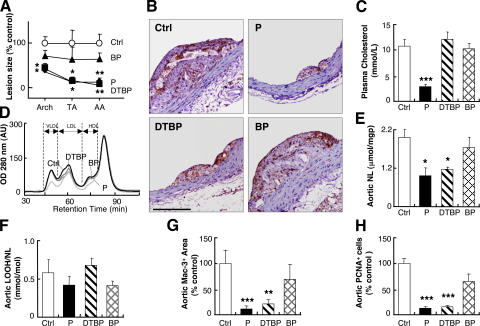
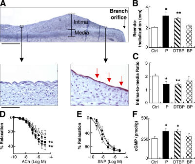
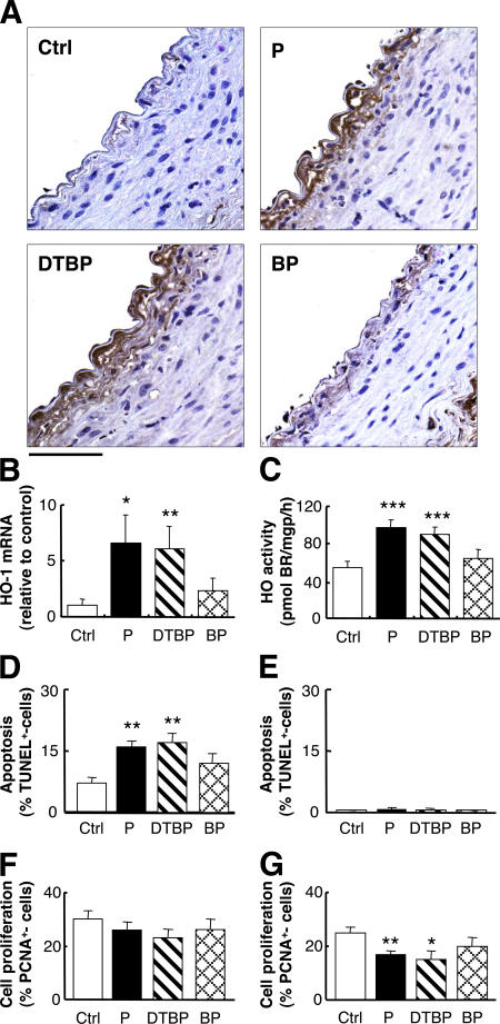
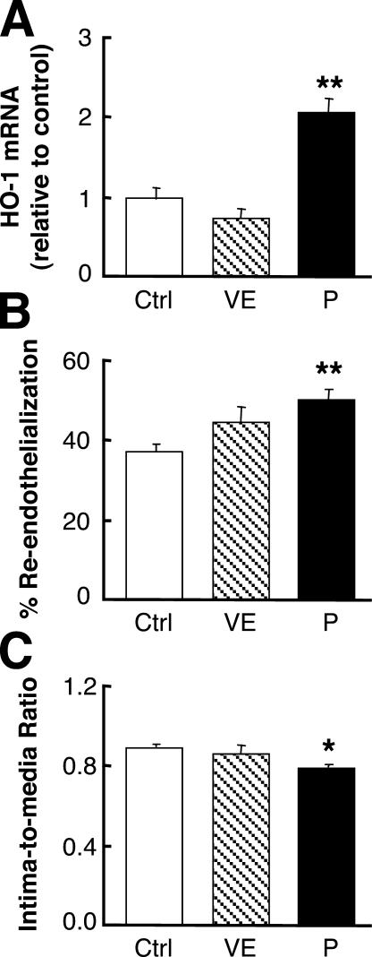
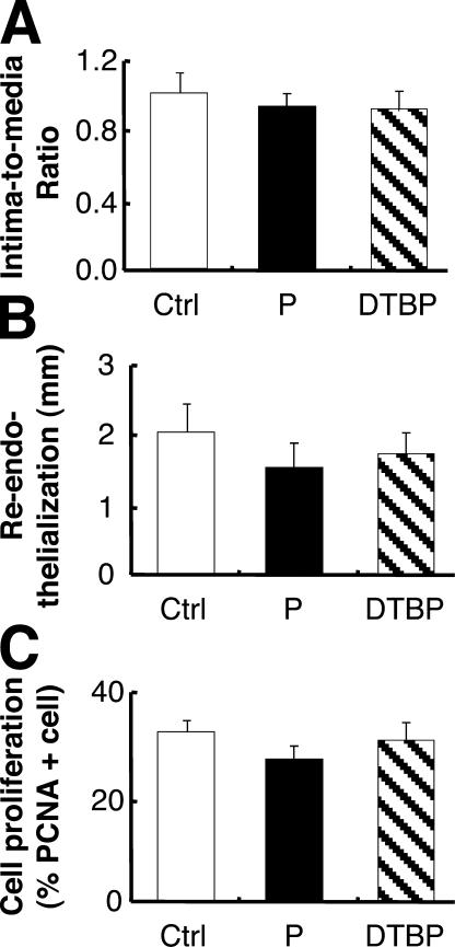
Oxidative stress is implicated in atherogenesis, yet most clinical trials with antioxidants, particularly vitamin E, have failed to protect against atherosclerotic diseases. A striking exception is probucol, which retards atherosclerosis in carotid arteries and restenosis of coronary arteries after angioplasty. Because probucol has in vitro cellular-protective effects independent of inhibiting lipid oxidation, we investigated the mode of action of probucol in vivo. We used three models of vascular disease: apolipoprotein E-deficient mice, a model of atherosclerosis; rabbit aortic balloon injury, a model of restenosis; and carotid injury in obese Zucker rats, a model of type 2 diabetes. Unexpectedly, we observed that the phenol moieties of probucol were insufficient, whereas its sulphur atoms were required for protection. Probucol and its sulphur-containing metabolite, but not a sulphur-free phenolic analogue, protected via cell-specific effects on inhibiting macrophage accumulation, stimulating reendothelialization, and inhibiting vascular smooth muscle cell proliferation. These processes were mediated via induction of heme oxygenase-1 (HO-1), an activity not shared by vitamin E. Our findings identify HO-1 as the molecular target of probucol. They indicate 2-electron rather than radical (1-electron) oxidants as important contributors to atherogenesis, and point to novel lead compounds for therapeutic intervention against atherosclerotic diseases.


Figures

Figure 1.
Figure 1.
Probucol and probucol dithiobisphenol, but not probucol bisphenol, inhibit atherosclerosis in Apoe−/− mice. (A) Lesion area in animals treated with probucol (P, ♦), probucol dithiobisphenol (DTBP, ▪), or probucol bisphenol (BP, ▴) compared with control (Ctrl, ○). n = 10 for each site, each using two, six, and three serial sections for arch, thoracic (TA), and abdominal aorta (AA), respectively. (B) Representative cross sections of abdominal aorta from control and the three treatment groups stained for macrophages indicating respective lesion size. Bar, 25 μm. (C) Plasma cholesterol, n = 10. (D) FPLC chromatograms of lipoproteins from pooled plasma (n = 10) of control, probucol, probucol dithiobisphenol, and probucol bisphenol animals. (E) Neutral lipids (NL, comprised of cholesterylesters and triglycerides) and (F) their hydroperoxides (LOOH) standardized to NL. n = 3 pools of five aortas per pool. (G) Average lesion area covered by Mac-3+ cells (i.e., macrophages) and (H) PCNA+ (i.e., proliferating) cells in the arch, thoracic, and abdominal aorta as compared with corresponding control. n = 3 for each site (three serial sections/site). *, P < 0.05; **, P < 0.01; ***, P < 0.001 compared with control.
Figure 2.
Figure 2.
Probucol and probucol dithiobisphenol, but not probucol bisphenol, inhibit intimal hyperplasia in rabbits in response to vessel injury. (A) Representative Verhoeff's hematoxylin-stained cross sections. Bar, 1,000 μm. (B) Intima-to-media ratio of vessels from control and drug-treated rabbits 6 wk after aortic balloon injury (eight serial sections per aortic segment, 100 μm apart). (C) Neutral lipids and (D) their hydroperoxides. (E) Time-dependent changes in plasma cholesterol of control rabbits or animals treated with probucol, probucol dithiobisphenol, or probucol bisphenol. All results are from six rabbits per group. Symbols and abbreviations are as described in the legend to Fig. 1.
Figure 3.
Figure 3.
Probucol and probucol dithiobisphenol, but not probucol bisphenol, promote functional reendothelialization in rabbit aortas after injury. (A) Representative longitudinal section with branch orifice (bar, 500 μm) showing denuded and CD-31+ reendothelialized aortic surface (red staining) distant and close to the branch orifice, respectively (bar, 25 μm), 6 wk after injury. (B) Reendothelialization assessed by the length (mm) of section covered by CD-31+ cells from the branch orifice for each group 6 wk after injury (three to six serial sections per segment, 100 μm apart). (C) Intima-to-media ratio determined by dividing the intimal over the medial area covered by an intact endothelium distal to the branch of the third lumbar artery (three to six serial sections per segment, 100 μm apart). (D–F) Aortic rings obtained proximally to the second lumbar arteries of the four groups were used for vessel ring studies and examined for relaxation after preconstriction in response to acetylcholine (ACh, D) and sodium nitroprusside (SNP, E), with results expressed as percentage of maximal relaxation. (F) Content of guanosine 3′,5′-cyclic monophosphate (cGMP) in aortic rings (obtained distally to the second lumbar arteries of the four groups) exposed to acetylcholine after preincubation in the presence of 3- isobutyl-1-methylxanthine and inhibitor of phosphodiesterases. All results are from six rabbits per group. Symbols and abbreviations are as described in the legend to Fig. 1.
Figure 4.
Figure 4.
Probucol and probucol dithiobisphenol, but not probucol bisphenol, induce HO-1 in balloon-injured rabbit aortas, and this is related to subsequent apoptosis and vascular smooth muscle cell proliferation. (A) Representative cross sections taken from control and treated rabbits 4 d after aortic balloon injury and stained for HO-1. Bar, 25 μm. n = 4 for each treatment (three serial sections/aorta). (B) HO-1 mRNA and (C) heme oxygenase activity in aortas taken from control and treated rabbits 4 d after injury. n = 8 per treatment. (D–G) Apoptosis and cell proliferation were assessed by the proportion of cells in corresponding aortic sections (n = 3 per aorta) obtained from differently treated animals (n = 4–6 per group) staining positive for TUNEL and PCNA, respectively, as described in Materials and methods. (D) Apoptosis 4 d after injury. (E) Apoptosis 42 d after injury. (F) Proliferation 4 d after injury. (G) Proliferation 42 d after injury. Symbols and abbreviations are as described in the legend to Fig. 1.
Figure 5.
Figure 5.
Vitamin E fails to inhibit intimal hyperplasia and does not promote reendothelialization or induce HO-1. (A) HO-1 mRNA assessed by real-time RT-PCR in rabbit aortic smooth muscle cells cultured for 24 h in the presence of vehicle (control), 50 μM probucol, or 50 μM α-tocopherol (VE). (B) Reendothelialization assessed by Evans blue staining for the three groups of rabbits shown in A 3 wk after injury. (C) Intima-to-media ratio of vessels from control rabbits and animals treated with probucol or α-tocopheryl acetate (VE) (n = 6 per group) 3 wk after aortic balloon injury (five serial sections per aortic segment, 100 μm apart). Results show mean ± SEM of a triplicate experiment performed twice with similar results obtained in both experiments. Symbols and abbreviations are as described in the legend to Fig. 1.
Figure 6.
Figure 6.
HO-1 is the molecular target for probucol and probucol dithiobisphenol. All animals (n = 6 per group) received tin(IV) protoporphyrin IX dichloride to block heme oxygenase. (A) Intima-to-media ratio of vessels 6 wk after aortic balloon injury (eight serial sections per segment, 100 μm apart). (B) Reendothelialization assessed by the length (mm) of section covered by CD-31+ cells from the branch orifice (three to six serial sections per segment, 100 μm apart), and (C) cell proliferation is indicated by PCNA+ cells 6 wk after injury. Symbols and abbreviations are as described in the legend to Fig. 1.

Comment in

References

    1. Steinberg, D., S. Parthasarathy, T.E. Carew, J.C. Khoo, and J.L. Witztum. 1989. Beyond cholesterol: modifications of low-density lipoprotein that increase its atherogenicity. N. Engl. J. Med. 320:915–924. - PubMed
    1. Stocker, R., and J.F. Keaney Jr. 2004. Role of oxidative modifications in atherosclerosis. Physiol. Rev. 84:1381–1478. - PubMed
    1. Upston, J.M., L. Kritharides, and R. Stocker. 2003. The role of vitamin E in atherosclerosis. Prog. Lipid Res. 42:405–422. - PubMed
    1. Hazell, L.J., and R. Stocker. 1997. α-Tocopherol does not inhibit hypochlorite-induced oxidation of apolipoprotein B-100 of low-density lipoprotein. FEBS Lett. 414:541–544. - PubMed
    1. Meagher, E.A., O.P. Barry, J.A. Lawson, J. Rokach, and G. FitzGerald. 2001. Effects of vitamin E on lipid peroxidation in healthy persons. JAMA. 285:1178–1182. - PubMed

Publication types

MeSH terms