. 2006 May;127(5):467-80.
doi: 10.1085/jgp.200509467. Epub 2006 Apr 10.

The polyamine binding site in inward rectifier K+ channels

Affiliations

The polyamine binding site in inward rectifier K+ channels

Harley T Kurata et al. J Gen Physiol. 2006 May.

Abstract

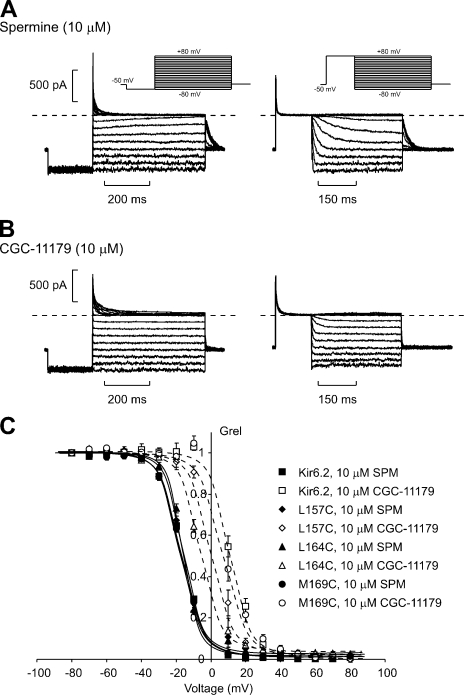
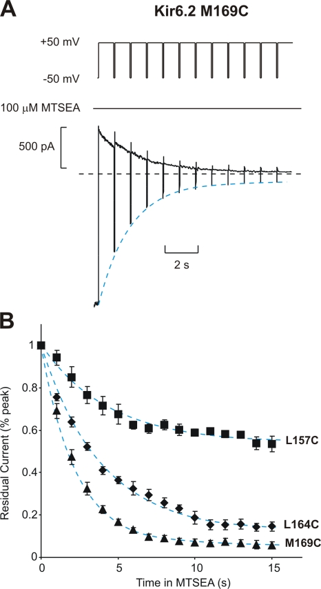
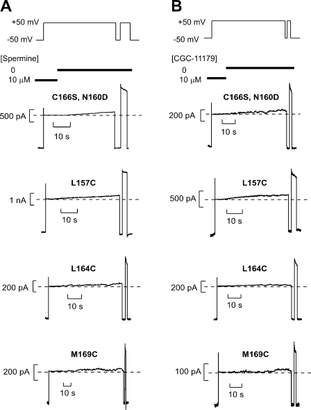
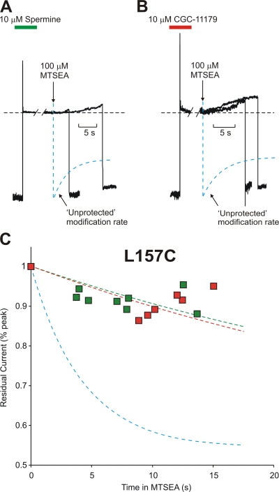
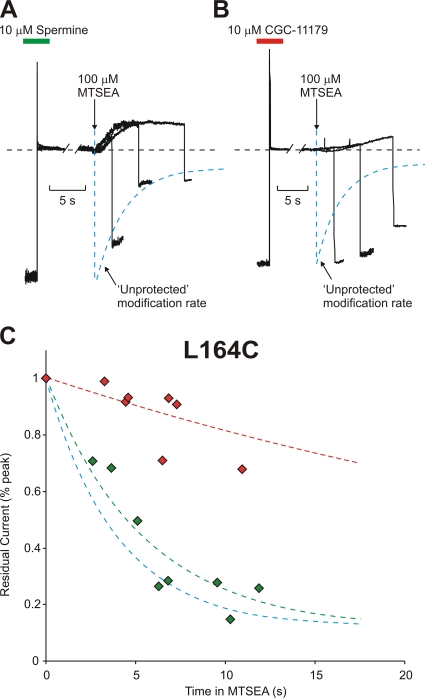
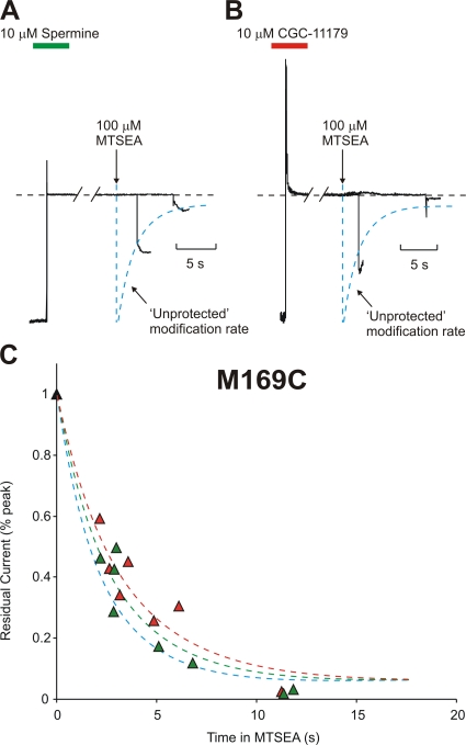
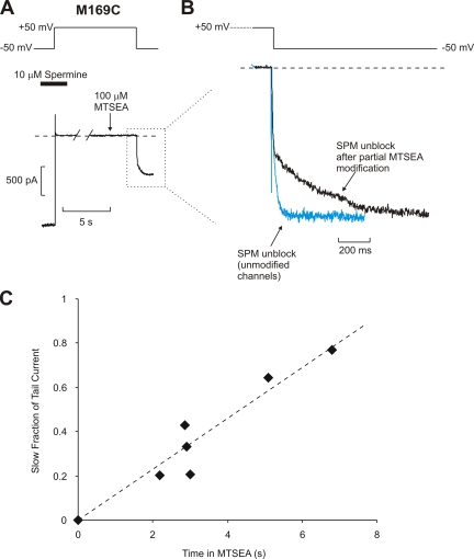
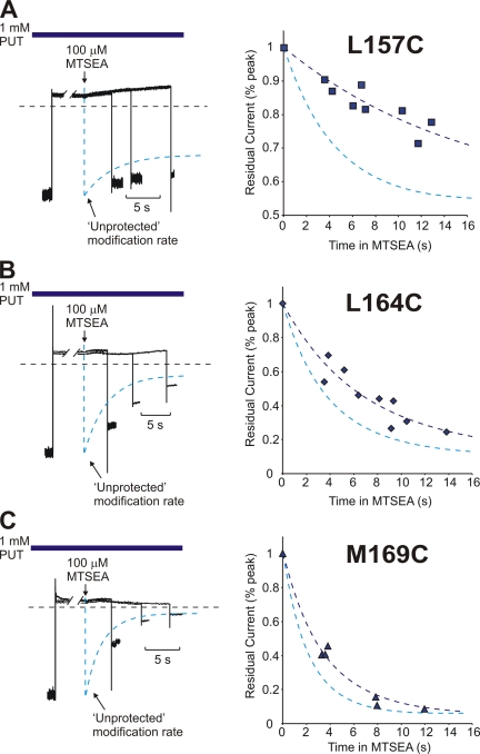
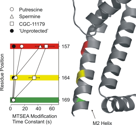
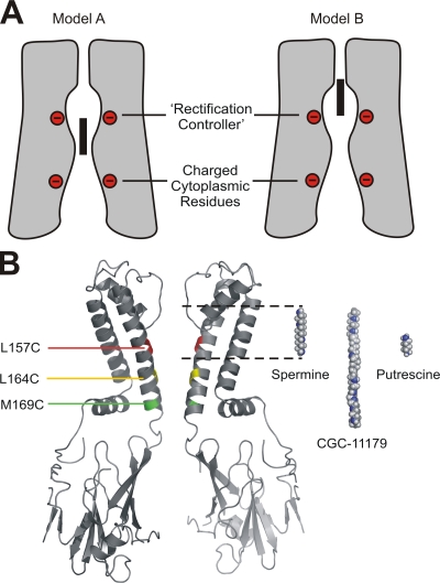
Strongly inwardly rectifying potassium channels exhibit potent and steeply voltage-dependent block by intracellular polyamines. To locate the polyamine binding site, we have examined the effects of polyamine blockade on the rate of MTSEA modification of cysteine residues strategically substituted in the pore of a strongly rectifying Kir channel (Kir6.2[N160D]). Spermine only protected cysteines substituted at a deep location in the pore, between the "rectification controller" residue (N160D in Kir6.2, D172 in Kir2.1) and the selectivity filter, against MTSEA modification. In contrast, blockade with a longer synthetic polyamine (CGC-11179) also protected cysteines substituted at sites closer to the cytoplasmic entrance of the channel. Modification of a cysteine at the entrance to the inner cavity (169C) was unaffected by either spermine or CGC-11179, and spermine was clearly "locked" into the inner cavity (i.e., exhibited a dramatically slower exit rate) following modification of this residue. These data provide physical constraints on the spermine binding site, demonstrating that spermine stably binds at a deep site beyond the "rectification controller" residue, near the extracellular entrance to the channel.


Figures

Figure 1.
Figure 1.
Blockade of Kir6.2[N160D] [C166S] channels by spermine and CGC-11179. (A and B) Spermine and CGC-11179 were applied at a concentration of 10 μM to the intracellular face of inside-out patches expressing Kir6.2[N160D][C166S] channels. Two protocols were used to quantify steady-state blocking parameters. In the left panels (blocking protocol), patches were held at −50 mV, pulsed for 200 ms to −80 mV, and then pulsed for 500 ms to voltages between 80 and +80 mV. In the right panels (unblocking protocol), patches were held at −50 mV, pulsed for 150 ms to +80 mV, and repolarized to voltages between +80 and −80 mV in 10-mV steps. (C) Steady-state currents at voltages between −80 and +80 mV were normalized to steady-state currents in the absence of blockers, for Kir6.2[N160D][C166S] and a number of cysteine-substituted channels (L157C, L164C, and M169C). Solid lines represent fitted Boltzmann functions for spermine block of each channel type, and dashed lines represent fitted Boltzmann functions for CGC-11179 block of each channel type.
Figure 2.
Figure 2.
MTSEA modification of cysteine residues substituted in the Kir6.2 pore. (A) Sample data of modification of Kir6.2 [N160D][C166S][M169C] by 100 μM MTSEA. To characterize the rate of MTSEA modification at +50 mV, patches were held at +50 mV after application of 100 μM MTSEA to the intracellular side of the patch, and pulsed for 30 ms to −50 mV at 1-s intervals. (B) Mean data illustrating the modification rates of Kir6.2[N160D][C166S][L157C] (τ = 4.3 ± 0.7 s; n = 5), [L164C] (τ = 3.9 ± 0.3 s; n = 4), and [M169C] (τ = 2.3 ± 0.2 s; n = 4), channels by 100 μM MTSEA, measured as described in A. Dashed blue lines (here and throughout the text) represent monoexponential fits to the decay of residual currents by MTSEA modification, in the absence of any applied blocker.
Figure 3.
Figure 3.
Slow polyamine unbinding from the pore of mutant Kir6.2 channels. Patches expressing Kir6.2[N160D][C166S] were pulsed to +50 mV in (A) 10 μM spermine or (B) 10 μM CGC-11179. With the patch held continuously at +50 mV, the bathing solution was then switched to a polyamine-free solution to observe the time course of dissociation of polyamines from channels in the patch. A voltage step to −50 mV is sufficient to rapidly unblock either spermine or CGC-11179 from the channel and demonstrates that prolonged blockade and holding of the membrane potential at +50 mV does not result in significant channel rundown. The absence of significant blockade in a subsequent pulse to +50 mV demonstrates that most polyamine has diffused away from each patch. Similar experiments were performed on the cysteine-substituted mutants L157C, L164C, and M169C. In the lower panels, the exact details of voltage pulses and timing of solution changes have been omitted but are similar to those illustrated in the top row.
Figure 4.
Figure 4.
Protection of residue 157C by spermine or CGC-11179 occupancy of the Kir6.2 pore. Patches expressing Kir6.2[N160D][C166S][L157C] were preblocked by voltage steps to +50 mV in either (A) 10 μM spermine or (B) CGC-11179. While held continuously at +50 mV, patches were moved into a polyamine-free solution and, where indicated by the downward arrow, exposed to a polyamine-free solution containing 100 μM MTSEA. After variable intervals in 100 μM MTSEA, patches were repolarized to −50 mV (to assess the extent of MTSEA modification) and immediately removed from the MTSEA-containing solution. The unprotected modification rate (dashed blue line) represents the rate of MTSEA modification of L157C channels in polyamine-free conditions (from Fig. 2 B), and is superimposed on the raw data for comparison. (C) Modification of channels preblocked with either CGC-11179 (red symbols, τ = 52 ± 11 s) or spermine (green symbols, τ = 44 ± 7 s) was measured in multiple patches after varying intervals in 100 μM MTSEA to determine the time course of modification when the pore is occupied by either polyamine. The unprotected modification time course of L157C is indicated by the blue line (τ = 4.2 ± 0.7 s). Preblocking with either spermine or CGC-11179 strongly protects against MTSEA modification at residue 157C.
Figure 5.
Figure 5.
Residue 164C is differentially protected by spermine or CGC-11179 occupancy of the Kir6.2 pore. Patches expressing Kir6.2[N160D][C166S][L164C] channels were preblocked by voltage steps to +50 mV in either (A) 10 μM spermine or (B) CGC-11179. As described in Fig. 4, patches were moved into a polyamine-free solution and exposed to a solution containing 100 μM MTSEA where indicated by the downward arrow. After variable intervals in 100 μM MTSEA, patches were repolarized to −50 mV and immediately removed from the MTSEA-containing solution. (C) Modification of channels preblocked with either CGC-11179 (red symbols, τ = 35 ± 8 s) or spermine (green symbols, τ = 5.4 ± 0.5 s) in multiple patches. The unprotected modification of L164C is indicated by the blue line (τ = 3.9 ± 0.5 s). Preblocking with spermine does not prevent MTSEA modification of 164C, while CGC-11179 protects strongly at this position.
Figure 6.
Figure 6.
Residue 169C is not protected by spermine or CGC-11179 occupancy of the Kir6.2 pore. Patches expressing Kir6.2 [N160D][C166S][M169C] were preblocked by voltage steps to +50 mV in either (A) 10 μM spermine or (B) CGC-11179 and exposed to a polyamine-free solution containing 100 μM MTSEA, as described in Figs. 4 and 5. (C) Modification of channels preblocked with either CGC-11179 (red symbols, τ = 3.3 ± 0.3 s) or spermine (green symbols, τ = 2.7 ± 0.2 s) in multiple patches. The time course of unprotected modification of M169C is indicated by the blue line (τ = 2.4 ± 0.3 s). Pore occupancy by either polyamine does not significantly alter the rate of cysteine modification at 169C.
Figure 7.
Figure 7.
MTSEA modification of M169C traps spermine in the Kir6.2 pore. (A) Sample data of a blocker protection experiment of Kir6.2[N160D][C166S][M169C] channels preblocked with spermine, collected as described in Figs. 4–6 . (B) Expanded data illustrating the tail currents observed in A upon repolarization to −50 mV (black trace). The blue trace, included for comparison, illustrates the rate of spermine unblock from unmodified M169C channels. The slow unblocking time course in modified M169C channels demonstrates that spermine remains bound in the pore during the modification step, and is effectively trapped by the introduction of positive charges at residue 169.
Figure 8.
Figure 8.
Protection of pore-lining cysteine residues by putrescine. Patches expressing (A) Kir6.2[N160D][C166S][L157C], (B) Kir6.2[N160D][C166S][L164C], or (C) Kir6.2[N160D][C166S][L169C] were blocked at +50 mV in 1 mM putrescine, exposed to 100 μM MTSEA for a variable interval (while continuously exposed to putrescine), and repolarized to −50 mV to determine the extent of MTSEA modification. Sample traces for each construct are presented in the lefthand panels, along with the unprotected MTSEA modification rates for comparison. Compiled data from multiple patches are presented in the righthand panels, and fit with a single exponential curve. Unprotected modification time courses are indicated by the dashed blue lines. At residue 157C, putrescine slowed the time constant of modification to 15.8 ± 1.3 s, from an unprotected time constant of 4.2 ± 0.7 s. At residue 164C in the presence of putrescine, the modification time constant was 7.1 ± 0.5 s, and the unprotected time constant was 3.9 ± 0.5 s. At residue 169C, putrescine slowed the modification time constant to 3.6 ± 0.3 s, from the unprotected time constant of 2.4 ± 0.3 s.
Figure 9.
Figure 9.
Spatial orientation of substituted cysteines in the Kir pore. Summary of the time constants of MTSEA modification (mean ± SEM) at residues 157C, 164C, and 169C, in the presence of putrescine, spermine, CGC-11179, or no blocker (unprotected), as measured in Figs. 2–8 . A representation of the M2 helix, based on the X-ray structure of KirBac1.1, is aligned with the plot to illustrate the relative positions of the substituted cysteine residues in the inner cavity.
Figure 10.
Figure 10.
The polyamine binding site in the Kir channel pore. (A) Cartoons to illustrate contrasting models of shallow (Model A) versus deep spermine binding (Model B). Red circles indicate rings of negative charges in the cytoplasmic domain (bottom circles) and the inner cavity (top circles) of strongly rectifying Kir channels. The black rectangle represents a spermine molecule in the Kir pore. (B) Using the KirBac1.1 crystal structure as a template, we have mapped the examined residues and colored them to reflect the protection profile by spermine and CGC-11179. Residue 157 (red) is protected against MTSEA modification by both spermine and CGC-11179 (see Fig. 4). Residue 164 (yellow) is protected by CGC-11179 but not by spermine (see Fig. 5). Residue 169 (green) is not protected by either polyamine (see Fig. 6). We have also aligned spermine, CGC-11179, and putrescine molecules with binding locations indicated by the observed protection profile. The head of spermine and CGC-11179 are placed near the entrance to the selectivity filter. The tail of spermine extends to the approximate location of the rectification controller residue (N160D in Kir6.2), while the considerably longer CGC-11179 molecule extends to the inner cavity entrance. Putrescine is located near the rectification controller residue.

References

    1. Chang, H.K., S.H. Yeh, and R.C. Shieh. 2003. The effects of spermine on the accessibility of residues in the M2 segment of Kir2.1 channels expressed in Xenopus oocytes. J. Physiol. 553:101–112. - PMC - PubMed
    1. Del Camino, D., M. Holmgren, Y. Liu, and G. Yellen. 2000. Blocker protection in the pore of a voltage-gated K+ channel and its structural implications. Nature. 403:321–325. - PubMed
    1. Dibb, K.M., T. Rose, S.Y. Makary, T.W. Claydon, D. Enkvetchakul, R. Leach, C.G. Nichols, and M.R. Boyett. 2003. Molecular basis of ion selectivity, block, and rectification of the inward rectifier Kir3.1/Kir3.4 K+ channel. J. Biol. Chem. 278:49537–49548. - PubMed
    1. Fakler, B., U. Brandle, E. Glowatzki, S. Weidemann, H.P. Zenner, and J.P. Ruppersberg. 1995. Strong voltage-dependent inward rectification of inward rectifier K+ channels is caused by intracellular spermine. Cell. 80:149–154. - PubMed
    1. Ficker, E., M. Taglialatela, B.A. Wible, C.M. Henley, and A.M. Brown. 1994. Spermine and spermidine as gating molecules for inward rectifier K+ channels. Science. 266:1068–1072. - PubMed

Publication types

MeSH terms