. 2006 Apr 10;173(1):83-93.
doi: 10.1083/jcb.200511149.

Lamina-associated polypeptide 2alpha regulates cell cycle progression and differentiation via the retinoblastoma-E2F pathway

Affiliations

Lamina-associated polypeptide 2alpha regulates cell cycle progression and differentiation via the retinoblastoma-E2F pathway

Daniela Dorner et al. J Cell Biol. .

Abstract

Lamina-associated polypeptide (LAP) 2alpha is a nonmembrane-bound LAP2 isoform that forms complexes with nucleoplasmic A-type lamins. In this study, we show that the overexpression of LAP2alpha in fibroblasts reduced proliferation and delayed entry into the cell cycle from a G0 arrest. In contrast, stable down-regulation of LAP2alpha by RNA interference accelerated proliferation and interfered with cell cycle exit upon serum starvation. The LAP2alpha-linked cell cycle phenotype is mediated by the retinoblastoma (Rb) protein because the LAP2alpha COOH terminus directly bound Rb, and overexpressed LAP2alpha inhibited E2F/Rb-dependent reporter gene activity in G1 phase in an Rb-dependent manner. Furthermore, LAP2alpha associated with promoter sequences in endogenous E2F/Rb-dependent target genes in vivo and negatively affected their expression. In addition, the expression of LAP2alpha in proliferating preadipocytes caused the accumulation of hypophosphorylated Rb, which is reminiscent of noncycling cells, and initiated partial differentiation into adipocytes. The effects of LAP2alpha on cell cycle progression and differentiation may be highly relevant for the cell- and tissue-specific phenotypes observed in laminopathic diseases.


Figures

Figure 1.
Figure 1.
Overexpression of LAP2α negatively affects cell cycle progression. (A) Stable HeLa Tet-on clones expressing myc-tagged LAP2α under the control of a doxycyclin-dependent promoter were grown in the absence or presence of doxycyclin and processed for immunofluorescence microscopy and immunoblot analyses of cell lysates. Cells grown permanently in the absence (−Dox) or presence (perm Dox) of doxycyclin or cells transferred to doxycyclin medium upon splitting at day 0 were seeded into plates, and cumulative cell numbers were determined. (B) Stable 3T3 cells expressing a LAP2α-GFP fusion or untransfected control cells were analyzed as in A. Cumulative cell numbers were analyzed in cultures grown in complete medium (10% FCS) or upon serum starvation in 0.5% FCS for 7 d before the addition of complete medium (0.5→10%). Graphs show one representative dataset from at least three independent experiments. Bars, 10 μm. (C) DNA flow cytometry of dense LAP2α-myc HeLa tet-on cultures grown in the absence or presence of doxycyclin and of 3T3 and LAP2α-GFP cells after 7 d of serum starvation (0.5% FCS) and at different time points after restimulation (10% FCS). Numbers at the bottom indicate percentages of cells in G1 and S phase.
Figure 2.
Figure 2.
Reduced LAP2α levels favor cell cycle progression. (A) HeLa cells were stably transfected with a LAP2α-specific shRNA vector construct (LAP2α RNAi) or with a construct targeting firefly luciferase (ctrl). Cells were analyzed by immunofluorescence microscopy using monoclonal anti-LAP2α antibody and by immunoblot analyses of total cell lysates using anti-LAP2 antibody (detecting both LAP2α and LAP2β). Bar, 10 μm. Note that the LAP2α RNAi immunofluorescence image was overexposed to detect cells. (B) Cumulative cell numbers were determined within 7 d of culture in complete medium (10% FCS) or in low serum medium (0.5% FCS). Graphs show the dataset of one of three independent clones showing the same phenotype. (C) DNA flow cytometry of control and LAP2α-deficient (LAP2α RNAi) cells grown in low serum medium for 4 d.
Figure 3.
Figure 3.
LAP2α binds to Rb at its COOH terminus and associates with Rb, lamin A/C, and E2F promoter sequences in vivo. (A) Schematic drawing of LAP2α and LAP2α fragments used for binding studies and localization of molecular domains and interaction regions with Rb and lamins A/C in the polypeptide. Numbers denote amino acid positions; +, interaction; −, no interaction of respective LAP2α fragments with Rb. Light gray boxes denote the constant LAP2 regions, including LEM-like and LEM domains (hatched). Medium gray denotes the α-specific region, whereas dark gray indicates the chromatin interaction domain. On the right, in vitro–translated 35S-labeled Rb was overlaid onto transblotted recombinant LAP2α fragments. A Ponceau S stain of the respective blot and an autoradiogram of the overlay are shown. Numbers on the right indicate molecular masses in kD. (B) HeLa cells stably expressing LAP2α-GFP were fixed in formaldehyde, mechanically lysed by sonication, and LAP2α was immunoprecipitated using monoclonal (m) or polyclonal (p) antibody to LAP2α. Control precipitations were performed with antibody-free medium (ctrl) and with preimmune serum (PI). Immunoblots of immunoprecipitates and respective inputs (IP, diluted 1:10) with antiserum to LAP2α, Rb, or monoclonal antibody to lamin A/C are shown. (C) Chromatin immunoprecipitation was performed from HeLa and 3T3 cells using monoclonal and polyclonal antibody to LAP2α, preimmune serum, or anti-acetylhistone H4 (H4), and E2F-dependent promoter sequences in the cyclin D1 and thymidine kinase (TK) genes were amplified by PCR. As a negative control, the presence of histone H4 or E-cadherin promoter sequences was tested. Ethidium bromide–stained DNA fragments in agarose gels are shown. IP, 1% input.
Figure 4.
Figure 4.
LAP2α reduces E2F transcription activity in an Rb-dependent manner. Asynchronous 3T3 cells (A), 3T3 cells synchronized by an overnight aphidicolin block and release from the block for different time periods (B), aphidicolin-arrested cells (G1 phase) and 4-h released cells (S phase; C), or TKO mouse fibroblasts lacking pocket proteins Rb, p107, and p130 (D) were used for E2F-reporter assays. Cells were transfected with an E2F-dependent luciferase reporter and a β-galactosidase expression construct (for normalization of transfection efficiencies) together with empty vectors (−) or expression vectors encoding E2F-1, Rb, LAP2α, and LAP2α N terminus (LAP2α-N). Reporter activity was measured 24 h after transfection. Relative activities were calculated by referring measured light units to that of the respective controls (reporter plasmids with empty vectors). Data represent mean values of at least three independent datasets, and SDs (error bars) are shown. DNA flow cytometry profiles of synchronized 3T3 cells at different stages of the cell cycle and immunoblot analysis of total cell lysates of untreated (−) or aphidicolin-treated 3T3 cells and of transformed HEK cells are shown in B.
Figure 5.
Figure 5.
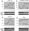
Overexpressed LAP2α affects the expression of endogenous E2F target genes. mRNA levels of the E2F target genes cyclin D1, thymidine kinase, and cyclin E and of actin as a control were determined by semiquantitative RT-PCR from control 3T3 cells (solid lines) or LAP2α-GFP–expressing 3T3 cells (dotted lines) harvested 0–24 h after serum addition to serum-starved cultures. Ethidium bromide–stained DNA fragments in agarose gels (bottom) were quantified by QuantiScan and plotted against incubation times (top).
Figure 6.
Figure 6.
LAP2α affects adipocyte differentiation. 3T3 F442A cells (control) or 3T3 F442A cells expressing LAP2α-GFP were grown in proliferation medium (P) or in differentiation medium containing insulin and T3 (D) for 10 d and were analyzed by phase-contrast microscopy (A), immunoblot analyses of total cell lysates (B), and oil red staining (C). White lines indicate that intervening lanes have been spliced out. Bars, 40 μm.

References

    1. Ait-Si-Ali, S., V. Guasconi, L. Fritsch, H. Yahi, R. Sekhri, I. Naguibneva, P. Robin, F. Cabon, A. Polesskaya, and A. Harel-Bellan. 2004. A Suv39h-dependent mechanism for silencing S-phase genes in differentiating but not in cycling cells. EMBO J. 23:605–615. - PMC - PubMed
    1. Arimura, T., A. Helbling-Leclerc, C. Massart, S. Varnous, F. Niel, E. Lacene, Y. Fromes, M. Toussaint, A.M. Mura, D.I. Keller, et al. 2005. Mouse model carrying H222P-Lmna mutation develops muscular dystrophy and dilated cardiomyopathy similar to human striated muscle laminopathies. Hum. Mol. Genet. 14:155–169. - PubMed
    1. Berger, R., L. Theodor, J. Shoham, E. Gokkel, F. Brok-Simoni, K.B. Avraham, N.G. Copeland, N.A. Jenkins, G. Rechavi, and A.J. Simon. 1996. The characterization and localization of the mouse thymopoietin/lamina-associated polypeptide 2 gene and its alternatively spliced products. Genome Res. 6:361–370. - PubMed
    1. Boguslavsky, R.L., C.L. Stewart, and H.J. Worman. 2006. Nuclear lamin A inhibits adipocyte differentiation: implications for Dunnigan-type familial partial lipodystrophy. Hum. Mol. Genet. 15:653–663. - PubMed
    1. Brehm, A., E.A. Miska, D.J. McCance, J.L. Reid, A.J. Bannister, and T. Kouzarides. 1998. Retinoblastoma protein recruits histone deacetylase to repress transcription. Nature. 391:597–601. - PubMed

Publication types

MeSH terms