Comparative Study
. 2006 May;16(5):584-94.
doi: 10.1101/gr.4843906. Epub 2006 Apr 10.

Evolutionary expansion and divergence in the ZNF91 subfamily of primate-specific zinc finger genes

Affiliations
Comparative Study

Evolutionary expansion and divergence in the ZNF91 subfamily of primate-specific zinc finger genes

Aaron T Hamilton et al. Genome Res. 2006 May.

Abstract

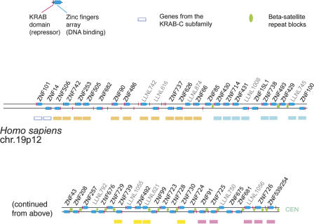
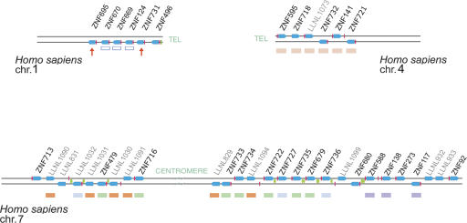
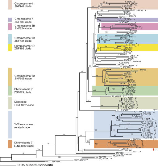
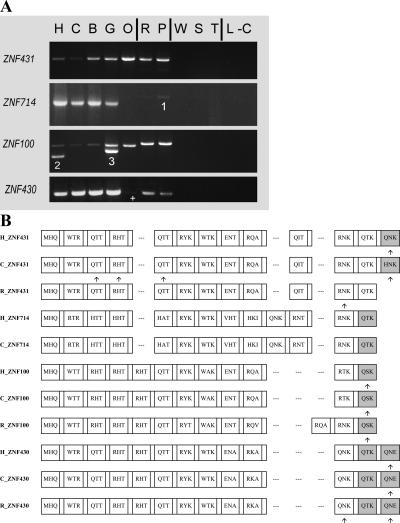
Most genes are conserved in mammals, but certain gene families have acquired large numbers of lineage-specific loci through repeated rounds of gene duplication, divergence, and loss that have continued in each mammalian group. One such family encodes KRAB-zinc finger (KRAB-ZNF) proteins, which function as transcriptional repressors. One particular subfamily of KRAB-ZNF genes, including ZNF91, has expanded specifically in primates to comprise more than 110 loci in the human genome. Genes of the ZNF91 subfamily reside in large gene clusters near centromeric regions of human chromosomes 19 and 7 with smaller clusters or isolated copies in other locations. Phylogenetic analysis indicates that many of these genes arose before the split between the New and Old World monkeys, but the ZNF91 subfamily has continued to expand and diversify throughout the evolution of apes and humans. Paralogous loci are distinguished by divergence within their zinc finger arrays, indicating selection for proteins with different regulatory targets. In addition, many loci produce multiple alternatively spliced transcripts encoding proteins that may serve separate and perhaps even opposing regulatory roles because of the modular motif structure of KRAB-ZNF genes. The tissue-specific expression patterns and rapid structural divergence of ZNF91 subfamily genes suggest a role in determining gene expression differences between species and the evolution of novel primate traits.


Figures

Figure 1.
Figure 1.
A diagram of the KRAB-ZNF cluster on HSA19p12 containing genes related to the ZNF91 subfamily. The map shows the physical order of the KRAB-zinc finger loci in the cluster, but is not scaled for genomic distances or relative size. Each finger-containing locus is represented by a blue arrow pointing 5′ to 3′ (left-to-right above the line, opposite strand, below). Loci that contain a KRAB have a red bar at the 5′ end of the symbol. Isolated red bars are KRAB-only pseudogene fragments (not labeled). Locus designations with the format “LLNLxxx” are from the new zinc finger catalog (Huntley et al. 2006) and are here used for pseudogene loci (light-gray text). Colored boxes beneath loci correspond to clade membership in the phylogeny (Fig. 3). In some cases, labeled genes in the cluster maps were not included in the phylogeny in Figure 3 because the spacer region used to create the phylogeny was modified or deleted from the locus, or because they were very divergent and not members of the ZNF91 subfamily, notably the “KRAB-C subfamily” loci (blue outline boxes). The relative positions of β-satellite repeat blocks between loci in this cluster are indicated by green ovals between the lines.
Figure 2.
Figure 2.
Diagrams of the KRAB-ZNF clusters on HSA1, HSA4, and HSA7 containing genes related to the ZNF91 subfamily. Maps show the physical order of the KRAB-zinc finger loci in the clusters, but are not scaled for genomic distance or relative size. Due to the spacing between loci, the HSA7 map shown could be defined as more than one cluster, but several clades of closely related loci (colored to match Fig. 3) have copies distributed across the region. Symbols and labels for genes and β-satellite repeats are the same as in Figure 1. The red arrows under the HSA1 cluster denote the two ZNF91 subfamily members in that cluster (the blue outline boxes denote KRAB-C subfamily genes). The HSA7 region shown also has a divergent gene at the p-telomeric end (ZNF713) included in the diagram although relationships between this gene and other KRAB-ZNF loci are unclear. Not shown are four clusters containing a total of 15 pseudogene loci on HSAY and three divergent loci (ZNF267, ZNF720, LLNL1128), which may be distant relatives of the ZNF91 subfamily, clustered together with unrelated genes on HSA16.
Figure 3.
Figure 3.
Neighbor-joining phylogeny of the ZNF91 subfamily based on the spacer and flanking sequences. The tree includes 116 loci (including outgroups); the single best tree is shown, and bootstrap values above 70% based on 1000 bootstrap replicates are indicated on branches. Gene names without a leading number are from the chromosome 19p12 cluster; in all other cases, the initial number indicates the chromosome from which the locus derives (e.g., 7_ZNF588 is from HSA7). Loci with the ZNF catalog designation LLNLxxx (Figs. 1, 2; Supplemental Table S1) are abbreviated “Lxxx.” Colored boxes define certain well-supported clades as designated to the left. For the clades including genes from the main clusters, the box colors match the colored bars under the individual member genes on the cluster maps in Figures 1 and 2. Note that the “Y-chromosome related clade” contains genes from the cluster on HSA7, but these are outnumbered by the pseudogene loci on HSAY. The red arrows indicate the ZNF91 subfamily members in the cluster on HSA1; the gray arrows indicate two nonclustered genes mentioned in the results section. Pseudogenes are marked with a ψ. Processed pseudogenes are indicated by “r” ahead of the locus name.
Figure 4.
Figure 4.
(A) PCR results using primers for selected members of the ZNF431 clade across a panel of primate genomic DNA. The samples are (H) human (Homo sapiens); (C) chimpanzee (Pan troglodytes); (B) bonobo (Pan paniscus); (G) gorilla (Gorilla gorilla); (O) orangutan (Pongo pygmaeus); (R) rhesus macaque (Macaca mulatta); (P) pigtailed macaque (Macaca nemestrina); (W) common woolly monkey (Lagothrix lagotricha); (S) black-handed spider monkey (Ateles geoffroyi); (T) red-chested mustached tamarin (Saguinus labiatus); (L) ring-tailed lemur (Lemur catta); and (−C) the negative control. Vertical lines distinguish the apes, Old World monkeys (the macaques), New World monkeys, and the lemur (a prosimian); see Supplemental Figure S1 for a simple phylogeny of the primates using the same letter codes. Unusual PCR products are numbered as follows: (1) a non-ZNF sequence; (2) LLNL745 (a pseudogene whose closest paralog is ZNF100); (3) a LLNL745-like sequence in gorilla. The + indicates a weak band that was confirmed to be present. Primers were designed from the known genomic sequences and therefore may not identically match all species, resulting in variation in PCR efficiency. LLNL745 may have been deleted in chimpanzee (Newman et al. 2005) and is not in the current chimpanzee genome assembly. (B) Zinc finger alignment hypothesis for the ZNF431 clade. The alignment includes the zinc finger array of the predicted protein for selected human paralogs (H) and their respective orthologs (if found) in chimpanzee (C) and rhesus (R) genomic data. Each box represents a zinc finger; gaps are added when one locus has added or deleted a zinc finger so that flanking fingers remain aligned. The amino acid codes inside each box (for positions −1, 3, and 6, relative to the start of the α-helix) are variable positions involved in sequence-specific DNA target recognition and binding. Degenerate fingers are shaded. Frameshifts in the genomic sequences are indicated by arrows under the fingers, but for the nonhuman primate sequences these are “repaired” to match the orthologous sequences due to the incomplete nature of the nonhuman sequence data. ZNF493 and ZNF738 are not shown due to high-sequence divergence and difficulty in alignment with the other members of the clade; these genes are depicted in Figure 5.
Figure 5.
Figure 5.
Zinc finger exon analysis for ZNF493 and ZNF738, two divergent genes from the ZNF431 clade. The alignments include the human predicted proteins and their orthologs determined from the chimpanzee and rhesus genomic data. Finger boxes, amino acid codes, predicted frameshifts, and degenerate finger shading are as in Figure 4B. The alignment for the predicted ZNF493 proteins (A) shows that the human version has two additional zinc finger motifs compared with both its chimpanzee and rhesus orthologs, indicating the change is an addition of fingers in the human. (B) The relative sizes of ZNF493 zinc finger exon PCR products from genomic DNA of human, chimpanzee, and bonobo is consistent with the human/chimpanzee difference in finger number; species designations are as in Figure 4A. (C) The chimpanzee ZNF738 protein’s differences with the human version, based on draft genomic sequence, include a deleted finger motif, a substitution that eliminates the stop codon seen in human, and a possible frameshift after the deleted finger. Although these differences have not been confirmed, any would change the predicted translation of the chimpanzee protein compared with the human ortholog.
Figure 6.
Figure 6.
Zinc finger alignment for the loci in the ZNF492 clade. The alignment includes the human paralogs and their orthologs if found in chimpanzee and rhesus genomic data. Finger boxes, amino acid codes, degenerate finger shading, and frameshifts are as in Figure 4B. The chimpanzee ZNF492/ZNF739 homolog finger-exon sequence is in a location and orientation matching human ZNF739 in the panTro1 draft, but there are assembly gaps around it and in the location where ZNF492 (if both are present) would be located. Newman et al. (2005) recorded a possible inversion in this region between human and chimpanzee. The location of the rhesus copy is unknown. Therefore, the presumed nonhuman ortholog sequences are not explicitly identified with either human locus name. The horizontal arrows below the rhesus ZNF492/ZNF739 protein homolog represent a finger in the protein alignment that is divergent from the same-numbered finger in the human and chimpanzee homologs. The rhesus ZNF492/ZNF739 protein’s fingers 8 and 9 are similar enough to fingers 10 and 11 that in this case, as an alternative to mutational change in the finger amino acid sequence, the pattern in this species could also be explained by a loss of fingers followed by an internal duplication of the aforementioned finger motifs, restoring the array to the same number of zinc finger motifs. The ZNF730 gene contains a stop codon after the 12th finger, eliminating several 3′-end ZNF motifs that are included in the ZNF492 and ZNF739 proteins. For chimpanzee ZNF730, the available genomic sequence is missing the first three finger motifs, but the cross-primate PCR results showed a similar band for chimpanzee and human (data not shown).
Figure 7.
Figure 7.
RT–PCR gels showing the expression patterns for (A) the KRAB-ZNF gene ZNF729 (a ZNF91 subfamily member on chromosome 19), (B) alternate splice variants of ZNF85 with and without the zinc fingers exon, and (C) alternate splice variants of ZNF43 with different 5′ ends and start sites. Diagrams at right indicate primer locations (arrows) designed to selectively amplify the depicted isoforms for ZNF43 and ZNF85. Notably, each of these isoforms has its first exon within a HERV70/ERV1 LTR repeat. Predicted 5′ ends on many (but not all) of the genes in the cluster overlap with the same type of LTR repeat sequence, which may indicate that the repeats are involved in driving gene expression, as has been shown previously for other genes including related zinc finger genes (Di Cristofano et al. 1995; Abrink et al. 1998). (D, bottom) Gels for two “housekeeping” genes selected as positive controls for the cDNA panels. Guide to the tissues included on the gels: (Adi) Adipose tissue; (Adr) Adrenal gland; (Bpl) Blood, peripheral leukocytes; (BoM) Bone marrow; (Br) Brain; (Brcb) Brain (cerebrum); (Bcll) Brain (cerebellum); (Bm) Brain (medulla oblongata); (Hrt) Heart; (Liv) Liver; (Lym) Lymph node; (MG) Mammary gland; (Pan) Pancreas; (Ov) Ovary; (Pla) Placenta; (Pro) Prostate gland; (Spl) Spleen; (SkM) Skeletal muscle; (Tes) Testis; (Thr) Thyroid; (Thm) Thymus; (−C) negative control. The zinc finger RT–PCR reactions were repeated under varying conditions, and the gels shown are for the 33-cycle experiments, which reveal tissues with present but low-abundance ZNF transcripts.

Similar articles

Cited by

References

    1. Abrink M., Larsson E., Hellman L., Larsson E., Hellman L., Hellman L. Demethylation of ERV3, an endogenous retrovirus regulating the Kruppel-related zinc finger gene H-plk, in several human cell lines arrested during early monocyte development. DNA Cell Biol. 1998;17:27–37. - PubMed
    1. Ayyanathan K., Lechner M.S., Bell P., Maul G.G., Schultz D.C., Yamada Y., Tanaka K., Torigoe K., Rauscher 3rd F.J., Lechner M.S., Bell P., Maul G.G., Schultz D.C., Yamada Y., Tanaka K., Torigoe K., Rauscher 3rd F.J., Bell P., Maul G.G., Schultz D.C., Yamada Y., Tanaka K., Torigoe K., Rauscher 3rd F.J., Maul G.G., Schultz D.C., Yamada Y., Tanaka K., Torigoe K., Rauscher 3rd F.J., Schultz D.C., Yamada Y., Tanaka K., Torigoe K., Rauscher 3rd F.J., Yamada Y., Tanaka K., Torigoe K., Rauscher 3rd F.J., Tanaka K., Torigoe K., Rauscher 3rd F.J., Torigoe K., Rauscher 3rd F.J., Rauscher 3rd F.J. Regulated recruitment of HP1 to a euchromatic gene induces mitotically heritable, epigenetic gene silencing: A mammalian cell culture model of gene variegation. Genes & Dev. 2003;17:1855–1869. - PMC - PubMed
    1. Bailey J.A., Yavor A.M., Massa H.F., Trask B.J., Eichler E.E., Yavor A.M., Massa H.F., Trask B.J., Eichler E.E., Massa H.F., Trask B.J., Eichler E.E., Trask B.J., Eichler E.E., Eichler E.E. Segmental duplications: Organization and impact within the current human genome project assembly. Genome Res. 2001;11:1005–1017. - PMC - PubMed
    1. Bailey J.A., Gu Z., Clark R.A., Reinert K., Samonte R.V., Schwartz S., Adams M.D., Myers E.W., Li P.W., Eichler E.E., Gu Z., Clark R.A., Reinert K., Samonte R.V., Schwartz S., Adams M.D., Myers E.W., Li P.W., Eichler E.E., Clark R.A., Reinert K., Samonte R.V., Schwartz S., Adams M.D., Myers E.W., Li P.W., Eichler E.E., Reinert K., Samonte R.V., Schwartz S., Adams M.D., Myers E.W., Li P.W., Eichler E.E., Samonte R.V., Schwartz S., Adams M.D., Myers E.W., Li P.W., Eichler E.E., Schwartz S., Adams M.D., Myers E.W., Li P.W., Eichler E.E., Adams M.D., Myers E.W., Li P.W., Eichler E.E., Myers E.W., Li P.W., Eichler E.E., Li P.W., Eichler E.E., Eichler E.E. Recent segmental duplications in the human genome. Science. 2002;297:1003–1007. - PubMed
    1. Bailey J.A., Liu G., Eichler E.E., Liu G., Eichler E.E., Eichler E.E. An Alu transposition model for the origin and expansion of human segmental duplications. Am. J. Hum. Genet. 2003;73:823–834. - PMC - PubMed

Publication types

LinkOut - more resources