. 2006 Apr 18;103(16):6386-91.
doi: 10.1073/pnas.0510691103. Epub 2006 Apr 10.

Signaling mediated by the dopamine D2 receptor potentiates circadian regulation by CLOCK:BMAL1

Affiliations

Signaling mediated by the dopamine D2 receptor potentiates circadian regulation by CLOCK:BMAL1

Irene Yujnovsky et al. Proc Natl Acad Sci U S A. .

Abstract

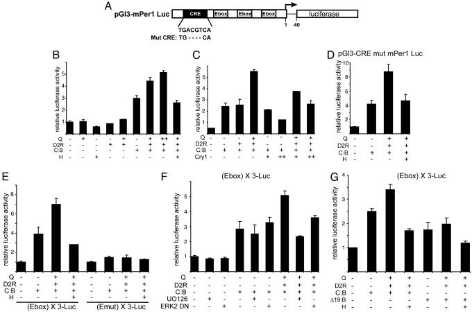
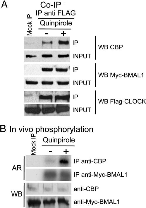
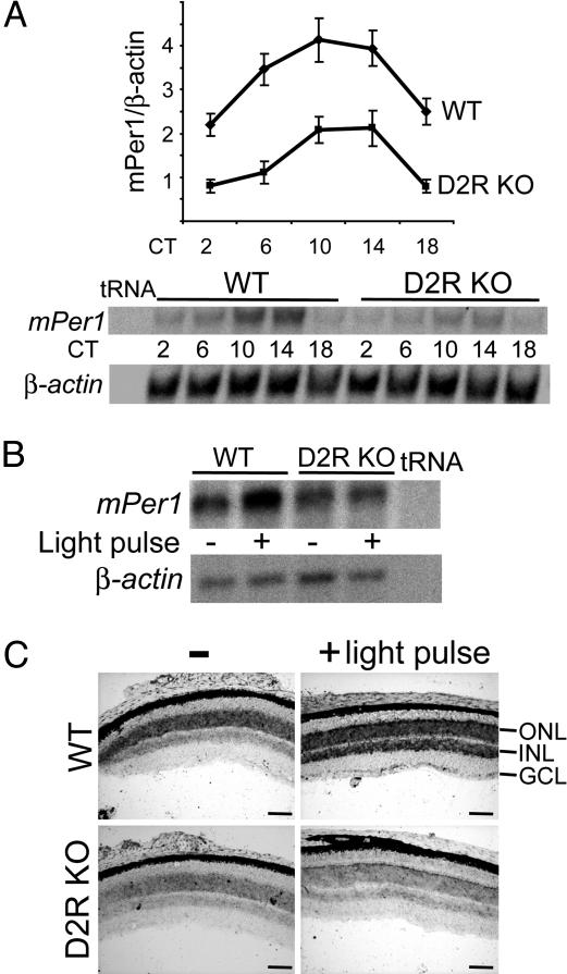
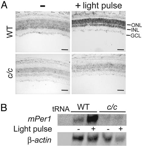
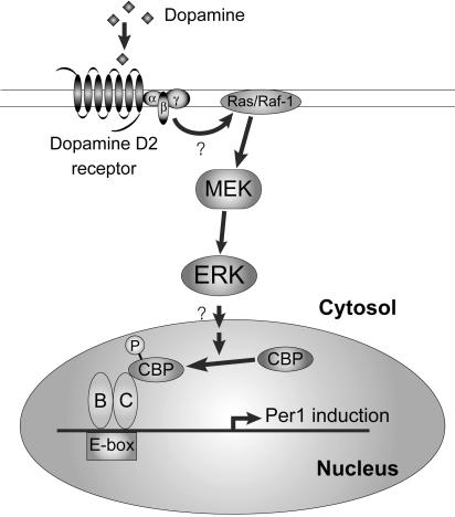
Environmental cues modulate a variety of intracellular pathways whose signaling is integrated by the molecular mechanism that constitutes the circadian clock. Although the essential gears of the circadian machinery have been elucidated, very little is known about the signaling systems regulating it. Here, we report that signaling mediated by the dopamine D2 receptor (D2R) enhances the transcriptional capacity of the CLOCK:BMAL1 complex. This effect involves the mitogen-activated protein kinase transduction cascade and is associated with a D2R-induced increase in the recruiting and phosphorylation of the transcriptional coactivator cAMP-responsive element-binding protein (CREB) binding protein. Importantly, CLOCK:BMAL1-dependent activation and light-inducibility of mPer1 gene transcription is drastically dampened in retinas of D2R-null mice. Because dopamine is the major catecholamine in the retina, central for the neural adaptation to light, our findings establish a physiological link among photic input, dopamine signaling, and the molecular clock machinery.


Conflict of interest statement

Conflict of interest statement: No conflicts declared.

Figures

Fig. 1.
Fig. 1.
D2R-mediated signaling increases CLOCK:BMAL1 transactivation potential on the mPer1 promoter. (A) Schematic representation of the mPer1 promoter. The upstream sequence of the mPer1 gene was fused to a luciferase reporter. The sequence of the CRE site and its deletion mutant are indicated below the box. (B) Effect of D2R activation on CLOCK:BMAL1-dependent transcription. CLOCK:BMAL1 (C:B, 100 ng) was cotransfected with D2R (100 ng) activated with increasing amounts of quinpirole (Q, 6 h, 1 μM and 10 μM) and with a construct containing the mPer1 promoter (pGL3-mPer1-Luc, 50 ng) in NG108-15 cells. When indicated, cells were pretreated with haloperidol (H, 10 μM) 1 h before quinpirole treatment. The total DNA amount was kept constant by adding empty vector as required. After normalization for transfection efficiency using β-galactosidase activity, reporter gene activities were expressed relative to those of a control transfected only with the empty vectors. All values are the mean ± SD (n = 3). Shown are the representative data of triplicate experiments. (C) Effect of D2R activation on CRY1-mediated repression. Experimental conditions were as in B except that increasing amounts of pcDNA3.1-hemagglutinin (HA)-tagged mCry1 (5 and 10 ng) were used. (D) D2R-dependent induction of CLOCK:BMAL1 was conserved in the absence of a functional CRE. Experimental conditions were as in B except that pGL3-CREmut mPer1 Luc was used as reporter gene construct. (E) The E box element is mediating D2R-induced activation of CLOCK:BMAL1. Experimental conditions were as in B except that reporter constructs containing three copies of the E box consensus sequence [pGL3 promoter (E box) X3 LUC] or its mutated form [pGL3 promoter (Emut) X3 LUC] were used. (F) D2R-induced activation of CLOCK:BMAL1 is mediated by the MAPK pathway. Experimental conditions were as in B except that the mitogen-activated ERK kinase inhibitor UO126 (20 μM) was applied to cells 1 h before quinpirole treatment or that pcDNA3-mERK2 Y185F (ERK2DN, 200 ng) was coexpressed when indicated. (G) D2R-induced signaling has no effect on the CLOCK-Δ19:BMAL1 complex. Experimental conditions were as in E except that the CLOCK-Δ19 expression vector was used when indicated (Δ19:B).
Fig. 2.
Fig. 2.
CBP recruitment to CLOCK:BMAL1. CBP phosphorylation levels increase in response to D2R activation. (A) Myc-mBMAL1 and FLAG-mCLOCK were coexpressed in NG108-15 cells together with D2R in the presence or absence of quinpirole (1 h, 10 μM). Cell extracts were subjected to immunoprecipitation (IP) with anti-Flag M2 antibody or with mouse IgG (Mock IP). Precipitates were immunoblotted with anti-CBP [Western blot CBP (WB CBP)], anti-Myc 9E10 antibody (WB Myc-BMAL1), or anti-Flag M2 (WB Flag-Clock). The whole-cell extracts were also immunoblotted with the same antibodies (Input). (B) In vivo phosphorylation of CBP and mBMAL1 was investigated in NG108-15 cells upon treatment with quinpirole. Shown are the autoradiography (AR) and the Western blot (WB).
Fig. 3.
Fig. 3.
Light-inducibility of mPer1 is reduced in the retina of D2R-null [knockout (KO)] mice. (A) RNA analysis of mPer1 expression in the eye of WT and D2R-null mice (D2R KO). (Upper) Quantitation by quantitative RT-PCR of mPer1 expression levels relative to those of β-actin at different circadian times (CT) in the eye of WT and D2R knockout mice. CT0 corresponds to “light on.” (Lower) RPA analysis of mPer1 and β-actin mRNA levels at each CT. In all experiments, a tRNA control was used. (B) Light regulation of mPer1 expression in the eye of WT and D2R knockout mice. RPA analysis of mPer1 and β-actin mRNA levels before and after a 30-min light pulse given at CT18. (C) Retinal expression patterns of mPer1 at CT18 before and after a 30 min light-pulse in WT and D2R-null mice. Retinal sections (10 μm) were examined for mPer1 RNA by in situ hybridization using antisense probes. ONL, Outer nuclear layer; INL, inner nuclear layer; GCL, ganglion cell layer. (Scale bar, 100 μm.)
Fig. 4.
Fig. 4.
Lack of mPer1 light-inducibility in the retina of clock/clock mutant mice. (A) Retinal expression patterns of mPer1 at CT18 before and after a 30-min light pulse in WT and clock/clock (c/c) mice. Retinal sections (10 μm) were examined for mPer1 RNA by in situ hybridization using an antisense probe. ONL, outer nuclear layer; INL, inner nuclear layer; GCL, ganglion cell layer. (Scale bar, 100 μm.) (B) Light regulation of mPer1 expression in the eye of WT and clock/clock mice. RPA analysis of mPer1 and β-actin before and after a 30-min light pulse given at CT18.
Fig. 5.
Fig. 5.
Proposed mechanism of D2R-mediated mPer1 induction. Activation of the dopamine D2R signaling cascade results in enhancement of CLOCK:BMAL1-driven transcription of clock genes. This signaling system involves the activation of MAPKs and the increased recruitment and phosphorylation of the transcriptional coactivator CBP to the CLOCK:BMAL1 complex. P, phosphate; B, BMAL1; C, CLOCK.

References

    1. Dunlap J. C. Cell. 1999;96:271–290. - PubMed
    1. Panda S., Hogenesch J. B., Kay S. A. Nature. 2002;417:329–335. - PubMed
    1. Schibler U., Sassone-Corsi P. Cell. 2002;111:919–922. - PubMed
    1. Young M. W., Kay S. A. Nat. Rev. Genet. 2001;2:702–715. - PubMed
    1. Cermakian N., Sassone-Corsi P. Nat. Rev. Mol. Cell Biol. 2000;1:59–67. - PubMed

Publication types

MeSH terms