. 2006 Apr 18;103(16):6166-71.
doi: 10.1073/pnas.0510496103. Epub 2006 Apr 10.

Hsp70 chaperones accelerate protein translocation and the unfolding of stable protein aggregates by entropic pulling

Affiliations

Hsp70 chaperones accelerate protein translocation and the unfolding of stable protein aggregates by entropic pulling

Paolo De Los Rios et al. Proc Natl Acad Sci U S A. .

Abstract

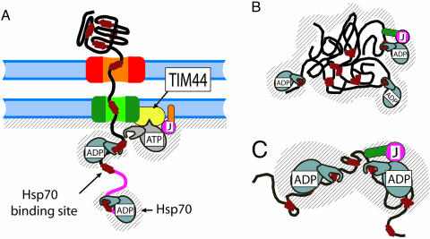
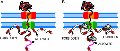
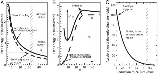
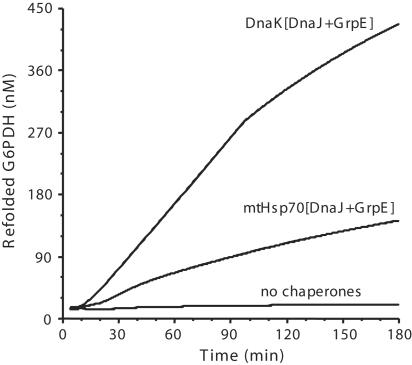
Hsp70s are highly conserved ATPase molecular chaperones mediating the correct folding of de novo synthesized proteins, the translocation of proteins across membranes, the disassembly of some native protein oligomers, and the active unfolding and disassembly of stress-induced protein aggregates. Here, we bring thermodynamic arguments and biochemical evidences for a unifying mechanism named entropic pulling, based on entropy loss due to excluded-volume effects, by which Hsp70 molecules can convert the energy of ATP hydrolysis into a force capable of accelerating the local unfolding of various protein substrates and, thus, perform disparate cellular functions. By means of entropic pulling, individual Hsp70 molecules can accelerate unfolding and pulling of translocating polypeptides into mitochondria in the absence of a molecular fulcrum, thus settling former contradictions between the power-stroke and the Brownian ratchet models for Hsp70-mediated protein translocation across membranes. Moreover, in a very different context devoid of membrane and components of the import pore, the same physical principles apply to the forceful unfolding, solubilization, and assisted native refolding of stable protein aggregates by individual Hsp70 molecules, thus providing a mechanism for Hsp70-mediated protein disaggregation.


Conflict of interest statement

Conflict of interest statement: No conflicts declared.

Figures

Fig. 1.
Fig. 1.
Schematic view of Hsp70’s role in protein translocation and in protein disaggregation and unfolding. (A) MtHsp70·ATP anchors to the pore-associated protein Tim44 and is, thus, ready to lock to a typical hydrophobic binding segment on an entering polypeptide (red rectangles). The nearby J domain of membrane-anchored PAM16/Tim16 (or PAM18/Tim14) (pink) triggers ATP hydrolysis as soon as a typical hydrophobic binding site exits from the pore. PAM16/18 contains a membrane-anchoring domain (orange). While locked onto the polypeptide, mtHsp70·ADP dissociates from Tim44. The shaded region is forbidden to a binding site on the polypeptide, upon association with mtHsp70. (B) Hsp70 interactions with a stable protein aggregate, as in A, with regions (hatched) of forbidden access for an exposed, chaperone-bound polypeptide loop in a misfolded polypeptide. The protein-binding domain (green) of a soluble J domain cochaperone (such as DnaJ, Hsp40) binds the aggregate and entraps freely diffusing Hsp70s within the entropic pulling region of the aggregated substrate by inducing with its J domain (pink) ATP hydrolysis and the locking of Hsp70 onto the polypeptide. (C) Close-up of the regions of mutually excluding volumes in the case of two Hsp70 molecules bound on either side of a misfolded region in the same misfolded polypeptide monomer.
Fig. 2.
Fig. 2.
Schematic view of the excluded volume effects without (A) and with (B) mtHsp70 bound to a translocating polypeptide. (A) Polypeptide conformations that occupy only the mitochondrial matrix space are allowed, whereas polypeptide conformations that would partially occupy the volume already taken by the membrane are forbidden. (B) Binding of mtHsp70 further forbids the polypeptide conformations, such that the chaperone would partially occupy the volume taken by the membrane.
Fig. 3.
Fig. 3.
Free-energy profiles without and with a bound Hsp70 molecule. (A) Free-energy profiles of polypeptide translocation and aggregate unfolding by action of Hsp70 were calculated as a function of a parameter n corresponding to the number of imported residues in the mitochondrial matrix (as in Fig. 1A, solid line) or of exposed flexible segments flanked by the aggregate on one side and the bound chaperone on the other (as in Fig. 1B, solid line for large aggregates and dashed line for small ones) or of flexible amino acids between two independent chaperones bound to the same misfolded polypeptide (as in Fig. 1C, dashed line). The horizontal dot-dashed line is the energy associated with thermal fluctuations. The vertical shaded region at residues 31–33 separates a region (to the left) where entropic pulling prevails from a region (to the right) where entropic pulling is least effective and where Brownian ratchet may still prevent protein backsliding. (B) Free-energy profile of unfolding for a native-like, misfolded or otherwise compact translocating protein in the absence (dashed line) or presence (solid line) of a polypeptide-bound (locked) Hsp70 chaperone on the matrix side of the membrane. The reaction coordinate n is the number of free residues at the preprotein N terminus available for translocation through the pore. (C) Acceleration of thermally driven unfolding as a function of the reduction of the free-energy barrier of unfolding, ΔG−ΔG70. The binding of Hsp70 at increasing distances from the excluded volume region (larger values of n in A) gives rise to a decreasing rate of pulling-biased spontaneous unfolding. However, as long as binding occurs in the entropic binding region (n < 30), some acceleration still occurs. All values of energies and rate accelerations are computed at T = 25°C.
Fig. 4.
Fig. 4.
Time-dependent reactivation of stable G6PDH aggregates. Stable G6PDH were formed at 52°C and reactivated at 30°C in the presence of bacterial DnaK or yeast mtHsp70, the bacterial cochaperones DnaJ, GrpE, and ATP.

References

    1. Nollen E. A. A., Morimoto R. I. J. Cell. Sci. 2002;115:2809–2816. - PubMed
    1. Sangster T. A., Lindquist S., Queitsch C. BioEssays. 2004;26:348–362. - PubMed
    1. Weiss Y. G., Maloyan A., Tazelaar J., Raj N., Deutschman C. S. J. Clin. Invest. 2002;110:801–806. - PMC - PubMed
    1. Picard D. Cell. Mol. Life Sci. 2002;59:1640–1648. - PMC - PubMed
    1. Neupert W., Brunner M. Nat. Rev. Mol. Cell Biol. 2002;3:555–565. - PubMed

Publication types

MeSH terms