. 2006 Apr 18;103(16):6321-5.
doi: 10.1073/pnas.0508593103. Epub 2006 Apr 10.

Bone marrow-derived cells fuse with normal and transformed intestinal stem cells

Affiliations

Bone marrow-derived cells fuse with normal and transformed intestinal stem cells

Adnan Z Rizvi et al. Proc Natl Acad Sci U S A. .

Abstract

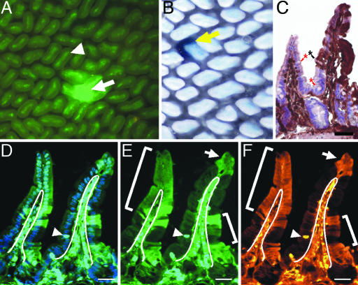
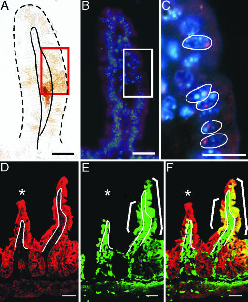
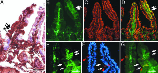
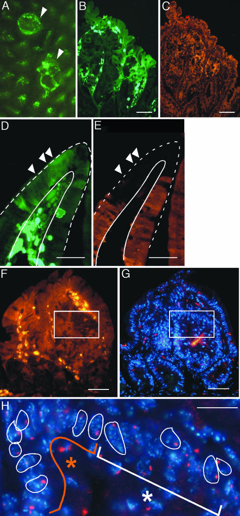
Transplanted adult bone marrow-derived cells (BMDCs) have been shown to adopt the phenotype and function of several nonhematopoietic cell lineages and promote tumorigenesis. Beyond its cancer enhancing potential, cell fusion has recently emerged as an explanation of how BMDCs regenerate diseased heptocytes, contribute to Purkinje neurons and skeletal and cardiac muscle cells, and participate in skin and heart regeneration. Although bone marrow-derived epithelial cells also have been observed in the intestine, fusion as a mechanism has not been investigated. Here, we show that transplanted BMDCs fuse with both normal and neoplastic intestinal epithelium. Long-term repopulation by donor-derived cells was detected in all principal intestinal epithelial lineages including enterocytes, goblet cells, Paneth cells, and enteroendocrine cells, suggesting that the fusion partners of the BMDCs are long-lived intestinal progenitors or stem cells. Fusion of BMDCs with neoplastic epithelium did not result in tumor initiation. Our findings suggest an unexpected role for BMDCs in both regeneration and tumorigenesis of the intestine.


Conflict of interest statement

Conflict of interest statement: No conflicts declared.

Figures

Fig. 1.
Fig. 1.
BMDCs incorporate into intestinal epithelium. (A) Whole-mount preparation of the distal small intestine of a lethally irradiated (12 Gy) recipient mouse transplanted with 1 × 10⁶ EGFP-expressing BMDCs, 6 months after transplantation. Arrow indicates EGFP-positive epithelial cells populating a crypt and an adjacent villus. Arrowhead indicates EGFP-positive nonepithelial cells that populate the villus core of all villi. (B) Whole-mount intestine from a β-gal-expressing bone marrow-transplanted recipient. Yellow arrow indicates β-gal-positive crypt and villus. (C) Tissue section from a transplanted distal small intestine stained with antibodies to EGFP, detected with DAB (brown), and counterstained with hematoxylin. The brown staining represents EGFP-positive epithelium, whereas the EGFP-negative epithelium appears pink/purple. (Controls for antibody staining are found in, which are published as supporting information on the PNAS web site.) EGFP-positive cells populate the epithelium of the right villus. The left villus is negative for EGFP expression. The black asterisk marks a differentiated goblet cell that is EGFP-positive, and the red asterisks denote EGFP-negative goblet cells. (DF) Tissue section of the distal small intestine of a recipient mouse transplanted with 500 EGFP-positive HSCs. Section in D is stained with Hoechst dye (blue) and visualized for EGFP expression (green) by direct fluorescence. (E) EGFP detected by direct fluorescence. EGFP-positive cells populate the villus-associated epithelium and are designated with white brackets and an arrow. White lines designate the boundary between the epithelium and lamina propria. Arrowhead indicates an intraepithelial lymphocyte. (F) Identical tissue section in D and E stained with antibodies to EGFP and detected with Cy3-conjugated secondary antibodies (orange). (Scale bars: 25 μm.)
Fig. 2.
Fig. 2.
Fusion between BMDCs and intestinal epithelium. Intestine from a male recipient transplanted with female donor EGFP-expressing BMDCs analyzed for EGFP expression and Y chromosome. (A) EGFP-stained intestinal epithelium detected with EGFP antibodies and DAB staining (brown). Red rectangle represents a region containing EGFP-positive cells. Epithelium is demarcated with solid and dashed black lines. (B) Y chromosome probe (red) and Hoechst-stained nuclei (blue) detected on the same tissue section in A. EGFP-expressing region is denoted by a white rectangle. The boxed region is magnified in C. Nuclei in C are outlined in white. (DF) Intestinal tissue section from a β-gal-expressing recipient transplanted with EGFP-positive BMDCs was analyzed for coexpression of β-gal and EGFP by confocal microscopy. White line indicates the boundary between the epithelium and lamina propria. White asterisk denotes a villus lacking EGFP epithelial expression. (D) β-gal (red) is uniformly expressed in the intestinal epithelium as detected with antibodies to β-gal and Cy5-conjugated secondary antibodies. (E) EGFP expression (green) on the same tissue section as D detected by direct fluorescence. (F) Merge of β-gal- and EGFP-stained tissue showing colocalization of markers for both donor and recipient populations (yellow). Epithelial cells expressing β-gal only appear red, and EGFP-positive lamina propria cells are green. (Scale bars: 25 μm.)
Fig. 3.
Fig. 3.
EGFP expression is detected in all four principal intestinal epithelial lineages. The intestines from EGFP-expressing BMDC-transplanted recipients were analyzed with markers for intestinal lineages at 6 months after transplantation. (A) Intestine stained with antibodies to EGFP, detected with DAB (brown), and counterstained with hematoxylin. The EGFP-negative epithelium on the right villus appears pink/purple, whereas the EGFP-positive epithelium on the left villus is dark brown. Black asterisk denotes an EGFP-positive goblet cell adjacent to enterocytes (arrows) on the same villus. (BD) Intestine stained with the enterocyte marker cytokeratin. (C) Cytokeratin staining (red) is uniformly distributed in villus-associated epithelium and is unaltered in EGFP-expressing cells (green; B). (D) Merged image from B and D showing colocalization of EGFP and cytokeratin (yellow). Arrows point to EGFP-expressing epithelium. (EG) Intestine stained with an enteroendocrine cell marker. (E) EGFP-expressing cells (green). White arrows point to four of the EGFP-expressing cells on the right villus. (F) Enteroendocrine cells identified with antibodies to serotonin (red) in the nuclear Hoechst dye-stained tissue (blue). Red arrow/arrowhead indicate the serotonin-positive cells. (G) Merged image; serotonin expression in EGFP-expressing enteroendocrine cells (yellow cell and red arrowhead) is similar to expression in WT enteroendocrine cells (red cell and red arrow). (Scale bars: 25 μm.)
Fig. 4.
Fig. 4.
BMDCs fuse with tumor epithelium. (A and B) EGFP-expressing BMDCs incorporate into intestinal adenomas (arrowhead) observed by intestinal whole-mount (A) and tissue (B) sections. (B and C) EGFP-expressing regions (green) have a normal expression pattern of the epithelial marker cytokeratin (orange) when compared to adjacent non-donor-derived areas. (D and E) However, donor-derived regions (green) do not express the intestinal epithelial differentiation marker Fabp (orange). White arrowheads mark several EGFP-expressing tumor epithelial cells. Villus epithelium is demarcated with white lines. (FH) BMDCs fuse with tumor epithelium. (F) Adenoma stained with antibodies for EGFP and Cy3-conjugated secondary antibodies (orange). White box contains both EGFP-positive and WT cells. (G) Adjacent tissue section in F stained with Hoechst nuclear dye (blue) and Y chromosome probe (red). (H) Higher magnification of boxed region. The orange asterisk represents donor-derived population, whereas the white asterisk represents the recipient population. Nuclei are outlined in white. (Scale bars: 25 μm.)

References

    1. Lagasse E., Connors H., Al-Dhalimy M., Reitsma M., Dohse M., Osborne L., Wang X., Finegold M., Weissman I. L., Grompe M. Nat. Med. 2000;6:1229–1234. - PubMed
    1. Rasulov M. F., Vasilchenkov A. V., Onishchenko N. A., Krasheninnikov M. E., Kravchenko V. I., Gorshenin T. L., Pidtsan R. E., Potapov I. V. Bull. Exp. Biol. Med. 2005;139:141–144. - PubMed
    1. Alvarez-Dolado M., Pardal R., Garcia-Verdugo J. M., Fike J. R., Lee H. O., Pfeffer K., Lois C., Morrison S. J., Alvarez-Buylla A. Nature. 2003;425:968–973. - PubMed
    1. Laflamme M. A., Myerson D., Saffitz J. E., Murry C. E. Circ. Res. 2002;90:634–640. - PubMed
    1. Nygren J. M., Jovinge S., Breitbach M., Sawen P., Roll W., Hescheler J., Taneera J., Fleischmann B. K., Jacobsen S. E. Nat. Med. 2004;10:494–501. - PubMed

Publication types

Substances