. 2006 Jul;235(7):1830-44.
doi: 10.1002/dvdy.20821.

Jag2-Notch1 signaling regulates oral epithelial differentiation and palate development

Affiliations

Jag2-Notch1 signaling regulates oral epithelial differentiation and palate development

Liam M Casey et al. Dev Dyn. 2006 Jul.

Abstract

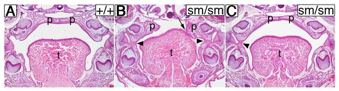
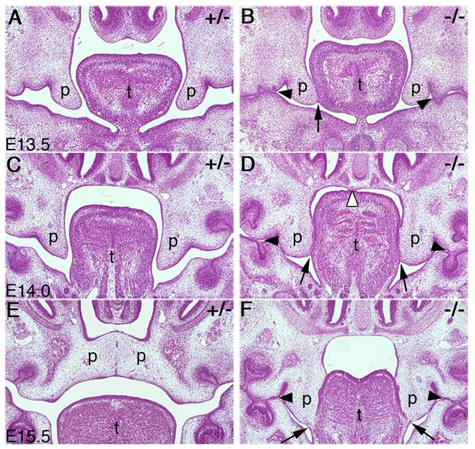
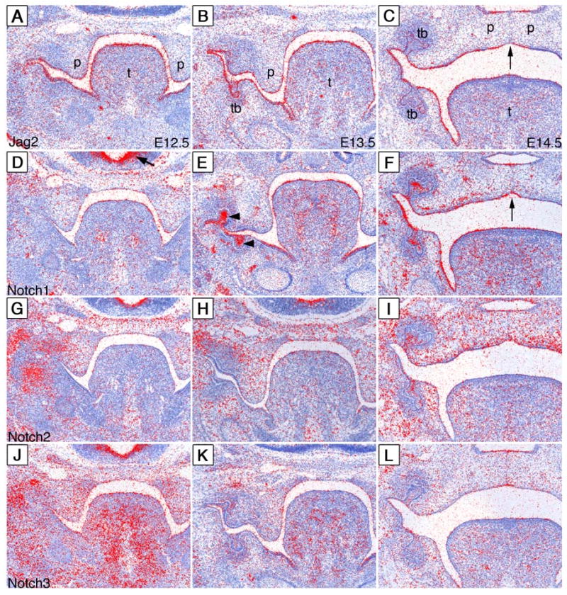
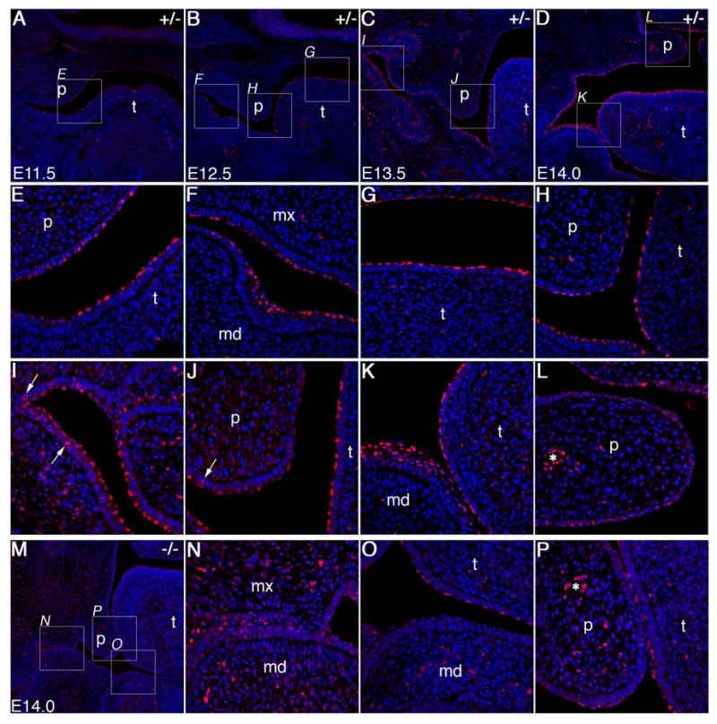
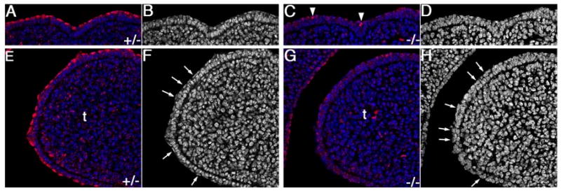
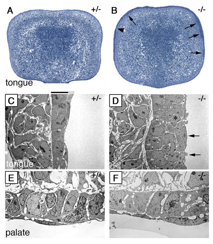
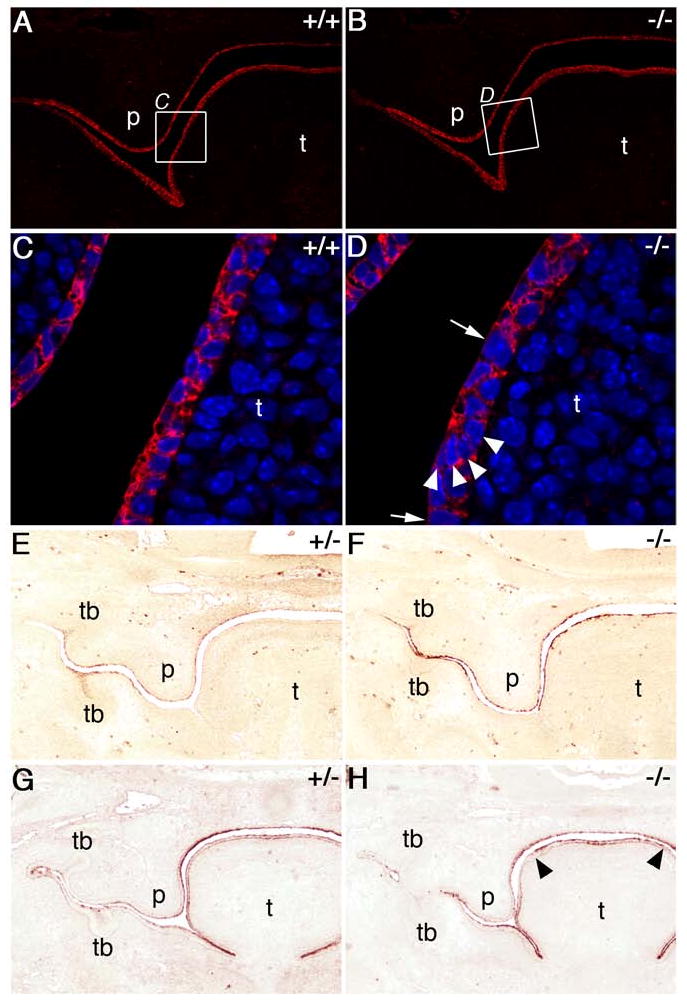
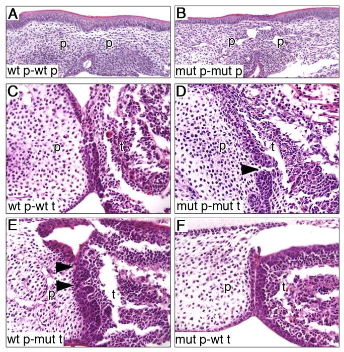
During mammalian palatogenesis, palatal shelves initially grow vertically from the medial sides of the paired maxillary processes flanking the developing tongue and subsequently elevate and fuse with each other above the tongue to form the intact secondary palate. Pathological palate-mandible or palate-tongue fusions have been reported in humans and other mammals, but the molecular and cellular mechanisms that prevent such aberrant adhesions during normal palate development are unknown. We previously reported that mice deficient in Jag2, which encodes a cell surface ligand for the Notch family receptors, have cleft palate associated with palate-tongue fusions. In this report, we show that Jag2 is expressed throughout the oral epithelium and is required for Notch1 activation during oral epithelial differentiation. We show that Notch1 is normally highly activated in the differentiating oral periderm cells covering the developing tongue and the lateral oral surfaces of the mandibular and maxillary processes during palate development. Oral periderm activation of Notch1 is significantly attenuated during palate development in the Jag2 mutants. Further molecular and ultrastructural analyses indicate that oral epithelial organization and periderm differentiation are disrupted in the Jag2 mutants. Moreover, we show that the Jag2 mutant tongue fused to wild-type palatal shelves in recombinant explant cultures. These data indicate that Jag2-Notch1 signaling is spatiotemporally regulated in the oral epithelia during palate development to prevent premature palatal shelf adhesion to other oral tissues and to facilitate normal adhesion between the elevated palatal shelves.


Figures

Fig. 1
Fig. 1
Jag2sm/sm homozygous mutants exhibit cleft palate and aberrant oral epithelial adhesions. Frontal sections of neonatal wildtype (A) and homozygous mutant (B, C) littermates from an intercross of Jag2+/sm heterozygotes. In the wildtype neonate the palatal shelves have fused to form the intact palate (A). In contrast, a Jag2sm/sm homozygous mutant had cleft palate with one palatal shelf adhered to the tongue (B, arrow) and bilateral adhesions between the maxillary and mandibular epithelia (B, arrowheads). Another mutant littermate showed an intact palate with a unilateral maxillary-mandibular adhesion (C, arrowhead). p, palatal shelf; t, tongue.
Fig. 2
Fig. 2
Palate development in the Jag2ΔDSL/ΔDSL mutants. Frontal sections of E13.5 to E15.5 heterozygous (A, C, E) and homozygous (B, D, F) embryos are shown. (A, B) At E13.5, the heterozygous and homozygous mutant embryos had similar sized palatal shelves but palate-tongue adhesion (B, closed arrow) and maxillary-mandibular adhesions (B, arrowheads) are beginning to form in the mutants. (C, D) At E14.0, palate-tongue adhesion in the mutants is often observed bilaterally and is more pronounced (D, arrows). The adhesion between the maxillary and mandibular epithelia is most pronounced in the regions of the mandibular molar tooth buds (D, closed arrowheads). The mutant shown in panel D also had aberrant adhesion between the tongue and the nasal septum (D, open arrowhead). (E, F) At E15.5, the wildtype and heterozygous embryos showed fusion of the palatal shelves and disappearance of the midline epithelial seam (E) whereas the homozygous mutant embryos exhibited bilateral palate-tongue fusion with degeneration of the most ventral aspect of the palate-tongue seam (F, arrows) while the maxillary-mandibular adhesion seam remained intact (F, arrowheads). p, palatal shelf; t, tongue.
Fig. 3
Fig. 3
Expression patterns of Jag2, Notch1, Notch2 and Notch3 mRNA during palate development. Radioactive in situ hybridization of E12.5-E14.5 embryos with cRNA probes to Jag2 (A-C) Notch1 (D-F), Notch2 (G-I), and Notch3 (J-L). mRNA signals are show in red color with the samples counterstained blue. (A) At E12.5, Jag2 mRNA is detected throughout the oral epithelium with the highest level of expression in the tongue epithelium. (B) Jag2 mRNA shows a similar distribution in the oral epithelium of E13.5 embryos with the additional expression in the developing tooth bud epithelium. (C) At E14.5, Jag2 mRNA is still detected in the tongue, mandibular and maxillary epithelia. Jag2 mRNA is also present in the oral and nasal epithelia of the palate but is absent from the epithelial seam between the fusing palatal shelves (arrow). (D) At E12.5, low levels of Notch1 mRNA are detected in the tongue and palatal epithelia, whereas strong Notch1 mRNA expression is detected in the ventricular zone of the brain (arrow). (E) At E13.5, Notch1 mRNA is highly expressed in the tongue epithelium, the tooth bud epithelium (arrowheads), mandibular epithelium and the lateral maxillary epithelia. Little Notch1 mRNA expression is detected in the palatal epithelium at this stage. (F) At E14.5, embryos continue to show significant amounts of Notch1 mRNA expression in the tongue and throughout the lateral oral epithelium. The epithelial seam between the fusing palatal shelves shows little Notch1 expression (arrow). (G-I) Notch2 mRNA is expressed throughout the craniofacial mesenchyme from E12.5 to E14.5 with the oral epithelial expression restricted to the tooth buds (I). (J) Notch3 mRNA is expressed abundantly in the craniofacial mesenchyme at E12.5 and is weakly detected in the oral epithelium at this stage (J). (K) By E13.5, Notch3 expression is dramatically reduced in mesenchyme and little expression is observed in the oral epithelium. (L) At E14.5, low levels of Notch3 mRNA are detected in the tooth bud and palatal epithelium. p, palatal shelf; t, tongue; tb, tooth bud.
Fig. 4
Fig. 4
Spatiotemporal patterns of Notch1 activation in the oral epithelium during palate development in Jag2 heterozygous and homozygous mutant embryos. Activated Notch1 staining is shown in red, with DAPI staining of nuclei shown in blue. (A) At E11.5, acitvated-Notch1 is detected specifically in the outer layer cells of oral epithelium covering the tongue primordium in wildtype and heterozygous embryos. A few periderm cells at the oral surface of the maxillary processes are also positively stained. The boxed region is shown in higher magnification in panel E. (B) At E12.5, activated-Notch1 staining is observed throughout the periderm layer of the tongue and mandibular oral epithelia. Significantly lower but detectable levels of activated Notch1 protein are also observed in the outer layer of the maxillary and palatal epithelia. Boxed areas are shown at higher magnification in panels F-H. (C) At E13.5, embryos show abundant activated-Notch1 protein in the nuclei of the periderm and suprabasal cells of lateral oral epithelium, tongue epithelium and lateral maxillary epithelium. Activated-Notch1 is detected in one to two cell layers of the lateral oral and tooth bud epithelium but only in a single cell layer of the tongue, the periderm layer. Boxed regions are shown at higher magnification in panels I and J. Cells in the basal cell layer are occasionally positive for activated-Notch1 (arrows in I and J). In contrast to the rest of the oral epithelia, very few periderm cells in the medial and distal epithelium of the palatal shelves are positive for activated Notch1 protein (J). (D) At E14.0, the pattern of activated-Notch1 staining is largely the same with strong staining in the periderm and suprabasal cell layers of the lateral oral epithelium and in the periderm cells of the tongue. Higher magnification micrographs of boxed regions are shown in panels K and L. The elevated pre-fusion palatal shelves had little activated Notch1 staining in the surface epithelium compared with the vascular endothelial cells (L, asterisk) or other regions of the oral epithelium which are highly positive (K). (M) Low magnification view of a frontal section through the anterior oral cavity of an E14.0 Jag2ΔDSL/ΔDSL mutant embryo showing significantly reduced levels of activated-Notch1 throughout the oral epithelium. Higher magnification views of the regions boxed in panel M reveals that cells at the surface of lateral oral epithelia (N), of the tongue (O, P), and of mandible (N, P) exhibited either low levels or no staining for activated Notch1 in the nuclei, although endothelial cells lining blood vessels in the mutant palate had strong staining for activated Notch1 comparable to that in the heterozygous palate (asterisks in L and P). md, mandible; mx, maxilla; p, palatal shelf; t, tongue.
Fig. 5
Fig. 5
Jag2 is required for Notch1 activation and proper periderm differentiation in the oral epithelium. Confocal micrographs of dorsal (A-D) and lateral (E-H) regions of the developing tongue in heterozygous (A, B, E, F) and homozygous mutant (C, D, G, H) embryos at E12. Even at this early stage of oral epithelial differentiation, Jag2ΔDSL/ΔDSL mutant embryos show dramatically reduced activated Notch1 staining in the tongue epithelium (C, G), compared with that in heterozygous littermates (A, E). Very few cells in the homozygous mutant tongue epithelium exhibited high levels of activated Notch1 staining (arrowheads in C) whereas almost all surface layer cells in the wildtype and heterozygous tongue showed high levels of nuclear staining for activated Notch1 (A, E). (B, D) Black-white images of the same fields shown in A and E, respectively, with nuclear DAPI staining shown in white. The wildtype and heterozygous mutant tongue epithelium at this stage consists of two layers with the surface layer being the flat periderm cells. (D, H) Black-white images of the same fields shown in C and G, respectively, with nuclear DAPI staining shown in white. The homozygous mutant tongue epithelium at this stage also consists of two layers but the nuclei of the surface layer cells are irregular and do not resemble the flat nuclei characteristic of the wildtype periderm cells (arrows in F and H). t, tongue.
Fig. 6
Fig. 6
Defects in epithelial organization in the Jag2ΔDSL/ΔDSL mutants. (A, B) Semi-thin (2μm) sections of E13.0 wildtype (A) and Jag2ΔDSL/ΔDSL mutant (B) tongue stained with toluidine blue. The wildtype tongue epithelium shows an even thickness over the entire circumference of the tongue. In contrast the mutant tongue shows uneven thickness with patches of thick (B, arrows) and thin epithelia (B, arrowhead). (C) Electron micrographs of the lateral tongue epithelium show that the wildtype tongue epithelium has a basal layer of cuboidal shaped cells covered by a continuous sheet of flattened periderm cells. (D) Jag2ΔDSL/ΔDSL mutant tongue epithelium is multiple cell layers thick in some regions and appears disorganized. The periderm layer of the mutant tongue appears discontinuous as some of these cells appear less flattened and lack long cellular processes (D, arrows). In some cases cells attached to the basal lamina are exposed to the surface in the mutant tongue epithelium (D, asterisk). (E, F) Electron micrographs of the epithelia at the tip of the palatal shelves showing no significant differences between the wildtype (E) and Jag2ΔDSL/ΔDSL mutant (F) palatal epithelia.
Fig. 7
Fig. 7
Analysis of epithelial differentiation markers in Jag2ΔDSL/ΔDSL homozygotes. Wildtype (A, C, E, G) and mutant (B, D, F, H) embryo sections immunostained for E-cadherin (A-D) or keratin-17 (E-H). A, B) At E13.0 E-cadherin is abundant throughout the oral epithelium of both wildtype and Jag2ΔDSL/ΔDSL-mutant embryos. C, D) Higher magnification views of the regions boxed in panels A and B showing that E-cadherin is detected in all cells of the oral epithelium but some regions of the mutant epithelium are one cell layer thick (D, arrows) while others are multiple cell layers thick with reduced E-cadherin staining between cells (D, arrowheads). E-H) E13.5 embryos fixed with bouin's fixative and immunostained for keratin-17 (E, F) or pan-cytokeratin (G, H). Wildtype embryos (E) show some keratin-17 expression in the stellate reticulum of the tooth buds, the periderm flanking the mandibular and maxillary tooth buds, and the periderm layer of the palate and nasal epithelium. At this stage the wildtype tongue epithelium is only weakly stained. In comparison, homozygous mutant littermates (F) show much stronger staining for keratin-17 throughout the oral epithelium, especially the tongue. Additionally, the mutant tongue epithelium appears irregular in thickness compared to wildtype tongue epithelium. Pan-cytokeratin staining of wildtype (G) and mutant (H) embryos shows that general keratinization is unaltered throughout much of the mutant oral epithelium except for the periderm cell layer on the dorsal side of the tongue which shows stronger staining compared to wildtype embryos (H, arrowheads). p, palate; t, tongue; tb, tooth bud.
Fig. 8
Fig. 8
Palate-tongue recombination organ culture assays. (A, B) Pairings of wildtype (A) and Jag2ΔDSL/ΔDSL homozygous mutant (B) palatal shelves both showed complete fusion and disappearance of the epithelial seam after 3 days in culture. (C-F) In different pairings of palate and tongue tissues all pairings show association of palate and tongue epithelia but only the mutant palate-mutant tongue (D) and wildtype palate-mutant tongue (E) pairings showed an irregular and discontinuous epithelial seam (arrowheads in D and E). mut, mutant; p, palatal shelf; t, tongue; wt, wildtype.
Fig. 9
Fig. 9
Expression analysis of Fgf10 and Fgfr2b in Jag2ΔDSL/ΔDSL mutant embryos. mRNA signals are shown in red on sections counterstained blue. (A, B) Jag2ΔDSL/+ heterozygous and Jag2ΔDSL/ΔDSL homozygous mutant littermates show comparable levels of Fgf10 mRNA expression in palatal and tongue mesenchyme at E13.5. (C-F) Fgfr2b mRNA is highly expressed throughout the oral epithelium of wildtype and Jag2ΔDSL/+ heterozygous embryos at E13.5 (C) and E14.5 (E) whereas its levels appear consistently reduced in the dorsal tongue epithelium of Jag2ΔDSL/ΔDSL homozygous mutants at these developmental stages (D, F). p, palatal shelf; t, tongue.
Fig. 10
Fig. 10
Analysis of Tgfβ3 signaling in Jag2ΔDSL/ΔDSL mutant embryos. (A, B) At E14.5, Tgfβ3 mRNA expression in the oral epithelia is restricted to the MEE of the palatal shelves in both Jag2ΔDSL/+ heterozygous and Jag2ΔDSL/ΔDSL homozygous mutant embryos, although the palatal shelves adhered to the lateral sides of the tongue (arrow in B) in the homozygous mutant. No Tgfβ3 mRNA expression was detected at the site of maxillary-mandibular adhesion (B, arrowhead) in the homozygous mutant. (C, D) Mmp13 mRNA expression overlaps with that of Tgfβ3 mRNA in the MEE of the palatal shelves in both Jag2ΔDSL/+ heterozygous and Jag2ΔDSL/ΔDSL homozygous mutant palatal shelves (D, arrow). Mmp13 is not detected at the site of maxillary-mandibular adhesion in the Jag2ΔDSL/ΔDSL mutant embryo (D, arrwohead). p, palatal shelf; t, tongue.
Fig. 11
Fig. 11
Cell death is detected in the palate-tongue epithelial seam but not in the maxillary-mandibular epithelial seam in the Jag2ΔDSL/ΔDSL mutant embryos. (A) In the Jag2ΔDSL/+ heterozygous embryo, a number of apoptotic cells are detected in the epithelial seam between the fusing palatal shelves (arrowheads). (B, C) Jag2ΔDSL/ΔDSL mutant embryos show numerous apoptotic cells in the palate-tongue epithelial seam (B, arrowheads) but not the maxillary-mandibular seam (C). Regions of ossification are stained non-specifically. mdm, mandibular molar; mxm, maxillary molar; oc, ossification center; p, palatal shelf; t, tongue.

References

    1. Akiyama M, Smith LT, Yoneda K, Holbrook KA, Hohl D, Shimizu H. Periderm cells form cornified cell envelope in their regression process during human epidermal development. J Invest Dermatol. 1999;112:903–909. - PubMed
    1. Alappat SR, Zhang Z, Suzuki K, Zhang X, Liu H, Jiang R, Yamada G, Chen Y. The cellular and molecular etiology of the cleft secondary palate in Fgf10 mutant mice. Dev Biol. 2005;277:102–113. - PubMed
    1. Artavanis-Tsakonas S, Rand MD, Lake RJ. Notch signaling: cell fate control and signal integration in development. Science. 1999;284:770–776. - PubMed
    1. Blavier L, Lazaryev A, Groffen J, Heisterkamp N, DeClerck YA, Kaartinen V. TGF-beta3-induced palatogenesis requires matrix metalloproteinases. Mol Biol Cell. 2001;12:1457–1466. - PMC - PubMed
    1. Cecconi F, Alvarez-Bolado G, Meyer BI, Roth KA, Gruss P. Apaf1 (CED-4 homolog) regulates programmed cell death in mammalian development. Cell. 1998;94:727–737. - PubMed

Publication types

MeSH terms