

Extended surgical resection for xanthogranulomatous cholecystitis mimicking advanced gallbladder carcinoma: A case report and review of literature
- PMID: 16610041
- PMCID: PMC4087666
- DOI: 10.3748/wjg.v12.i14.2293
Extended surgical resection for xanthogranulomatous cholecystitis mimicking advanced gallbladder carcinoma: A case report and review of literature
Abstract
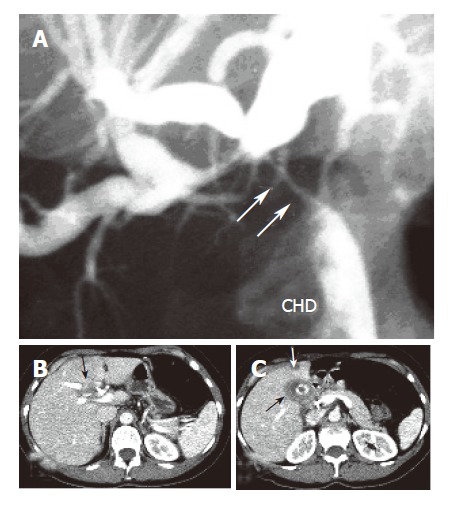
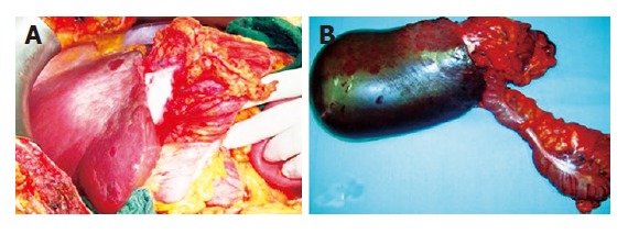
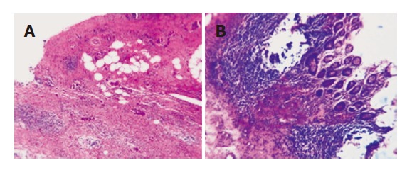
Xanthogranulomatous cholecystitis (XGC) is a destructive inflammatory disease of the gallbladder, rarely involving adjacent organs and mimicking an advanced gallbladder carcinoma. The diagnosis is usually possible only after pathological examination. A 46 year-old woman was referred to our center for suspected gallbladder cancer involving the liver hilum, right liver lobe, right colonic flexure, and duodenum. Brushing cytology obtained by endoscopic retrograde cholangiography (ERC) showed high-grade dysplasia. The patient underwent an en-bloc resection of the mass, consisting of right lobectomy, right hemicolectomy, and a partial duodenal resection. Pathological examination unexpectedly revealed an XGC. Only six cases of extended surgical resections for XGC with direct involvement of adjacent organs have been reported so far. In these cases, given the possible coexistence of XGC with carcinoma, malignancy cannot be excluded, even after cytology and intraoperative frozen section investigation. In conclusion, due to the poor prognosis of gallbladder carcinoma on one side and possible complications deriving from highly aggressive inflammatory invasion of surrounding organs on the other side, it seems these cases should be treated as malignant tumors until proven otherwise. Clinicians should include XGC among the possible differential diagnoses of masses in liver hilum.
Figures
References
-
- Kwon AH, Matsui Y, Uemura Y. Surgical procedures and histopathologic findings for patients with xanthogranulomatous cholecystitis. J Am Coll Surg. 2004;199:204–210. - PubMed
-
- Furuta A, Ishibashi T, Takahashi S, Sakamoto K. MR imaging of xanthogranulomatous cholecystitis. Radiat Med. 1996;14:315–319. - PubMed
-
- Enomoto T, Todoroki T, Koike N, Kawamoto T, Matsumoto H. Xanthogranulomatous cholecystitis mimicking stage IV gallbladder cancer. Hepatogastroenterology. 2003;50:1255–1258. - PubMed
-
- Pinocy J, Lange A, König C, Kaiserling E, Becker HD, Kröber SM. Xanthogranulomatous cholecystitis resembling carcinoma with extensive tumorous infiltration of the liver and colon. Langenbecks Arch Surg. 2003;388:48–51. - PubMed
-
- Maeda T, Shimada M, Matsumata T, Adachi E, Taketomi A, Tashiro Y, Tsuneyoshi M, Sueishi K, Sugimachi K. Xanthogranulomatous cholecystitis masquerading as gallbladder carcinoma. Am J Gastroenterol. 1994;89:628–630. - PubMed
Publication types
MeSH terms
LinkOut - more resources
Full Text Sources
Medical
Research Materials
