

Changes in growth factor and cytokine expression in biliary obstructed rat liver and their relationship with delayed liver regeneration after partial hepatectomy
- PMID: 16610056
- PMCID: PMC4087684
- DOI: 10.3748/wjg.v12.i13.2053
Changes in growth factor and cytokine expression in biliary obstructed rat liver and their relationship with delayed liver regeneration after partial hepatectomy
Abstract
Aim: To study the effects of obstructive jaundice on liver regeneration after partial hepatectomy.
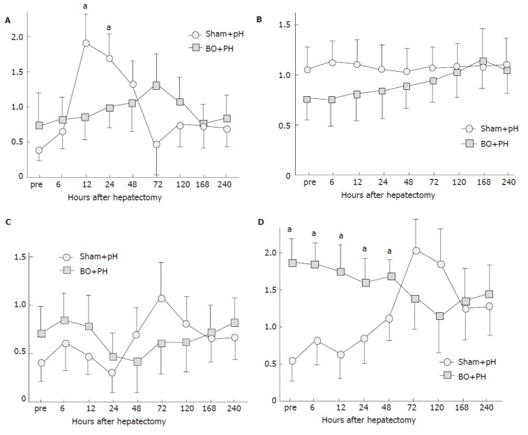
Methods: Hepatocyte growth factor (HGF), its receptor, c-Met, vascular endothelial growth factor (VEGF) and transforming growth factor-beta1 (TGF-beta1) mRNA expression in both liver tissue and isolated liver cells were investigated after biliary obstruction (BO) by quantitative reverse-transcription polymerase chain reaction (RT-PCR) using a LightCycler. Immunohistochemical staining for desmin and alpha-smooth muscle actin (alpha-SMA) was also studied. Regenerating liver weight and proliferating cell nuclear antigen (PCNA) labeling index, and growth factor expression were then evaluated after 70% hepatectomy with concomitant internal biliary drainage in BO rats or sham-operated rats.
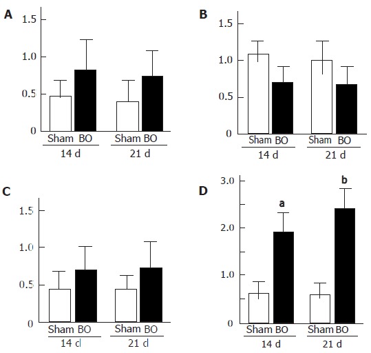
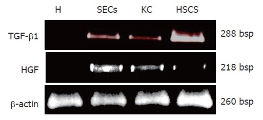
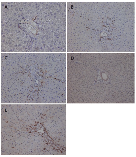
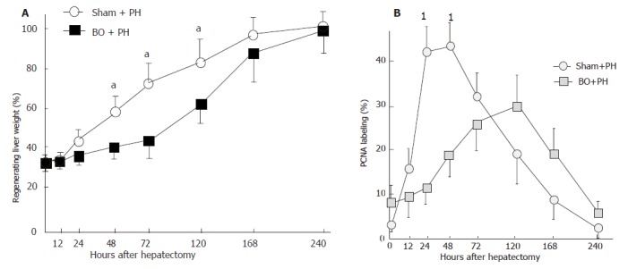
Results: Hepatic TGF-beta1 mRNA levels increased significantly 14 days after BO, and further increased with duration of cholestasis. Meanwhile, HGF and VEGF tended to increase, but was not significant. In cell isolates, TGF-beta1 mRNA was found mainly in the hepatic stellate cell (HSC) fraction. Immunohistochemical studies revealed an increased number of HSCs (desmin-positive cells) and activated HSCs (alpha-SMA-positive cells) in portal areas after BO. In a hepatectomy model, liver regeneration was delayed in BO rats, as compared to sham-operated rats. TGF-beta1 mRNA was significantly up-regulated up to 48 h after hepatectomy, and the earlier HGF mRNA peak was lost in BO rats.
Conclusion: BO induces HSCs proliferation and activation, leading to up-regulation of TGF-beta1 mRNA and suppression of HGF mRNA in livers. These altered expression patterns may be strongly involved in delayed liver regeneration after hepatectomy with obstructive jaundice.
Figures
References
-
- Miyazaki M, Ito H, Nakagawa K, Ambiru S, Shimizu H, Shimizu Y, Kato A, Nakamura S, Omoto H, Nakajima N, et al. Aggressive surgical approaches to hilar cholangiocarcinoma: hepatic or local resection. Surgery. 1998;123:131–136. - PubMed
-
- Boerma EJ. Research into the results of resection of hilar bile duct cancer. Surgery. 1990;108:572–580. - PubMed
-
- Aronson DC, Chamuleau RA, Frederiks WM, Bosman DK, Oosting J. The effect of extrahepatic cholestasis on liver regeneration after partial hepatectomy in the rat. Liver. 1995;15:242–246. - PubMed
-
- Mizuno S, Nimura Y, Suzuki H, Yoshida S. Portal vein branch occlusion induces cell proliferation of cholestatic rat liver. J Surg Res. 1996;60:249–257. - PubMed
MeSH terms
Substances
LinkOut - more resources
Full Text Sources
Miscellaneous
