. 2006 Apr 12:2:12.
doi: 10.1186/1746-6148-2-12.

Degenerative suspensory ligament desmitis as a systemic disorder characterized by proteoglycan accumulation

Affiliations

Degenerative suspensory ligament desmitis as a systemic disorder characterized by proteoglycan accumulation

Jaroslava Halper et al. BMC Vet Res. .

Abstract

Background: Degenerative suspensory ligament desmitis (DSLD) is a debilitating disorder thought to be limited to suspensory ligaments of Peruvian Pasos, Peruvian Paso crosses, Arabians, American Saddlebreds, American Quarter Horses, Thoroughbreds, and some European breeds. It frequently leads to persistent, incurable lameness and need to euthanize affected horses. The pathogenesis remains unclear, though the disease appears to run in families. Treatment and prevention are empirical and supportive, and not effective in halting the progression of the disease. Presently, the presumptive diagnosis of DSLD is obtained from patient signalment and history, clinical examination, and ultrasonographic examination of clinically affected horses, and is confirmed at post mortem examination. Presently, there are no reliable methods of diagnosing DSLD in asymptomatic horses. The goal of this study was to characterize and define the disorder in terms of tissue involvement at the macroscopic and microscopic levels.

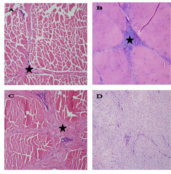
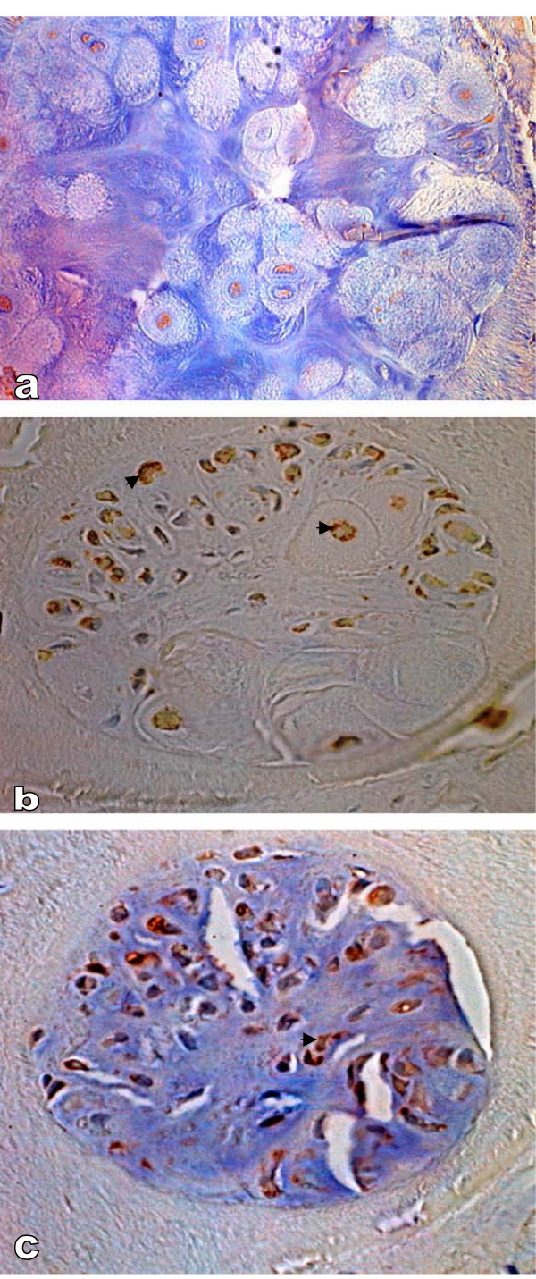
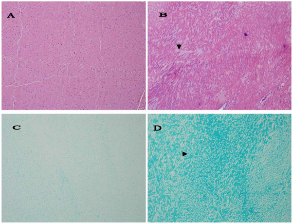
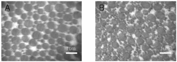
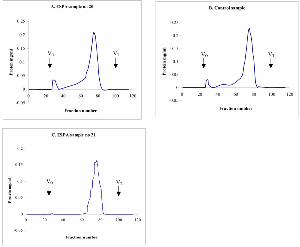
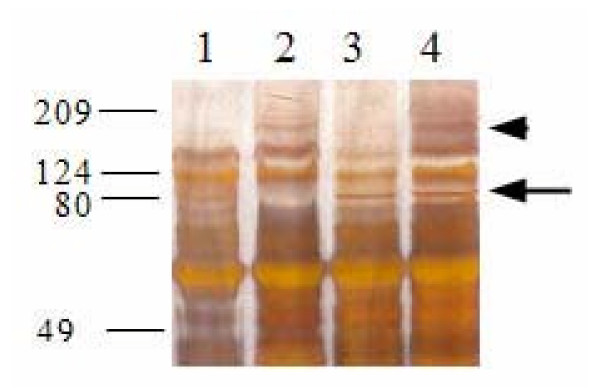
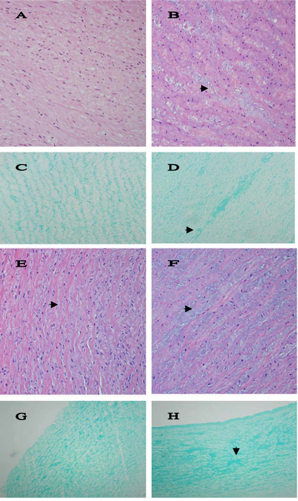
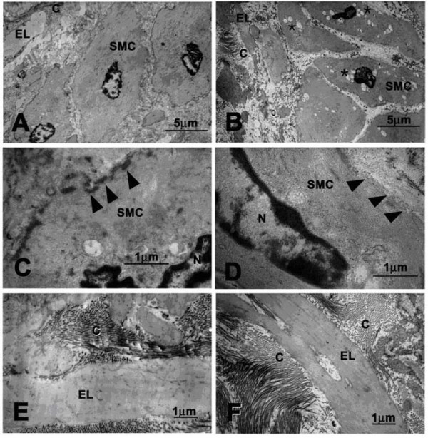
Results: We examined tissues and organs from 28 affected horses (22 Peruvian Pasos, 6 horses of other breeds) and from 8 control horses. Histopathological examination revealed the presence of excessive amounts of proteoglycans in the following tissues removed from DSLD-affected horses: suspensory ligaments, superficial and deep digital flexor tendons, patellar and nuchal ligaments, cardiovascular system, and sclerae. Electron microscopy demonstrated changes in diameters of collagen fibrils in the tendon, and in smooth muscle cells of the media of the aorta compatible with increased cell permeability in DSLD-affected cells. Separation of tendon extracts by gel chromatography revealed the presence of additional proteoglycan(s) in extracts from affected, but not control extracts.

Conclusion: This study demonstrates for the first time that DSLD, a disease process previously thought to be limited to the suspensory ligaments of the distal limbs of affected horses, is in fact a systemic disorder involving tissues and organs with significant connective tissue component. Abnormal accumulation of proteoglycans between collagen and elastic fibers rather than specific collagen fibril abnormalities is the most prominent histological feature of DSLD. Because of this observation and because of the involvement of many other tendons and ligaments beside the suspensory ligament, and of non-ligamentous tissue we, therefore, propose that equine systemic proteoglycan accumulation or ESPA rather than DSLD is a more appropriate name for this condition.


Figures

Figure 1
Figure 1
Comparison of normal and DSLD-affected tendons. A. Only thin septa (*) separate bundles of collagen and elastic fibers in a normal tendon. Hematoxylin & eosin, magnification × 200. B. A section of DSLD-affected tendon reveals PG deposits (*) between collagen fibers and in septa. Hematoxylin & eosin, magnification × 200. C. A section of tendon from a horse without DSLD shows the presence of fibrosis or scar tissue (*) between collagen fibers and in septa. Hematoxylin & eosin, magnification × 200. D. A proliferative lesion found in one DSLD case consists of swirls of active fibroblasts in young, well vascularized tissue. Hematoxylin & eosin, magnification × 200.
Figure 2
Figure 2
Immunostaining of metaplastic cartilage. A. Immunostaining reveals very little decorin, magnification × 500. B. The cartilage reveals the presence of aggrecan (▶) in the cytoplasm, magnification × 400. C. The same chondrocytes were also positive for biglycan (▶), magnification × 400. Countestain: hematoxylin.
Figure 3
Figure 3
Histopathological changes in nuchal ligament. A. Only thin septa separate bundles of collagen and elastic fibers in a normal nuchal ligament. Hematoxylin & eosin, magnification × 200 (× 200). B. In DSLD – affected tissue streaks of proteoglycans (▶) separate bundles of collagen and elastic fibers. Hematoxylin & eosin, magnification × 200. C. Alcian blue stains very lightly normal nuchal ligament, magnification × 200. D. PGs accumulated among bundles stain intensively with alcian blue (▶) in DSLD-affected nuchal ligament.
Figure 4
Figure 4
Electron micrographs of normal and DSLD-affected tendon. A. A cross-section of normal tendon reveals that most collagen fibrils have fairly large diameters. B. A marked increase in small collagen fibrils was observed in cross-sections of DSLD-affected tendon.
Figure 5
Figure 5
Sepharose CL-2B chromatography of PGs in extracts from normal and DSLD-affected tendons. Guanidium HCl extracts from midmetacarpal portions of SDFTs from 2 affected horses (no. 20 and 21, Table 1) and from one control horse (no. 1, Table 3) were separated on a molecular sieve Sepharose CL-2B column (1.3 × 110 cm, equilibrated and eluted in 4 M guanidine HCl) at 0.1 ml/min. The majority of PGs eluted in one wide peak (fractions 66–81).
Figure 6
Figure 6
SDS-PAGE of Sepharose CL-2B peaks. Aliquots of pooled Sepharose CL-2B fractions were precipitated with 100% ethanol, dissolved in 15 μl of 100 mM Tris buffer, adjusted to pH 8.0 with concentrated acetic acid and digested with 20 mU of chondroitinase ABC at 37°C for 24 h. Chondroitinase ABC digested samples and non-digested replicate aliquots were separated on silver stained 10% SDS-polyacrylamide gels. Lane 1: untreated sample from control SDFT no. 1; lane 2: untreated sample from DSLD SDFT no.8 (Table 1); lane 3: chondroitinase ABC treated sample from control SDFT no. 1; lane 4: chondroitinase ABC treated sample from SDFT no. 8 (Table 1).
Figure 7
Figure 7
Histopathological changes in arteries. A. The media of normal aortic arch shows orderly aligned elastic fibers and very little PGs between them. Hematoxylin & eosin, magnification × 200. B. Small pools of PGs (▶) separate fibers and cells in the media of DSLD-affected aortic arch. Hematoxylin & eosin, magnification × 200. C. Alcian blue stains material aligned closely with elastic fibers in normal arch, magnification × 200. D. Pools of PGs (▶) stain strongly with alcian blue in the media of DSLD-affected arch, magnification × 200. E. The media of normal coronary artery shows orderly aligned elastic fibers separated by thin layers of PGs (▶) between them. Hematoxylin & eosin, magnification × 200 (× 200). F. Small pools of PGs (▶) separate fibers and cells in the media of DSLD-affected coronary artery. Hematoxylin & eosin, magnification × 200. G. Alcian blue stains material aligned closely with elastic fibers in the media of normal coronary artery, magnification × 100. H. Pools of PGs (▶) stain strongly with alcian blue in the media of DSLD-affected coronary artery, magnification × 100.
Figure 8
Figure 8
Ultrastructural features of normal and DSLD-affected aorta. A. Normal smooth muscle cell from media from a healthy aorta. B. A smooth muscle cell from the media of DSLD-affected aorta reveals the presence of numerous cytoplasmic vacuoles (*). C. normal smooth muscle cell has a well defined cell membrane (▶). D. The cell membrane of a diseased media is disrupted and missing in places (▶). E and F. Organization of collagen fibrils and elastic fibers is similar in normal (E) and DSLD aorta (F). SMC: smooth muscle cell, EL: elastic lamina, C: collagen fibrils.

References

    1. Dowling BA, Dart AJ, Hodgson DR, Smith RKW. Superficial digital flexor tendonitis in the horse. Equine Vet J. 2000;32:369–378. - PubMed
    1. Genovese RL. Prognosis of superficial digital flexor tendon and suspensory ligament injuries. Proc Am Assoc Equine Pract. 1993. pp. 17–19.
    1. Goodship AE. The pathophysiology of flexor tendon injury in the horse. Equine Vet Educ. 1993;5:23–29.
    1. Peloso JG, Mundy GD, Cohen ND. Prevalence of, and factors associated with musculoskeletal racing injuries of Thoroughbreds. J Am Vet Med Assoc. 1994;204:620–626. - PubMed
    1. Bramlage LR, Hogan PM. Career results of 137 Thoroughbred racehorses that have undergone superior check ligament desmotomy for treatment of tendinitis. Proc Am Assoc Equine Pract. 1996. pp. 162–163.

LinkOut - more resources