. 2006 Feb;8(2):104-11.
doi: 10.1593/neo.05622.

The role of c-KIT in tumorigenesis: evaluation in canine cutaneous mast cell tumors

Affiliations

The role of c-KIT in tumorigenesis: evaluation in canine cutaneous mast cell tumors

Joshua D Webster et al. Neoplasia. 2006 Feb.

Abstract

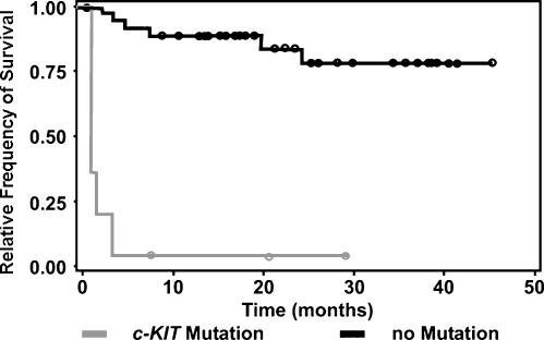
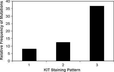
The c-KIT proto-oncogene has been implicated in the pathogenesis of several neoplastic diseases, including gastrointestinal stromal tumors and mastocytosis in humans, and mast cell tumors (MCTs) in canines. Cutaneous MCTs are common neoplasms in dogs and have a variable biologic behavior. The goal of this study was to define the prognostic significance of c-KIT mutations identified in canine MCTs and the associations between c-KIT mutations, KIT localization, and KIT expression levels. Microdissection and polymerase chain reaction were performed on 60 MCTs to identify c-KIT mutations. Anti-KIT antibodies were used for immunohistochemical evaluation of KIT localization. Forty-two MCTs were included in a tissue microarray, and KIT expression was quantified using immunofluorescence. Canine MCTs with c-KIT mutations were significantly associated with an increased incidence of recurrent disease and death. c-KIT mutations were also significantly associated with aberrant protein localization; however, the level of KIT expression did not correlate with either c-KIT mutations or changes in protein localization. Considering the high prevalence of canine MCTs and the central role of c-KIT in the tumorigenesis of certain tumors, canine MCTs are an excellent model for characterizing the role of c-KIT in neoplastic diseases and is a potential target for novel therapeutic agents in clinical trials.


Figures

Figure 1
Figure 1
LCM of neoplastic canine cutaneous MCTs (original magnification, x 10). LCM was performed using archival formalin-fixed paraffin-embedded tissue sections. DNA was extracted from captured cells, and PCR amplification was performed to identify c-KIT mutations. (A) Hematoxylin-stained section of MCT prior to microdissection. (B) Section of MCT following microdissection. (C) Laser capture microdissected cells adhered to cap.
Figure 2
Figure 2
A 2% agarose gel of PCR-amplified c-KIT exon 11 and intron 11 from LCM-extracted DNA from canine MCTs. L: 100-bp ladder; M: heterozygous for normal allele (191 bp) and mutant allele (250 bp), with an upper band representing heterodimerization of normal and mutant alleles; N: 191-bp homozygous normal allele; NC: negative control (no template).
Figure 3
Figure 3
Sections of canine cutaneous MCTs (skin) stained with anti-KIT antibodies and counterstained with hematoxylin (original magnification, x 100, oil) representing three patterns of KIT localization identified in neoplastic canine mast cells. (A) KIT staining pattern I, consisting of perimembrane protein localization. (B) KIT staining pattern II, consisting of focal to stippled cytoplasmic staining. (C) KIT staining pattern III, consisting of diffuse cytoplasmic staining.
Figure 4
Figure 4
Kaplan-Meier survival curve: relative frequency of survival versus time in months for canine cutaneous MCT patients with and without identified c-KIT mutations. The presence of duplication mutation in the c-KIT proto-oncogene was significantly associated with a decreased survival duration [P = .0068, HR = 6.23 (1.66–23.40)].
Figure 5
Figure 5
Correlation between ITD c-KIT mutations and KIT protein localization in canine MCTs. A significant association was found between the presence of c-KIT mutations and the cellular localization of KIT in canine MCTs (P = .046). Seven of nine (77.8%) MCTs with ITD c-KIT mutations had aberrant KIT localization in neoplastic MCTs.

References

    1. Chan PM, Ilangumaran S, La Rose J, Chakrabartly A, Rottapel R. Autoinhibition of the KIT receptor tyrosine kinase by cystolic juxtamembrane region. Mol Cell Biol. 2003;23:3067–3078. - PMC - PubMed
    1. Yarden Y, Kuang W, Yang-Feng T, Coussens L, Munemitsu S, Dull TJ, Chen E, Schlessinger J, Francke U, Ullrich A. Human proto-oncogene c-kit: a new cell surface receptor tyrosine kinase for an unidentified ligand. EMBOJ. 1987;6:3341–3351. - PMC - PubMed
    1. Yarden Y, Escobedo JA, Kuang WJ, Yang-Feng TL, Daniel TO, Tremble PM, Chen EY, Ando ME, Harkins RN, Francke U, et al. Structure of the receptor for platelet-derived growth factor helps define a family of closely related growth factor receptors. Nature. 1986;323:226–232. - PubMed
    1. Huang E, Nocka K, Beler DR, Chu TY, Buck J, Lahm HW, Wellner D, Leder P, Besmer P. The hematopoietic growth factor KL is encoded by the Sl locus and is the ligand of the c-kit receptor, the gene product of the W locus. Cell. 1990;63:225–233. - PubMed
    1. Nocka K, Buck J, Levi E, Besmer P. Candidate ligand for the c-kit transmembrane kinase receptor: KL, a fibroblast derived growth factor stimulates mast cells and erythroid progenitors. EMBO J. 1990;9:3287–3294. - PMC - PubMed

Publication types

LinkOut - more resources