. 2006 Feb;8(2):143-52.
doi: 10.1593/neo.05697.

Impact of extracellular acidity on the activity of P-glycoprotein and the cytotoxicity of chemotherapeutic drugs

Affiliations

Impact of extracellular acidity on the activity of P-glycoprotein and the cytotoxicity of chemotherapeutic drugs

Oliver Thews et al. Neoplasia. 2006 Feb.

Abstract

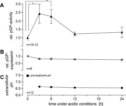
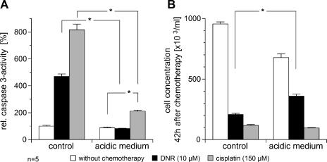
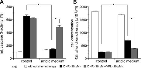
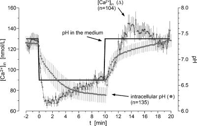
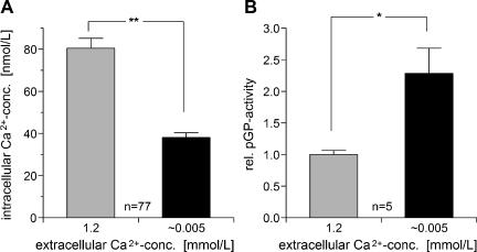
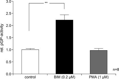
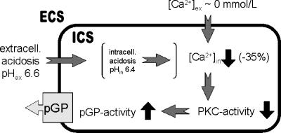
The expression and activity of P-glycoprotein (pGP) play a role in the multidrug resistance of tumors. Because solid-growing tumors often show pronounced hypoxia or extracellular acidosis, this study attempted to analyze the impact of an acidic environment on the expression and activity of pGP and on the cytotoxicity of chemotherapeutic agents. For this, prostate carcinoma cells were exposed to an acidic extracellular environment (pH 6.6) for up to 24 hours. pGP activity was more than doubled after 3 to 6 hours of incubation in acidic medium, whereas cellular pGP expression remained constant, indicating that increased transport rate is the result of functional modulation. In parallel, the cytotoxic efficacy of daunorubicin showed pronounced reduction at low pH, an effect that was reversible on coincubation with a pGP inhibitor. A reduction of intracellular Ca2+ concentration by 35% under acidic conditions induced a higher transport rate of pGP, an effect comparable to that found on inhibition of protein kinase C (PKC). These data indicate that pGP activity is increased by low extracellular pH presumably as a result of lowered intracellular calcium levels and inhibition of PKC. These findings may explain the reduced cytotoxicity of chemotherapeutic agents in hypoxic/acidic tumors.


Figures

Figure 1
Figure 1
(A) Activity of pGP (described by the ratio of rhodamine-123 efflux rates in the presence or absence of VPL, respectively) and (B) relative pGP expression in AT1 cells kept under acidic conditions for up to 24 hours. (C) Extracellular pH during this period. Values at t = 0 hour were measured at pH 7.47 (pretreatment pH of normal cell culture medium) after which the medium was changed to pH 6.6. Values are expressed as mean ± SEM. *P < .05; n, number of experiments.
Figure 2
Figure 2
Cytotoxic activity of DNR (10 µM) and cisplatin (150 µM) measured by (A) the drug-induced caspase 3 activity and (B) the repopulation of tumor cells 42 hours after chemotherapy under acidic (pH 6.6) and control (pH 7.4) conditions. Values are expressed as mean ± SEM of five experiments. *P < .05.
Figure 3
Figure 3
Cytotoxic activity of DNR (10 µM) measured by (A) the DNR-induced caspase 3 activity and (B) the repopulation of tumor cells 42 hours after chemotherapy under acidic (pH 6.6) and control (pH 7.4) conditions. In addition, the impact of pGP inhibition by VPL (10 µM) on DNR cytotoxicity under these conditions is shown. Values are expressed as mean ± SEM of five experiments. *P < .05.
Figure 4
Figure 4
Time course of intracellular pH (●) and intracellular Ca2+ concentration (Δ) after a rapid change of extracellular pH in the medium. Values are expressed as mean ± SEM (n, number of cells investigated).
Figure 5
Figure 5
Impact of extracellular Ca2+ concentration on (A) intracellular Ca2+ concentration [Ca2+]in (n, number of cells investigated) and (B) on the activity of pGP (described by the ratio of rhodamine-123 efflux rates in the presence or absence of VPL, respectively; n, number of experiments). Values are expressed as mean ± SEM. *P < .05; **P < .001.
Figure 6
Figure 6
Impact of PKC inhibition by BIM (0.2 µM) or activation by PMA (1 µM) on the activity ofpGP (described by the ratio of rhodamine-123 efflux rates in the presence or absence of VPL). Values are expressed as mean ± SEM. *P < .05; n, number of experiments.
Figure 7
Figure 7
Model of the mechanisms by which an acidic extracellular microenvironment may affect pGP activity. ECS, extracellular space; ICS, intracellular space (for details, see text).

References

    1. Vaupel P, Kallinowski F, Okunieff P. Blood flow, oxygen and nutrient supply, and metabolic microenvironment of human tumors: a review. Cancer Res. 1989;49:6449–6465. - PubMed
    1. Höckel M, Vaupel P. Tumor hypoxia: definitions and current clinical, biological, and molecular aspects. J Natl Cancer Inst. 2001;93:266–276. - PubMed
    1. Bush RS, Jenkin RD, Allt WE, Beale FA, Bean H, Dembo AJ, Pringle JF. Definitive evidence for hypoxic cells influencing cure in cancer therapy. Br J Cancer Suppl. 1978;37:302–306. - PMC - PubMed
    1. Gray LH, Conger AD, Ebert M, Hornsey S, Scott OCA. The concentration of oxygen dissolved in tissues at the time of irradiation as a factor in radiotherapy. Br J Radiol. 1953;26:638–648. - PubMed
    1. Henderson BW, Fingar VH. Relationship of tumor hypoxia and response to photodynamic treatment in an experimental mouse tumor. Cancer Res. 1987;47:3110–3114. - PubMed

Publication types

MeSH terms

Substances

LinkOut - more resources