. 2006 Mar;8(3):173-80.
doi: 10.1593/neo.05625.

Epstein-Barr virus infection alters cellular signal cascades in human nasopharyngeal epithelial cells

Affiliations

Epstein-Barr virus infection alters cellular signal cascades in human nasopharyngeal epithelial cells

Angela Kwok Fung Lo et al. Neoplasia. 2006 Mar.

Abstract

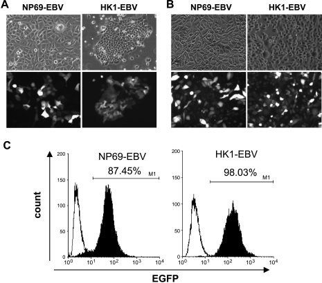
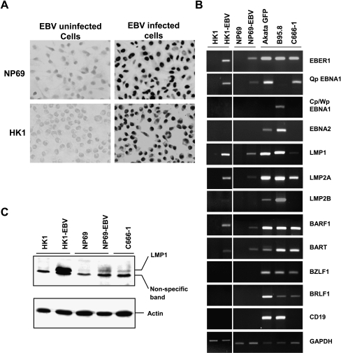
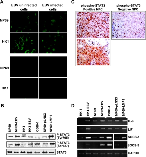
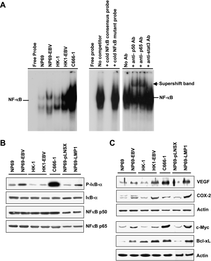
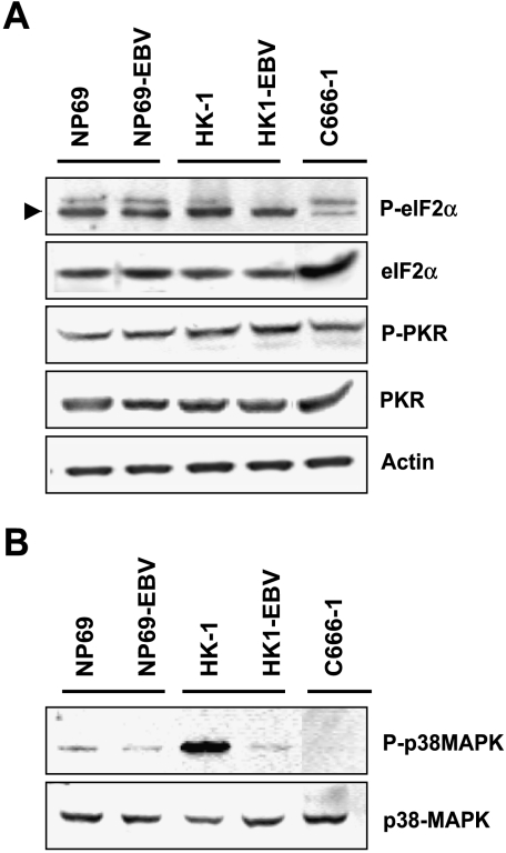
Epstein-Barr virus (EBV) latent infection is a critical event in nasopharyngeal carcinoma (NPC) tumorigenesis. EBV-encoded genes have been shown to be involved in immune evasion and in the regulation of various cellular signaling cascades. To elucidate the roles of EBV in NPC development, stable infection of EBV in nasopharyngeal epithelial cell lines was established. Similar to primary tumors of NPC, these infected cells exhibited a type II EBV latency expression pattern. In this study, multiple cellular signaling pathways in EBV-infected cells were investigated. We first demonstrated that in vitro EBV infection resulted in the activation of STAT3 and NFkappaB signal cascades in nasopharyngeal epithelial cells. Increased expression of their downstream targets (c-Myc, Bcl-xL, IL-6, LIF, SOCS-1, SOCS-3, VEGF, and COX-2) was also observed. Moreover, EBV latent infection induced the suppression of p38-MAPK activities, but did not activate PKR cascade. Our findings suggest that EBV latent infection is able to manipulate multiple cellular signal cascades to protect infected cells from immunologic attack and to facilitate cancer development.


Figures

Figure 1
Figure 1
Detection of EGFP-neorEBV in nasopharyngeal epithelial cells. Neomycin-resistant cells at (A) passage 5 and at (B) passage 20 postinfection exhibited a fluorescent signal under fluorescence microscopy, indicating EGFP-neorEBV infection (original magnification, x40). (C) Fluorescence-activated cell sorter analysis of the EGFP expression of epithelial cells at passage 23 postinfection. Filled curves represent the fluorescence intensity of EBV-infected cells, whereas black lines denote the fluorescence intensity of uninfected cells.
Figure 2
Figure 2
EBV latency expression pattern in nasopharyngeal epithelial cells. (A) In situ RNA hybridization of EBER in EGFP-neorEBV-infected cells. (B) Semiquantitative RT-PCR analysis of EBV-associated transcripts and CD19 in EBV-infected nasopharyngeal epithelial cells. GAPDH was used as loading control. (C) Western blot analysis of LMP1 protein expression in EBV-infected cells.
Figure 3
Figure 3
Activation of STAT pathways in EBV-infected nasopharyngeal epithelial cells. (A) Immunofluorescent staining with STAT3-specific antibody (original magnification, x100; upper panel). Lower panel shows negative controls in which no primary antibody was added. (B) Western blot analysis of STAT3 activation using phosphorylation-specific antibodies. Actin was included as loading control. (C) Immunohistochemical staining for phospo-STAT3. Left: Positive staining of phospho-STAT3 was exhibited in two primary NPC samples (original magnification, x400). Right: A case of NPC showed negative staining of phospho-STAT3. Positive staining of lymphocytes as internal positive control (original magnification, x400). (D) Expression of STAT3-associated genes in EBV-infected nasopharyngeal epithelial cells. Semiquantitative RT-PCR analysis of IL-6, LIF, SOCS-1, and SOCS-3.
Figure 4
Figure 4
Effects of EBV latent infection on the NFκB pathway in nasopharyngeal epithelial cells. (A) EMSA showing the kinetics of NFκB binding activity in nuclear extracts isolated from EBV-infected and uninfected cells (left panel). The specificity of binding was determined by competition experiments in which a nuclear extract isolated from C666-1 cells was analyzed in the presence of a 50-fold excess of an unlabelled NFκB consensus probe or an NFκB mutant probe (right panel, lanes 1–4). The activation of p50-NFκB and p65-NFκB complexes was examined by supershift analysis using antibodies against p50 and p65. Antibody against STAT3 was used as negative control (right panel, lanes 5–8). (B) Western blot analysis of NFκB activity using antibody specific for the phosphorylated form of IκB. The total levels of IκB, p50-NFκB, and p60-NFκB were also examined. (C) Western blot analysis of NFκB downstream targets (VEGF and COX-2) associated with cell invasion, and of c-Myc and Bcl-xL in EBV-infected nasopharyngeal epithelial cells.
Figure 5
Figure 5
Western blot analysis of the activity of (A) PKR and (B) p38-MAPK pathways in EBV-infected nasopharyngeal epithelial cells with antibodies specific for the phosphorylated forms of eIF-2α, PKR, and p38-MAPK.

Similar articles

Cited by

References

    1. Lo KW, To KF, Huang DP. Focus on nasopharyngeal carcinoma. Cancer Cell. 2004;5:423–428. - PubMed
    1. Young LS, Rickinson AB. Epstein-Barr virus: 40 years on. Nat Rev Cancer. 2004;4:757–768. - PubMed
    1. Young LS, Murray PG. Epstein-Barr virus and oncogenesis: from latent genes to tumours. Oncogene. 2003;22:5108–5121. - PubMed
    1. Portis T, Longnecker R. Epstein-Barr virus, (EBV) LMP2A mediates B-lymphocyte survival through constitutive activation of the Ras/PI3K/Akt pathway. Oncogene. 2004;23:8619–8628. - PubMed
    1. Scholle F, Bendt KM, Raab-Traub N. Epstein-Barr virus LMP2A transforms epithelial cells, inhibits cell differentiation, and activates Akt. J Virol. 2000;74:10681–10689. - PMC - PubMed

Publication types

MeSH terms

LinkOut - more resources