Comparative Study
. 2006 Mar;8(3):214-22.
doi: 10.1593/neo.05769.

Nanoparticle imaging of integrins on tumor cells

Affiliations
Comparative Study

Nanoparticle imaging of integrins on tumor cells

Xavier Montet et al. Neoplasia. 2006 Mar.

Abstract

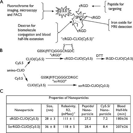
Nanoparticles 10 to 100 nm in size can deliver large payloads to molecular targets, but undergo slow diffusion and/or slow transport through delivery barriers. To examine the feasibility of nanoparticles targeting a marker expressed in tumor cells, we used the binding of cyclic arginine-glycine-aspartic acid (RGD) nanoparticle targeting integrins on BT-20 tumor as a model system. The goals of this study were: 1) to use nanoparticles to image alpha(V)beta3 integrins expressed in BT-20 tumor cells by fluorescence-based imaging and magnetic resonance imaging, and, 2) to identify factors associated with the ability of nanoparticles to target tumor cell integrins. Three factors were identified: 1) tumor cell integrin expression (the alpha(V)beta3 integrin was expressed in BT-20 cells, but not in 9L cells); 2) nanoparticle pharmacokinetics (the cyclic RGD peptide cross-linked iron oxide had a blood half-life of 180 minutes and was able to escape from the vasculature over its long circulation time); and 3) tumor vascularization (the tumor had a dense capillary bed, with distances of <100 microm between capillaries). These results suggest that nanoparticles could be targeted to the cell surface markers expressed in tumor cells, at least in the case wherein the nanoparticles and the tumor model have characteristics similar to those of the BT-20 tumor employed here.


Figures

Figure 1
Figure 1
Design, synthesis, and properties of nanoparticles. (A) schematic diagram of nanoparticle components and functions. (B) Synthesis of nanoparticles. DTT linearizes the disulfide-linked cRGD, linearizing the peptide to IRGD. (C) Physical properties of the nanoparticles used in this study.
Figure 2
Figure 2
Expression of αvβ3 on tumor cells. (A) Dual-wavelength FACS analysis of the binding of cRGD and anti-αvβ3 to BT-20 tumor. BT-20 cells bind anti-αvβ3 and cRGD, but not the control. (B) Uptake of the cRGD-CLIO nanoparticle by BT-20 cells. DTT treatment linearized the peptide and reduced affinity. (C) Binding of the cRGD to BT-20 and 9L cells determined as cell-associated fluorescein from fluorescein immunoassay. Values of apparent affinity constant (Kd) and binding per 1,000,000 cells are shown.
Figure 3
Figure 3
Molecular specificity of the cRGD-CLIO(Cy5.5) nanoparticle in vivo by dual-channel tissue FRI. A mixture of cRGD-CLIO(Cy5.5) and scrRGD-CLIO(Cy3.5) was injected. (A) Cy3.5 channel fluorescence of dissected tissues. (B) Cy5.5 channel tissue fluorescence. (C) Ratio of tissue fluorescence in the Cy5.5 and Cy3.5 channels. Only the BT-20 tumor has a high ratio of Cy5.5/Cy3.5 fluorescence. The BT-20 tumor was different from all other tissues and from the 9L tumor at P < .001.
Figure 4
Figure 4
Imaging the accumulation of the cRGD-CLIO(Cy5.5) nanoparticle by fluorescence and magnetic resonance. (A) White light and fluorescence reflectance images of implanted BT-20 tumors (two per animal). (B) Time dependence of tumor fluorescence determined by fluorescence reflectance, as shown in (A). (C) FMT images at indicated depths. Relative nanoparticle concentration in each plane. (D) MR imaging of nanoparticle accumulation in the tumor. Tumors are presented as colorized T2 maps superimposed over a T2-weighted MR image (TR = 2000; TE = 50) at 24 hours postinjection. Values are average tumor T2 values ± 1 SD.
Figure 5
Figure 5
Nanoparticle uptake by tumor cells of the BT-20 tumor. (A) The distribution of cRGD-CLIO(Cy5.5) within the tumor by iron stain or Cy5.5 fluorescence. After nanoparticle injection, iron and fluorescence are broadly distributed throughout the tumor. (B) Distribution of CD31 (endothelial cells) at low magnification. The tumor is highly vascularized. (C) Distribution of CD31 (endothelial cells), CD11b (macrophages), and αvβ3 by immunohistochemistry. There are a few macrophages in the tumor (CD11b). However, αvβ3 expressed in tumor cells is broadly distributed throughout the tumor, such as iron or Cy5.5 fluorescence from the nanoparticle.
Figure 6
Figure 6
Intravital microscopy of the exposed sigmoid colon after injection of the cRGD-CLIO(Cy5.5) nanoparticle or the cRGD. (A) Vessel and interstitial fluorescence 30 minutes after injection of cRGD-CLIO(Cy5.5). Nanoparticles are confined to the vasculature (vertical arrows). (B) Vessel and interstitial fluorescence at 120 minutes after nanoparticle injection. Nanoparticles are present in both the interstitium (horizontal arrows) and the vasculature (vertical arrows). (C) Time dependence of vessel fluorescence and interstitial fluorescence after injection of cRGD-CLIO(CY5.5). Images from (A) and (B) plus additional time points were used. (D) Vessel and interstitial fluorescence 4 minutes after injection of the cRGD. The peptide is already present in both the interstitium (horizontal arrow) and the vasculature (vertical arrow). (E) Vessel and interstitial fluorescence at 8 minutes after peptide injection. (F) Time dependence of vessel fluorescence and interstitial fluorescence after injection of the cRGD. Images from (D) and (E) plus additional time points were used.

References

    1. Ferrari M. Cancer nanotechnology: opportunities and challenges. Nat Rev Cancer. 2005;5:161–171. - PubMed
    1. NCI/NIH . Cancer Nanotechnology Plan, A Strategic Initiative to Transform Clinical Oncology and Basic Research through Directed Application of Nanotechnology. Washington, DC: 2004. pp. 8–9. ( http://nano.cancer.gov/)
    1. Moghimi SM, Hunter AC, Murray JC. Nanomedicine: current status and future prospects. FASEB J. 2005;19:311–330. - PubMed
    1. Reynolds F, O'Loughlin T, Weissleder R, Josephson L. Method of determining nanoparticle core weight. Anal Chem. 2005;77:814–817. - PubMed
    1. Zhao M, Kircher MF, Josephson L, Weissleder R. Differential conjugation of tat peptide to superparamagnetic nanoparticles and its effect on cellular uptake. Bioconjug Chem. 2002;13:840–844. - PubMed

Publication types

MeSH terms

LinkOut - more resources