. 2006 Jun;17(6):2799-810.
doi: 10.1091/mbc.e05-05-0450. Epub 2006 Apr 12.

Members of the NIMA-related kinase family promote disassembly of cilia by multiple mechanisms

Affiliations

Members of the NIMA-related kinase family promote disassembly of cilia by multiple mechanisms

Dorota Wloga et al. Mol Biol Cell. 2006 Jun.

Abstract

The genome of Tetrahymena thermophila contains 39 loci encoding NIMA-related kinases (NRKs), an extraordinarily large number for a unicellular organism. Evolutionary analyses grouped these sequences into several subfamilies, some of which have orthologues in animals, whereas others are protist specific. When overproduced, NRKs of three subfamilies caused rapid shortening of cilia. Ultrastructural studies revealed that each NRK triggered ciliary resorption by a distinct mechanism that involved preferential depolymerization of a subset of axonemal microtubules, at either the distal or proximal end. Overexpression of a kinase-inactive variant caused lengthening of cilia, indicating that constitutive NRK-mediated resorption regulates the length of cilia. Each NRK preferentially resorbed a distinct subset of cilia, depending on the location along the anteroposterior axis. We also show that normal Tetrahymena cells maintain unequal length cilia. We propose that ciliates used a large number of NRK paralogues to differentially regulate the length of specific subsets of cilia in the same cell.


Figures

Figure 1.
Figure 1.
Phylogeny of NRKs. Amino acid sequences of kinase domains of NRKs and other serine-threonine kinases were aligned and a neighbor-joining tree prepared using HsCdk2 as an outgroup. Numbers above branches represent bootstrap support values above 50%. The multiple sequence alignment is shown in Supplemental Figure S6. Novel motifs found downstream of the kinase domain of Cnk2p type NRKs are shown in Supplemental Figure S7.
Figure 2.
Figure 2.
Overexpressed NRKs colocalize with cilia and cortical microtubular organelles. GFP fluorescent confocal images in cells overproducing Nrk1p-GFP (A and B), GFP-Nrk17p (C), GFP-Nrk30p (D and G), and Nrk2p-GFP (E and F). Expression of transgenes was induced with 2.5 μg/ml CdCl2 for 2–4 h. oa, oral apparatus; noa, new oral apparatus; bb, basal bodies; df, deep fiber; tm, transverse microtubules; c, cilia; ct, ciliary tips, f, cytoplasmic fibers, lm, longitudinal microtubule bundles, pm, postciliary microtubules.
Figure 3.
Figure 3.
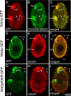
Nrk1p and Nrk2p GFP fusion proteins preferentially localize to cilia and cortical microtubules. Immunofluorescent confocal images of cells overproducing Nrk1p-GFP (A–C), Nrk2p-GFP (D–F), and Nrk2p-K35R-GFP (G–I). Fluorescence of GFP is shown in A (red) and D and G (green). Top row, an Nrk1p-GFP–overproducing cell was processed for double immunofluorescence using anti-GFP antibodies (red in A) and TAP952 antibodies against monoglycylated tubulins (B). The TAP 952 antibody labels strongly new assembling cilia. Note that Nrk1p-GFP accumulates preferentially in TAP952-positive new cilia (C). Middle row, an Nrk2p-GFP–overproducing cell was subjected to double immunofluorescence with anti-GFP antibodies (D) and anti-total tubulin antibodies SG (E). Note an accumulation of Nrk2p-GFP at the tips of a subset of cilia located mainly in the ventral and anterior region of the cell (see F). Arrowheads indicate the absence of GFP label at tips of posterior cilia (F). Bottom row, an Nrk2p-K35R-GFP–overproducing cell labeled by anti-GFP antibodies (G) and total tubulin antibodies (H). The arrows point out a similar localization of Nrk2p-K35R-GFP at ciliary tips (ct) compared with Nrk2p. However, cilia of Nrk2p-K35R-GFP do not undergo shortening (I).
Figure 4.
Figure 4.
Overproduction of NRKs causes ciliary resorption. Immunofluorescence confocal images of cells after 4 h of treatment with cadmium, which are either wild-type (A) or overproduce Nrk1p-GFP (B), Nrk2p-GFP (C), Nrk2p-K35R-GFP (D), or lack both NRK1 and NRK2 genes (E). (F) Graph that shows the average length of cilia as a function of time of treatment with cadmium either in wild-type or overproducing cells. Between 30 and 268 cilia were measured on multiple cells for each time point. Because many cilia were completely resorbed, and only visible cilia were measured, results at later time points are overestimated for GFP-Nrk17p, Nrk2p-GFP, and GFP-Nrk30p. (G) A histogram showing an average length of cilia in a growing population of cells that are either wild type, overproduce GFP (negative control), or lack NRK1, NRK2, or both genes. Between 150 and 260 cilia of multiple cells were measured for each strain.
Figure 5.
Figure 5.
A time-course study of the distribution of shortening cilia in cells overproducing Nrk2p-GFP (A–E), GFP-Nrk17p (F–J), and GFP-Nrk30p (K–O). Cells were treated with 2.5 μg/ml cadmium chloride for the time indicated and fixed and labeled by immunofluorescence with anti-total tubulin SG antibodies and anti-α-tubulin 12G10 antibodies.
Figure 6.
Figure 6.
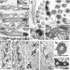
Transmission electron microscopy of controls and cells overproducing NRKs. (A–D) Longitudinal sections of cilia of control cells with the Nrk2p-K35R-GFP transgene but without cadmium treatment (A) or wild-type cells treated with cadmium (B) or cadmium-treated Nrk2p-GFP–overproducing cells (C) and cadmium-treated Nrk2p-K35R-GFP–overproducing cells (D). (E) Cross-section of normal anterior adoral membranelle from NRK-2-dead kinase (without cadmium) showing normally occurring inclusions in these cilia. Note that inclusions are present but they are limited to only one row of cilia in the oral membranelle (F) Cross-sections of a locomotory cilium in the Nrk2p-K35R-GFP–overproducing cell. (G) Cross-section of a group of cilia at the level of ciliary tips in cells overproducing Nrk2p-GFP. Note displacements of axonemal microtubules (arrows). (H) Longitudinal section of a cilium from a Nrk2p-K35R-GFP–overproducing cell treated with cadmium. Note the lateral location of the electron-dense deposit (arrows). (I) Section of a control cell cortex treated with cadmium. (J–N) Images showing sections of the cortical regions of Nrk2p-GFP cells (J, K, M, and N) and Nrk2p-K35R-GFP cells (L) all treated with cadmium. Note electron-dense deposits around microtubules of basal bodies as well as associated microtubule bundles; these are asymmetrically localized in Nrk2p-GFP cells (K, arrows) and Nrk2p-K35R-GFP cells (L). Bar, 0.2 μm.
Figure 7.
Figure 7.
Longitudinal sections of cilia of cells with the Nrk2p-GFP transgene with cadmium induction showing progressive shortening (A, B, D, and E). A cross-section of resorbing cilia of Nrk2p-GFP–overproducing cells is shown in C. Large arrows in A, B, and D show electron-dense deposits near the tips. Small arrows in B show IFT particle-like structures. The arrowhead in D shows an electron-dense deposit near the basal body. Bar, 0.2 μm
Figure 8.
Figure 8.
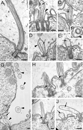
(A–F) Shortening of cilia in GFP-Nrk30p–overproducing cells. (A) Control cell that carries the GFP-Nrk30p transgene but without cadmium induction. (B–E) Images showing progressive shortening of cilia in GFP-Nrk30p cells treated with cadmium. Arrowheads point at budges of ciliary membrane. Arrows show materials that seem to be breakdown products of the depolymerizing axonemes. (F) Cross-section of a shortening cilium from a GFP-Nrk30p–overproducing cells that lacks a central pair. (G–J) Shortening of cilia in GFP-Nrk17p–overproducing cells. (G) Cross-sections of several cilia (arrowheads) away from the cortical surface. Note the lack of the central pair and one or more of peripheral doublets or lack the entire axoneme cross-section. Furthermore, there is noticeable swelling of the cilium and accumulation of breakdown products. (H and I) Longitudinal sections showing gaps at the proximal end of the central pair, whereas peripheral doublets remain intact in the same area. (J) Cilia with completely depolymerized sets of axonemal structures in different stages of resorption. Bar, 0.2 μm.

Similar articles

Cited by

References

    1. Barsel S. E., Wexler D. E., Lefebvre P. A. Genetic analysis of long-flagella mutants of Chlamydomonas reinhardtii. Genetics. 1988;118:637–648. - PMC - PubMed
    1. Berman S. A., Wilson N. F., Haas N. A., Lefebvre P. A. A novel MAP kinase regulates flagellar length in Chlamydomonas. Curr. Biol. 2003;13:1145–1149. - PubMed
    1. Bradley B. A., Quarmby L. M. A NIMA-related kinase, Cnk2p, regulates both flagellar length and cell size in Chlamydomonas. J. Cell Sci. 2005;118:3317–3326. - PubMed
    1. Bradley B. A., Wagner J. J., Quarmby L. M. Identification and sequence analysis of six new members of the NIMA-related kinase family in Chlamydomonas. J. Eukaryot. Microbiol. 2004;51:66–72. - PubMed
    1. Brown J. M., Fine N. A., Pandiyan G., Thazhath R., Gaertig J. Hypoxia regulates assembly of cilia in suppressors of Tetrahymena lacking an intraflagellar transport subunit gene. Mol. Biol. Cell. 2003;14:3192–3207. - PMC - PubMed

Publication types

MeSH terms

LinkOut - more resources