

Diffusion tensor imaging of tract involvement in children with pontine tumors
- PMID: 16611765
- PMCID: PMC8133969
Diffusion tensor imaging of tract involvement in children with pontine tumors
Abstract
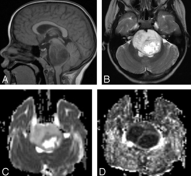
Background and purpose: Conventional MR imaging permits subcategorization of brain stem tumors by location and focality; however, assessment of white matter tract involvement by tumor is limited. Diffusion tensor imaging (DTI) is a promising method for visualizing white matter tract tumor involvement supratentorially. We investigated the ability of DTI to visualize and quantify white matter tract involvement in pontine tumors.
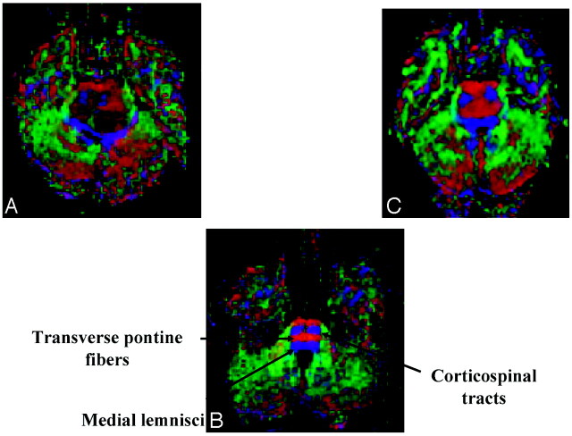
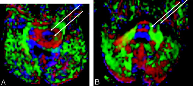
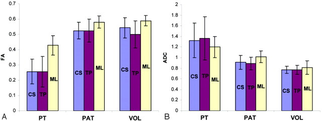
Methods and materials: DTI data (echo-planar, 1.5T) were retrospectively analyzed in 7 patients with pontine tumors (6 diffuse, 1 focal), 4 patient controls, and 5 normal volunteers. Fractional anisotropy (FA) and apparent diffusion coefficient (ADC) were calculated from the diffusion tensor in 6 regions of interest: bilateral corticospinal tracts, transverse pontine fibers, and medial lemnisci. Relationships between FA and ADC values and results of the neurologic examinations were evaluated.
Results: The corticospinal tracts and transverse pontine fibers were affected more often than the medial lemnisci. The DTI parameters (FA and ADC) were significantly altered in all tracts of patients with pontine tumors (P < .05), compared with those values in the control groups. A marginally significant (P = .057) association was seen between the severity of cranial nerve deficit and decreased FA.
Conclusion: DTI provided superior visualization and quantification of tumor involvement in motor, sensory, and transverse pontine tracts, compared with information provided by conventional MR imaging. Thus, DTI may be a sensitive measure of tract invasion. Further prospective studies are warranted to assess the ability of DTI to delineate tumor focality and improve risk stratification in children with pontine tumors.
Figures
Similar articles
-
Diffusion tensor imaging and white matter tractography in patients with brainstem lesions.Acta Neurochir (Wien). 2007 Nov;149(11):1117-31; discussion 1131. doi: 10.1007/s00701-007-1282-2. Epub 2007 Aug 23. Acta Neurochir (Wien). 2007. PMID: 17712509
-
DTI assessment of the brainstem white matter tracts in pediatric BSG before and after therapy: a report from the Pediatric Brain Tumor Consortium.Childs Nerv Syst. 2011 Jan;27(1):11-8. doi: 10.1007/s00381-010-1323-7. Epub 2010 Nov 4. Childs Nerv Syst. 2011. PMID: 21052693 Free PMC article.
-
Quantification of Corticospinal Tracts with Diffusion Tensor Imaging in Brainstem Surgery: Prognostic Value in 14 Consecutive Cases at 3T Magnetic Resonance Imaging.World Neurosurg. 2015 Jun;83(6):1006-14. doi: 10.1016/j.wneu.2015.01.045. Epub 2015 Mar 5. World Neurosurg. 2015. PMID: 25749578
-
The role of diffusion tensor imaging and fractional anisotropy in the evaluation of patients with idiopathic normal pressure hydrocephalus: a literature review.Neurosurg Focus. 2016 Sep;41(3):E12. doi: 10.3171/2016.6.FOCUS16192. Neurosurg Focus. 2016. PMID: 27581308 Review.
-
Diffusion-weighted MR of the brain: methodology and clinical application.Radiol Med. 2005 Mar;109(3):155-97. Radiol Med. 2005. PMID: 15775887 Review. English, Italian.
Cited by
-
Application of technical strategies for surgical management of adult intrinsic pontine gliomas: a retrospective series.Int J Clin Exp Med. 2015 Apr 15;8(4):5175-85. eCollection 2015. Int J Clin Exp Med. 2015. PMID: 26131091 Free PMC article.
-
Impact of DTI tractography on surgical planning for resection of a pediatric pre-pontine neurenteric cyst: a case discussion and literature review.Childs Nerv Syst. 2015 Mar;31(3):457-63. doi: 10.1007/s00381-014-2587-0. Epub 2014 Nov 19. Childs Nerv Syst. 2015. PMID: 25407831 Review.
-
Advanced imaging in paediatric neuroradiology.Pediatr Radiol. 2009 Jun;39 Suppl 3:456-63. doi: 10.1007/s00247-009-1230-9. Pediatr Radiol. 2009. PMID: 19440766 Review. No abstract available.
-
NTMS based tractography and segmental diffusion analysis in patients with brainstem gliomas: Risk stratification and clinical potential.Brain Spine. 2024 Feb 9;4:102753. doi: 10.1016/j.bas.2024.102753. eCollection 2024. Brain Spine. 2024. PMID: 38510608 Free PMC article.
-
Diffuse intrinsic pontine glioma-current status and future strategies.Childs Nerv Syst. 2011 Sep;27(9):1391-7. doi: 10.1007/s00381-011-1468-z. Epub 2011 Apr 30. Childs Nerv Syst. 2011. PMID: 21533575 Review.
References
-
- Barkovich AJ. Pediatric neuroimaging. In: Barkovich AJ. Intracranial, Orbital, and Neck Tumors of Childhood. 3rd ed. Philadelphia, Pa: Lippincott Williams & Wilkins;2000. :462–70
-
- Lesniak MS, Klem JM, Weingart J, et al. Surgical outcome following resection of contrast-enhanced pediatric brainstem gliomas. Pediatr Neurosurg 2003;39:314–22 - PubMed
-
- Barkovich AJ, Krischer J, Kun LE, et al. Brain stem gliomas: a classification system based on magnetic resonance imaging. Pediatr Neurosurg 1991;16:73–83 - PubMed
-
- Gauvain KM, McKinstry RC, Mukherjee P, et al. Evaluating pediatric brain tumor cellularity with diffusion-tensor imaging. AJR Am J Roentgenol 2001;177:449–54 - PubMed
-
- Tien RD, Felsberg GJ, Friedman H, et al. MR imaging of high-grade cerebral gliomas: value of diffusion-weighted echoplanar pulse sequences. AJR Am J Roentgenol 1994;162:671–77 - PubMed
Publication types
MeSH terms
LinkOut - more resources
Full Text Sources