

Measuring longitudinal white matter changes: comparison of a visual rating scale with a volumetric measurement
- PMID: 16611781
- PMCID: PMC8134007
Measuring longitudinal white matter changes: comparison of a visual rating scale with a volumetric measurement
Abstract
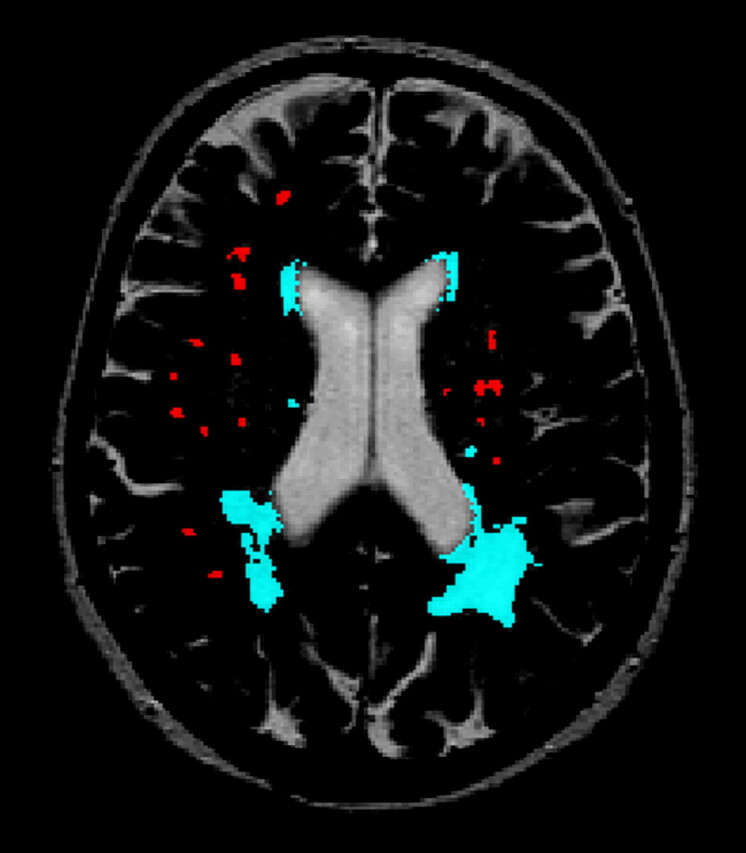
Background and purpose: Detection of longitudinal changes in white matter hyperintensities (WMH) by using visual rating scales is problematic. We compared a widely used visual rating scale with a volumetric method to study longitudinal white matter changes.
Methods: WMH were assessed with the visual Scheltens scale and a volumetric method in 100 elderly subjects aged 70-81 years for whom repetitive MR images were available with an interval of 33 (SD, 1.4) months. Reliability was determined by intraclass correlation coefficients. To examine the sensitivity of both the visual and volumetric method, we calculated Spearman rank correlations of WMH ratings and volume measurements with age.
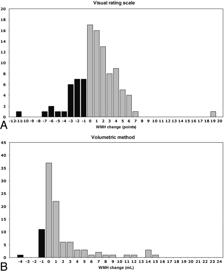
Results: Reliability of the visual rating scale was good, whereas reliability of the volumetric measurement was excellent. For baseline measurements of WMH, we found weaker associations between WMH and age when assessed with the visual scale (r = 0.20, P = .045) than with the volumetric method (r = 0.31, P = .002). Longitudinal evaluation of WMH assessments showed regression in 26% of the subjects when analyzed with the visual rating scale against 12% of the subjects when using volumetric measurements. Compared with the visual rating, the correlation between progression in WMH and age was twice as high when using the volumetric measurement (r = 0.19, P = .062 and r = 0.39, P < .001, respectively).
Conclusion: Volumetric measurements of WMH offer a more reliable, sensitive, and objective alternative to visual rating scales in studying longitudinal white matter changes.
Figures

References
-
- De Groot JC, De Leeuw FE, Oudkerk M, et al. Cerebral white matter lesions and cognitive function: the Rotterdam Scan Study. Ann Neurol 2000;47:145–51 - PubMed
-
- Whitman GT, Tang Y, Lin A, et al. A prospective study of cerebral white matter abnormalities in older people with gait dysfunction. Neurology 2001;57:990–94 - PubMed
-
- Awad IA, Spetzler RF, Hodak JA, et al. Incidental subcortical lesions identified on magnetic resonance imaging in the elderly. I. Correlation with age and cerebrovascular risk factors. Stroke 1986;17:1084–89 - PubMed
-
- Ylikoski A, Erkinjuntti T, Raininko R, et al. White matter hyperintensities on MRI in the neurologically nondiseased elderly: analysis of cohorts of consecutive subjects aged 55 to 85 years living at home. Stroke 1995;26:1171–77 - PubMed
Publication types
MeSH terms
LinkOut - more resources
Full Text Sources
Other Literature Sources
Medical