

MR imaging assessment of brain and cervical cord damage in patients with neuroborreliosis
- PMID: 16611786
- PMCID: PMC8133998
MR imaging assessment of brain and cervical cord damage in patients with neuroborreliosis
Abstract
Background and purpose: Neuroborreliosis is frequently indistinguishable from multiple sclerosis (MS) on both clinical and radiologic grounds. By using MR imaging, we assessed "occult" brain white matter (WM), brain gray matter (GM), and cervical cord damage in patients with neuroborreliosis in an attempt to achieve a more accurate picture of tissue damage in these patients, which might contribute to the diagnostic work-up.
Methods: We studied 20 patients with neuroborreliosis and 11 sex- and age-matched control subjects. In all subjects, we acquired dual echo, T1-weighted, diffusion tensor (DT) and magnetization transfer (MT) MR imaging scans of the brain and fast short-tau inversion recovery and MT MR imaging scans of the cervical cord. T2-visible lesion load was measured by using a local thresholding segmentation technique. Mean diffusivity and fractional anisotropy histograms of the brain and cervical cord MT ratio histograms were produced. Normalized brain volumes (NBV) were measured by using SIENAx.
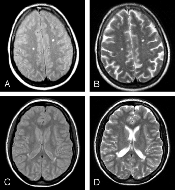
Results: Brain T2-visible lesions were detected in 12 patients, whereas no occult damage in the normal-appearing WM and GM was disclosed by using MT and DT MR imaging. No macroscopic lesions were found in the cervical cord, which was also spared by occult pathology. NBV did not differ between patients with neuroborreliosis and control subjects.
Conclusion: This study shows that, contrary to what happens in MS, occult brain tissue damage and cervical cord pathology are not frequent findings in patients with neuroborreliosis. These observations might be useful in the diagnostic work-up of patients with neuroborreliosis and T2 brain lesions undistinguishable from those of MS.
Figures

References
-
- Stanek G, Strle F. Lyme borreliosis. Lancet Neurol 2003;362:1639–47 - PubMed
-
- Logigian EL, Kaplan RF, Steere AC. Chronic neurologic manifestations of Lyme disease. N Engl J Med 1990;323:1438–44 - PubMed
-
- Morgen K, Martin R, Stone RD, et al. FLAIR and magnetization transfer imaging of patients with post-treatment Lyme disease syndrome. Neurology 2001;57:1980–85 - PubMed
-
- Logigian EL, Johnson KA, Kijewski MF, et al. Reversible cerebral hypoperfusion in Lyme encephalopathy. Neurology 1997;49:1661–70 - PubMed
-
- Halperin JJ, Luft BJ, Anand AK, et al. Lyme neuroborreliosis: central nervous system manifestations. Neurology 1989;39:753–59 - PubMed
MeSH terms
LinkOut - more resources
Full Text Sources
Medical