

Pitfalls in lactate measurements at 3T
Affiliations
- PMID: 16611787
- PMCID: PMC8133981
Item in Clipboard
Pitfalls in lactate measurements at 3T
AJNR Am J Neuroradiol.
2006 Apr.
Abstract
In clinical MR spectroscopy at higher field strengths, lactate may show reduced or absent signal intensity at an echo time of 144 ms. Although this false-negative result may be predicted from theory, experimental verification and clinical impact have not been fully established. Using scanners from 3 major vendors, spectra from phantoms and patients demonstrate the lactate signal loss and potential error in interpretation. Strategies are discussed to overcome, or at least alleviate, this problem.
Figures
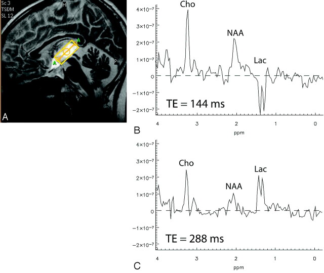
Single-voxel spectra acquired at 1.5T from the brain of a patient with a high-grade glioma by using PRESS localization. B, TE = 144 ms. C, TE = 288 ms. Cho indicates choline; Lac, lactate; NAA, N-acetylaspartate.
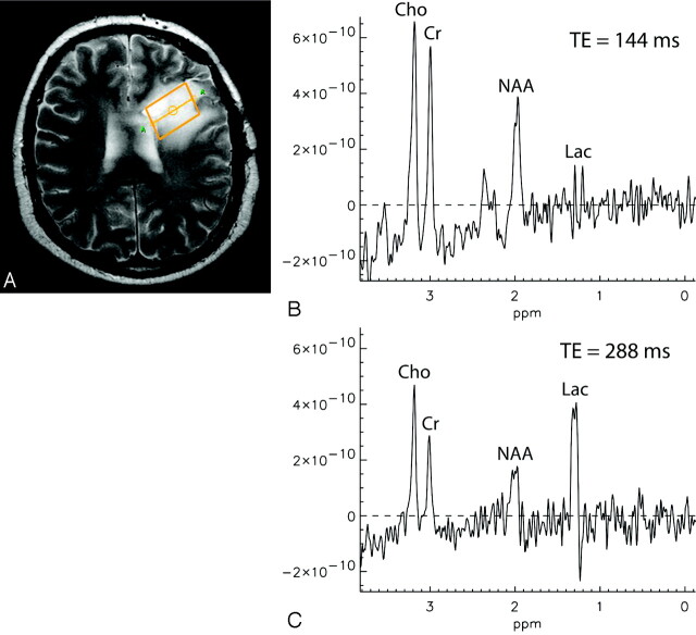
Single-voxel spectra acquired at 3T from the brain of a patient with a grade III glioma by using PRESS localization. B, TE = 144 ms. C, TE = 288 ms. Cr indicates creatine.
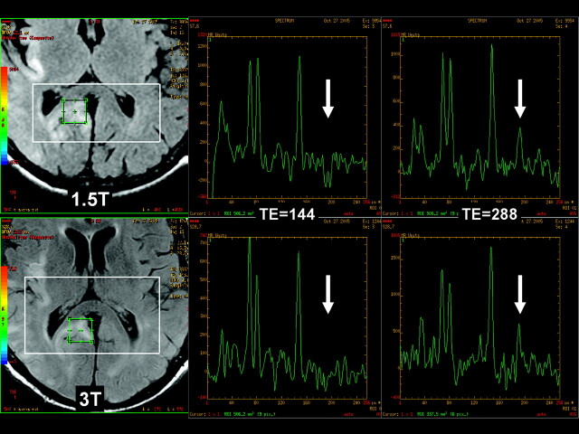
Multivoxel spectra acquired 1 hour apart at 1.5T and 3T from the same region in the brain of a patient with MELAS, by using standard PRESS localization with TE = 144 ms and TE = 288 ms. An inverted lactate doublet is clearly visible at 1.5T, but not at 3T (arrows). Upright lactate peaks at TE = 288 are seen equally well at both field strengths (arrows).
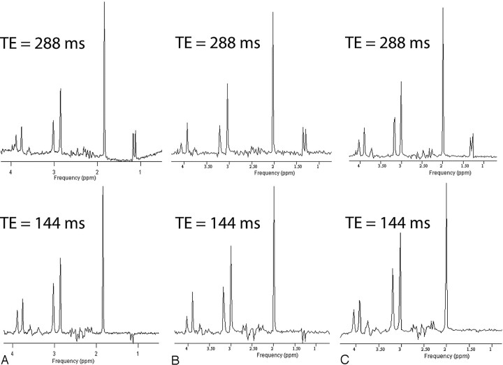
Proton spectra acquired from a standard brain metabolite phantom containing 5 mmol/L of lactate. The measurements were performed on 3 3T MR imaging scanners from 3 different vendors (Philips Medical Systems, GE Healthcare, and Siemens Medical Solutions). Single-voxel MR spectra (VOI size = 2 × 2 × 2 cm3, PRESS localization) were acquired from the same volume once with TE = 144 ms and once with TE = 288 ms. Radio-frequency pulse bandwidths for the selective refocusing pulses vary between vendors in the range of 874–2300 Hz.
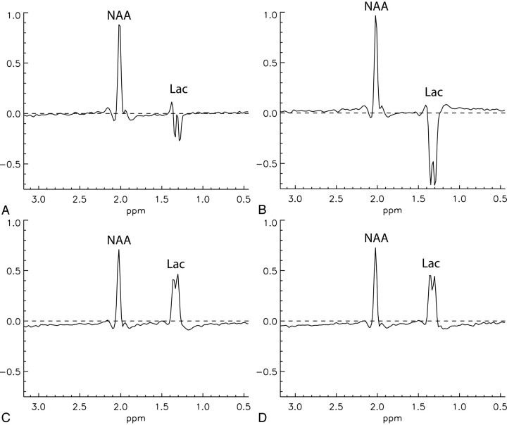
Spectra from an MRSI imaging dataset acquired at 3T from a phantom containing 10 mmol/L of NAA and 20 mmol/L of lactate without PRESS localization. A, TE = 144 ms, with refocusing pulse gradient. B, TE = 144 ms, without refocusing pulse gradient. C, TE = 288 ms, with refocusing pulse gradient. D, TE = 288 ms, without refocusing pulse gradient.
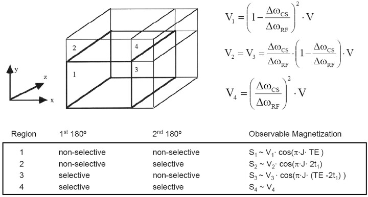
Partial volumes and their coupling evolution for a single-voxel PRESS experiment: The 90° excitation pulse is applied with a gradient in the z direction, whereas the 2 refocusing pulses are applied with gradients in the x and y direction, respectively. The size of the partial volumes is determined by the chemical shift displacement. The signal phase of the magnetization is determined by TE, the scalar coupling constant (J) of lactate, and the time interval (t1) between the excitation pulse and the first refocusing pulse.
References
-
- de Graaf R. In Vivo NMR Spectroscopy: Principles and Techniques. Chichester, UK: John Wiley and Sons Ltd;1998. :53–57
-
- Allen PS, Thompson RB, Wilman AH. Metabolite-specific NMR spectroscopy in vivo. NMR Biomed 1997;10:435–44 - PubMed
-
- Yablonskiy DA, Neil JJ, Raichle ME, et al. Homonuclear J coupling effects in volume localized NMR spectroscopy: pitfalls and solutions. Magn Reson Med 1998;39:159–78 - PubMed
-
- Kelley DAC, Lawrence LW, Star-Lack JM. Lactate detection at 3T: compensating J coupling effects with BASING. J Magn Reson Imaging 1999;9:732–37 - PubMed
-
- Star-Lack J, Spielman D, Adalsteinsson E, et al. In vivo lactate editing with simultaneous detection of choline, creatine, NAA, and lipid singlets at 1.5 T using PRESS excitation with applications to the study of brain and head and neck tumors. J Magn Reson 1998;133:243–54 - PubMed
Publication types
MeSH terms
Substances
LinkOut - more resources
Full Text Sources
Medical