

Intrasubject reproducibility of functional MR imaging activation in language tasks
- PMID: 16611797
- PMCID: PMC8133984
Intrasubject reproducibility of functional MR imaging activation in language tasks
Abstract
Background and purpose: The purpose of this study was to examine the reproducibility of functional MR imaging (fMRI) activation (volume and laterality) within both inferior frontal and temporoparietal regions of interest for both receptive and expressive language tasks.
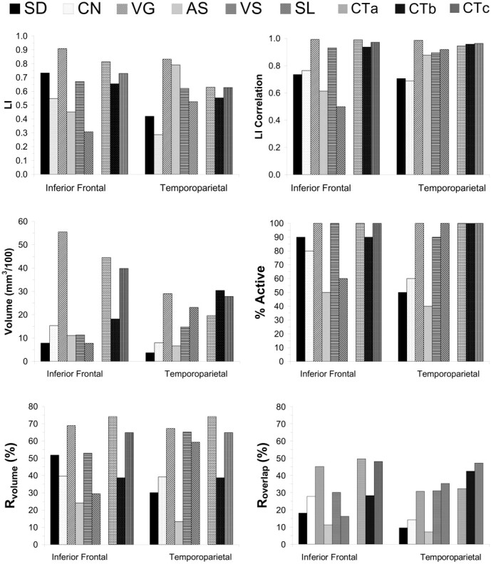
Methods: Ten healthy volunteers participated in fMRI experiments for 6 language tasks: verb generation, confrontation naming, semantic decision making, visual sentence comprehension, auditory sentence comprehension, and story listening. Each subject was scanned during 2 separate sessions separated by a minimum of 4 weeks. Laterality of activation was defined by laterality indices (LIs), which were calculated by 2 methods: one method based on the measured volume of activation and the other method based on the F statistic of the activation. Reproducibility was calculated by using concurrence ratios for the volume of activation (R(overlap), R(volume)) and test-retest correlation for LIs.
Results: All tasks generated reproducible LIs within at least one of the regions of interest, but verb generation produced the highest test-retest correlations (r = 0.99) within both regions of interest. Verb generation was associated with the highest average concurrence ratios within the inferior frontal region of interest (R(overlap) = 45.2; R(volume) = 70.9). In general, the concurrence ratios were lower within the temporoparietal region of interest compared with the inferior frontal region of interest. LIs calculated with F statistics were more reproducible than the LIs calculated by activation volume.
Conclusion: fMRI is able to provide reproducible LIs in both inferior frontal and temporoparietal regions for assessing hemispheric dominance in language processing. The volume of activation, especially within the temporoparietal regions, is less reproducible than the laterality of activation, so the former should be used with caution.
Figures
References
-
- Binder JR, Swanson SJ, Hammeke TA, et al. Determination of language dominance using functional MRI: a comparison with the Wada test. Neurology 1996;46:978–84 - PubMed
-
- Springer JA, Binder JR, Hammeke TA, et al. Language dominance in neurologically normal and epilepsy subjects: a functional MRI study. Brain 1999;122:2033–46 - PubMed
-
- Benson RR, FitzGerald DB, LeSueur LL, et al. Language dominance determined by whole brain functional MRI in patients with brain lesions. Neurology 1999;52:798–809 - PubMed
-
- Ramsey NF, Sommer IE, Rutten GJ, et al. Combined analysis of language tasks in fMRI improves assessment of hemispheric dominance for language functions in individual subjects. Neuroimage 2001;13:719–33 - PubMed
MeSH terms
LinkOut - more resources
Full Text Sources
Medical