. 2006 Apr 12;26(15):4118-25.
doi: 10.1523/JNEUROSCI.0303-06.2006.

Separation of mu-opioid receptor desensitization and internalization: endogenous receptors in primary neuronal cultures

Affiliations

Separation of mu-opioid receptor desensitization and internalization: endogenous receptors in primary neuronal cultures

Seksiri Arttamangkul et al. J Neurosci. .

Abstract

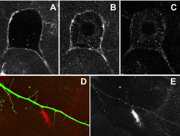
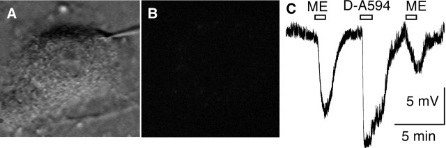
A close relationship between desensitization and internalization of mu-opioid receptors (MORs) has been proposed based on differential actions of series of agonists. The role that these two processes have in the development of tolerance and dependence to opioids has been a controversial subject that has been studied in a variety of model systems. Here, we examine desensitization and internalization of endogenous MORs simultaneously in primary cultures of locus ceruleus neurons using fluorescently tagged peptide agonists. With the use of two fluorescent opioid peptides, dermorphin-Bodipy Texas Red and dermorphin-Alexa594 (Derm-A594), desensitization was measured electrophysiologically and trafficking was followed by the accumulation of intracellular fluorescent puncta. Blocking endocytosis with concanavalin A eliminated the accumulation of fluorescent puncta but desensitization induced by Derm-A594 was unaffected. Likewise, after treatment with concanavalin A, there was no change in either desensitization or recovery from desensitization induced by [Met]5enkephalin. The results demonstrate that desensitization and the recovery from desensitization are not dependent on receptor internalization and suggest that the activity of endogenous MORs in primary neurons can be modulated at the level of the plasma membrane.


Figures

Figure 1.
Figure 1.
LC neurons expressing GFP. A, The LC is found next to the fourth ventricle (10×). B, An enlarged image showing the dense and compact structure of the nucleus (20×). C, Colocalization of GFP and anti-tyrosine hydroxylase (mouse monoclonal) labeled with Texas Red-conjugated secondary antibody. D, A single neuron in culture for 9 d.
Figure 2.
Figure 2.
Effectors sensitive to the activation of MORs. A, Activation of GIRK conductance. Whole-cell recordings were made in a high-potassium (10 mm) solution (V-hold −60 mV, step to −100 mV). Inward currents were measured at −100 mV. The superimposed traces show the currents triggered by Derm-BTR (D-BTR; 1 μm) and UK14304 (UK; 10 μm). The bar graph summarizes the currents induced by opioid agonists normalized to the current induced by UK14304. Both Derm-BTR (1 μm) and morphine (10 μm) were partial agonists in this assay. B, Inhibition of voltage-dependent calcium (barium) conductance. Whole-cell recordings were made and the inward current induced by a depolarizing step from −60 to −20 mV was measured. Representative traces show the inhibition caused by Derm-BTR (0.1 μm). The bar graph summarizes the inhibition of calcium (barium) currents as percentage of control. Derm-BTR caused the same maximal inhibition as ME (30 μm) in this assay. C, Recurrent synapses (autapses) mediated by glutamate are inhibited by opioids. The superimposed traces show the current induced during depolarization of cells from −60 to −10 mV followed by an EPSC. The EPSCs were inhibition by Derm-BTR (1 μm). The bar graph summarizes the inhibition of EPSCs by ME (10 μm), Derm-BTR (1 μm), UK 14304 (10 μm), and NBQX, an AMPA antagonist (10 μm). The results indicate that LC cells release glutamate when maintained in an isolated cell culture and opioids inhibit that release. Error bars indicate SE.
Figure 3.
Figure 3.
Internalization of Derm-BTR (450 nm). A, Binding of Derm-BTR (450 nm) to receptors on the plasma membrane was observed as a fluorescent ring along plasma membrane (15°C). The temperature was then increased to 35°C within 1 min and internalization was monitored for 45 min. B, Image of the same cell taken after incubation for 30 min. C, After washing the Derm-BTR with a hypertonic acid solution (0.5 m NaCl in acetate buffer, pH 4), extracellular fluorescence outside the cell was removed and fluorescent puncta were observed in the soma. D, E, Fluorescent puncta containing Derm-BTR found in a GFP-positive process.
Figure 4.
Figure 4.
Time course of Derm-BTR internalization and desensitization. A, A representative experiment showing the block of Derm-BTR (D-BTR; 100 nm) internalization by naloxone (10 μm; top row). Naloxone was washed from the bath and Derm-BTR (100 nm) was reapplied to demonstrate the presence of MORs on this cell. Images were taken with a confocal microscope at 2 min intervals. To reduce photobleaching, a single frame was collected at each of 5 z-planes. Only nonspecific staining was found in the presence of naloxone. B, Fluorescent intensity in the cytoplasm increased over the first 4–5 min and reached a plateau at 10 min. The fluorescence was normalized to the intensity measured at 10 min. C, A representative experiment showing the peak and decline of the potassium current induced by Derm-BTR (1 μm) during a 10 min application. The experiment was done in a high-potassium solution as in Figure 2A. The inward current was measured during a voltage step from −60 to −100 mV applied once every 20 s. The current is plotted as an outward current to better illustrate the decline in the current. Error bars indicate SE.
Figure 5.
Figure 5.
Desensitization and internalization in the same neuron. A, Intracellular recording and two-photon microscopy were done simultaneously during treatment with Derm-A594 (D-A594). The top left shows an image of a neuron take with infrared illumination and the same neuron after incubation with Derm-A594 (6 μm, 10 min, 5 min wash; right). The trace to the right is the recording of the membrane potential of that neuron. ME (10 μm) and Derm-A594 hyperpolarized this neuron. The hyperpolarization induced by Derm-A594 peaked and declined during a 10 min application. After washing Derm-A594, UK14304 caused a maximal hyperpolarization. B, The blockade of the hyperpolarization and internalization by β-CNA (1 μm).
Figure 6.
Figure 6.
Desensitization by a short pulse of Derm-A594. A, Image of a representative cell where desensitization resulting from a short application of Derm-A594 was measured. B, Fluorescence image taken with a two-photon microscope showing the lack of any puncta after a 2 min treatment with Derm-A594 (6 μm). C, Recording of membrane potential from the neuron shown in A and B. Desensitization was determined by comparing the decrease in the amplitude of the hyperpolarization induced by ME (300 nm), before and after a short pulse (2 min) of Derm-A594 (D-A594; 6 μm).
Figure 7.
Figure 7.
MOR desensitization is not changed after blockade of internalization. A, Images of a neuron showing the appearance of fluorescent puncta in a control experiment (top). B, Images of cell after treatment with con A. No fluorescent puncta were found. C, Recording of membrane potential from a cell treated with con A. All cells were initially tested with ME (10 μm). Con A (160–200 μg/ml) was superfused for 20 min. Derm-A594 (D-A594; 6 μm, in con A) induced membrane hyperpolarization similar in amplitude to that of ME. D, Summarized data showing the amplitude of the hyperpolarization induced by Derm-A594 and UK14304 (UK) in the absence and presence of con A. E, shows that the decline of the hyperpolarization induced by Derm-A594 was the same in the absence and presence of con A. Error bars indicate SE.
Figure 8.
Figure 8.
MOR recovery from desensitization is independent of internalization. A, A representative trace showing the recovery from desensitization induced by ME (10 μm). ME (300 nm) was tested before and 5 and 20 min after application of ME (10 μm, 5 min). The α2-adrenoceptor agonist UK14304 (1 μm) was applied at the end of the experiment. B, The recovery from desensitization induced by ME (10 μm) was not changed after treatment with concanavalin A (160 μg/ml, preincubated for 20 min). C, Summarized data showing the recovery from desensitization. Left, A short (2 min) application of ME (10 μm; gray bars) and Derm-A594 (D-A594; 6 μm; black bars) decreased the hyperpolarization induced by ME (300 nm) to ∼40% of control 5 min after washout. After 20 min, significant recovery was observed. Right, ME (10 μm) was perfused for 5 min and the hyperpolarization induced by ME (300 nm) was tested after 5 and 20 min. The time course or extent of recovery was not changed by concanavalin A (white bars) compared with controls (gray bars). Error bars indicate SE.

References

    1. Alvarez VA, Arttamangkul S, Dang VC, Salem A, Whistler JL, von Zastrow M, Grandy DK, Williams JT (2002). μ-Opioid receptors: ligand-dependent activation of potassium conductance, desensitization and internalization. J Neurosci 22:5769–5776. - PMC - PubMed
    1. Arttamangkul S, Alvarez-Maubecin V, Thomas G, Williams JT, Grandy DK (2000). Binding and internalization of fluorescent opioid peptide conjugates in living cells. Mol Pharmacol 58:1570–1580. - PubMed
    1. Bailey CP, Connor M (2005). Opioids: cellular mechanisms of tolerance and physical dependence. Curr Opin Pharmacol 5:60–68. - PubMed
    1. Bailey CP, Kelly E, Henderson G (2004). Protein kinase C activation enhances morphine-induced rapid desensitization of mu-opioid receptors in mature rat locus ceruleus neurons. Mol Pharmacol 66:1592–1598. - PubMed
    1. Beaumont V, Hepworth MB, Luty JS, Kelly E, Henderson G (1998). Somatostatin receptor desensitization in NG108–15 cells. A consequence of receptor sequestration. J Biol Chem 273:33174–33183. - PubMed

Publication types

Substances