. 2006 May;80(9):4380-7.
doi: 10.1128/JVI.80.9.4380-4387.2006.

Base pairing between the 5' half of epsilon and a cis-acting sequence, phi, makes a contribution to the synthesis of minus-strand DNA for human hepatitis B virus

Affiliations

Base pairing between the 5' half of epsilon and a cis-acting sequence, phi, makes a contribution to the synthesis of minus-strand DNA for human hepatitis B virus

Teresa M Abraham et al. J Virol. 2006 May.

Abstract

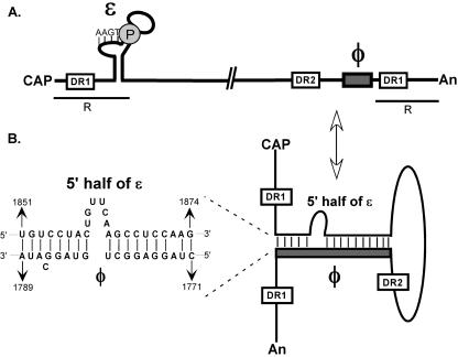
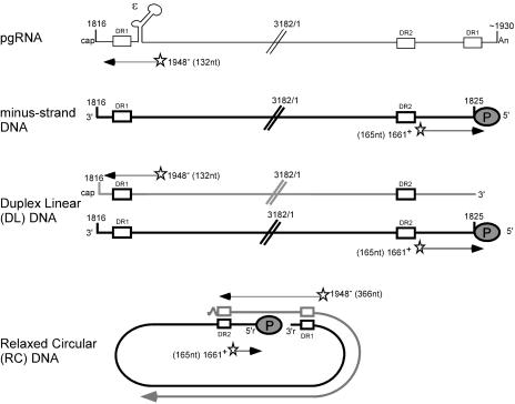
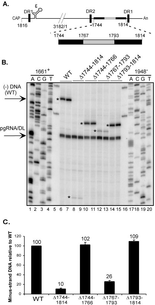
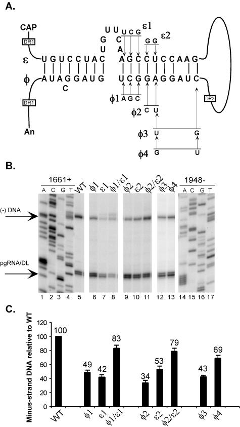
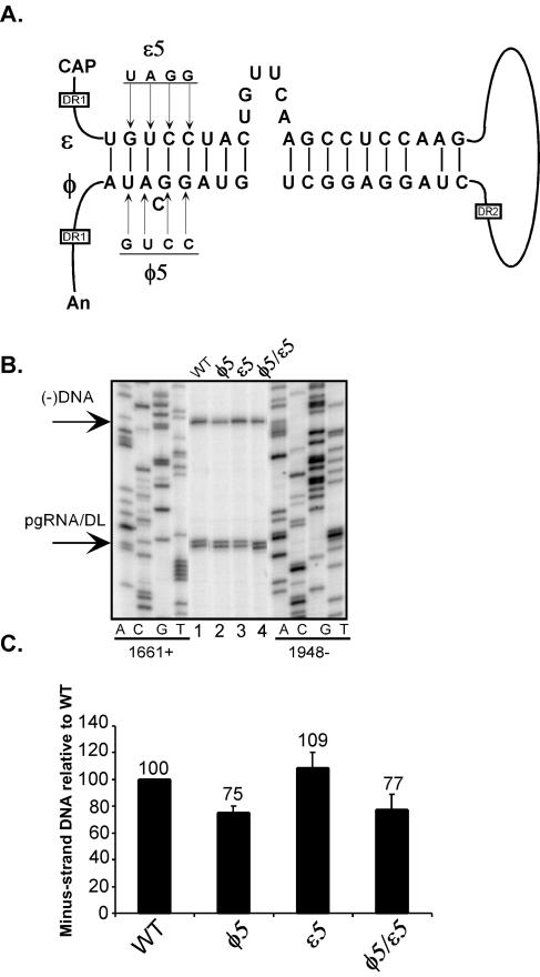
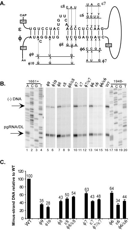
Synthesis of minus-strand DNA of human hepatitis B virus (HBV) can be divided into three phases: initiation of DNA synthesis, the template switch, and elongation of minus-strand DNA. Although much is known about minus-strand DNA synthesis, the mechanism(s) by which this occurs has not been completely elucidated. Through a deletion analysis, we have identified a cis-acting element involved in minus-strand DNA synthesis that lies within a 27-nucleotide region between DR2 and the 3' copy of DR1. A subset of this region (termed Phi) has been hypothesized to base pair with the 5' half of epsilon (H. Tang and A. McLachlan, Virology, 303:199-210, 2002). To test the proposed model, we used a genetic approach in which multiple sets of variants that disrupted and then restored putative base pairing between the 5' half of epsilon and phi were analyzed. Primer extension analysis, using two primers simultaneously, was performed to measure encapsidated pregenomic RNA (pgRNA) and minus-strand DNA synthesized in cell culture. The efficiency of minus-strand DNA synthesis was defined as the amount of minus-strand DNA synthesized per encapsidation event. Our results indicate that base pairing between phi and the 5' half of epsilon contributes to efficient minus-strand DNA synthesis. Additional results are consistent with the idea that the primary sequence of phi and/or epsilon also contributes to function. How base pairing between phi and epsilon contributes to minus-strand DNA synthesis is not known, but a simple speculation is that phi base pairs with the 5' half of epsilon to juxtapose the donor and acceptor sites to facilitate the first-strand template switch.


Figures

FIG. 1.
FIG. 1.
Model of dynamic conformation of pgRNA during minus-strand DNA synthesis. (A) The location of the cis-acting sequence, Φ, between DR2 and the 3′ copy of DR1. Nascent minus-strand DNA covalently linked to the P protein (gray circle) is shown on the bulge of ɛ, the encapsidation signal. The 5′ end of pgRNA is at nt 1816. The poly(A) tail begins approximately at nt 1930. The 115 nt terminal redundancy of pgRNA is shown by R. (B) A conformation of pgRNA where the 5′ half of ɛ base pairs with Φ to facilitate minus-strand DNA synthesis. The inset shows the 19 nt of Φ that base pair with the 5′ half of ɛ. Indicated are the nucleotide coordinates. The left part of the Φ/ɛ base-paired structure is defined as nucleotides that fall to the left of the bulge of ɛ. The right part of the Φ/ɛ base-paired structure is defined as nucleotides that fall to the right of the bulge of ɛ. The pgRNA is not drawn to scale. DR1, direct repeat sequence 1; DR2, direct repeat sequence 2. ɛ, encapsidation signal; Φ, cis-acting sequence on pgRNA.
FIG. 2.
FIG. 2.
Schematic representation of dual-PE assay. pgRNA is depicted by a thin dark line, with its salient features and nucleotide positions of the 5′ and 3′ ends indicated. Minus-strand DNA is depicted by a thick dark line, and plus-strand DNA is depicted by a gray line. The P protein covalently linked to the 5′ end of minus-strand DNA is illustrated as a gray oval. The stars represent end-labeled primers. Oligonucleotide 1948 anneals to position 1948 on the pgRNA. AMV reverse transcriptase extends to the 5′ end of pgRNA, resulting in a 132-nt product. Oligonucleotide 1948 also anneals to plus-strand DNA at position 1948. In the case of DL, it results in a 132-nt product, which comigrates with pgRNA. In the case of RC, extension of oligonucleotide 1948 results in a 366-nt product. Oligonucleotide 1661+ anneals at position 1661 on minus-strand DNA and extends to the 5′ end, resulting in a 165-nt product.
FIG. 3.
FIG. 3.
Deletion analysis between nt 1744 to 1814 defines boundaries of cis-acting sequence for minus-strand DNA synthesis. (A) The positions of the deletion variants are indicated on the pgRNA. Φ is located between nt 1767 and 1793. A total of 23 nt was deleted upstream of Φ, and 21 nt were deleted downstream of Φ. (B) Dual-PE analysis of pgRNA and minus-strand DNA levels of deletion variants shown in panel A. The asterisks indicate the positions at which the 5′ ends of minus-strand DNA for the WT and the deletion variants migrate. Because the deletions were located downstream of where oligonucleotide 1661+ annealed to minus-strand DNA, primer extension reactions resulted in products that reflect the size of the deletion. The positions of 5′ ends of pgRNA and minus-strand DNA (WT) are indicated by arrows on the left. Lanes 1 to 4 and 17 to 20, sequencing ladder of WT HBV DNA; 5 and 16, plasmid DNA control; 6 and 7, WT; 8 and 9, Δ1744-1814; 10 and 11, Δ1744-1766; 12 and 13, Δ1767-1793; 14 and 15, Δ1793-1814. (C) Efficiency of minus-strand DNA synthesis relative to a WT reference. The encapsidation competency of a virus is defined as the sum of pgRNA and minus-strand DNA. Therefore, the ability of a virus to synthesize minus-strand DNA is determined as the level of minus-strand DNA normalized to the sum of pgRNA and minus-strand DNA levels. The efficiency of minus-strand DNA synthesis for each variant was normalized to that of the WT standard. The numbers above each bar indicate the mean values of minus-strand DNA synthesis. The error bars represent standard deviations from analysis of replicative intermediates isolated from at least six independent transfections of each variant.
FIG. 4.
FIG. 4.
Base pairing between Φ and 5′ half of ɛ contributes to minus-strand DNA synthesis. (A) Putative base pairing between 19 nt of Φ and the 5′ half of ɛ on the pgRNA. Substitutions designed to disrupt base pairing are shown. Variants that restore base pairing combine the corresponding ɛ and Φ substitutions. Variants Φ3 and Φ4 change nucleotides on Φ alone. The positions of the DR sequences on pgRNA are indicated. The diagram is not drawn to scale. (B) Dual-PE assay of pgRNA and minus-strand DNA levels of substitution variants shown in panel A. The position of the 5′ ends of pgRNA and minus-strand DNA are indicated by arrows on the left. Lanes 1 to 4 and 14 to 17, sequencing ladders generated from WT HBV DNA; 5, WT; 6, Φ1; 7, ɛ1; 8, Φ1/ɛ1; 9, Φ2; 10, ɛ2; 11, Φ2/ɛ2; 12, Φ3; 13, Φ4. (C) The efficiency of minus-strand DNA synthesis was calculated by dividing the level of minus-strand DNA by the sum of minus-strand DNA plus pgRNA. The denominator represents the total encapsidation events. The numbers above each bar indicate the mean values of minus-strand DNA synthesis. The error bars indicate standard deviations from analysis of replicative intermediates isolated from at least six independent transfections of each variant.
FIG. 5.
FIG. 5.
The left part of Φ does not base pair with ɛ to contribute to minus-strand DNA synthesis. (A) Putative base pairing between Φ and the 5′ half of ɛ. Nucleotide substitutions that disrupt base pairing are indicated. Variants that restore base pairing combine the corresponding ɛ and Φ substitutions. (B) Dual-PE assay of pgRNA and minus-strand DNA levels of substitution variants shown in panel A. The positions of the 5′ ends of minus-strand DNA and pgRNA are indicated by arrows on the left. Sequencing ladders generated from WT HBV DNA, using individual primers, are indicated. Lanes 1, WT; 2, Φ5; 3, ɛ5; 4, Φ5/ɛ5. (C) Graphic representation of the amount of minus-strand DNA normalized to the total encapsidation events. The numbers above each bar indicate the mean values of minus-strand DNA synthesis. The error bars indicate standard deviations from analysis of replicative intermediates isolated from at least six independent transfections of each variant.
FIG. 6.
FIG. 6.
Base pairing between Φ and the 5′ half of ɛ is not sufficient for minus-strand DNA synthesis. (A) Putative base pairing between Φ and the 5′ half of ɛ on pgRNA is shown. Nucleotide substitutions that disrupt base pairing are indicated. Variants that restore base pairing combine the corresponding ɛ and Φ substitutions. Variants Φ9 and Φ10 only mutate nucleotides on Φ. (B) Dual-PE assay of pgRNA and minus-strand DNA levels of substitution variants shown in part A. The positions of the 5′ end of minus-strand DNA and pgRNA are indicated by arrows on the left. Lanes 1 to 4 and 17 to 20, sequencing ladders generated from WT HBV DNA; 5, Φ9; 6, Φ10; 7, Φ8; 8, ɛ8; 9, Φ8/ɛ8; 10, Φ7; 11, ɛ7; 12, Φ7/ɛ7; 13, Φ6; 14, ɛ6; 15, Φ6/ɛ6; 16, WT. (C) Graphic representation of the amount of minus-strand DNA normalized to the total encapsidation events. The numbers above each bar indicate the mean values of minus-strand DNA synthesis. The error bars indicate standard deviations from analysis of replicative intermediates isolated from at least six independent transfections of each variant.

Similar articles

Cited by

References

    1. Bartenschlager, R., and H. Schaller. 1992. Hepadnaviral assembly is initiated by polymerase binding to the encapsidation signal in the viral RNA genome. EMBO J. 11:3413-3420. - PMC - PubMed
    1. Beasley, R. P. 1988. Hepatitis B virus: the major etiology of hepatocellular carcinoma. Cancer 61:1942-1956. - PubMed
    1. Chen, Y., and P. Marion. 1996. Amino acids essential for RNase H activity of hepadnaviruses are also required for efficient elongation of minus-strand viral DNA. J. Virol. 70:6151-6156. - PMC - PubMed
    1. Cristofari, G., C. Bampi, M. Wilhelm, F.-X. Wilhelm, and J.-L. Darlix. 2002. A 5′-3′ long-range interaction in Ty1 RNA controls its reverse transcription and retrotransposition. EMBO J. 21:4368-4379. - PMC - PubMed
    1. Ganem, D., and R. Schneider. 2001. Hepadnaviridae: the viruses and their replication, p. 2923-3036. In D. M. Knipe, P. M. Howley, D. E. Griffin, R. A. Lamb, M. A. Martin, B. Roizman, and S. E. Straus (ed.), Fields virology, 4th ed. Lippincott Williams & Wilkins, Philadelphia, Pa.

Publication types

LinkOut - more resources