. 2006 May;80(9):4396-405.
doi: 10.1128/JVI.80.9.4396-4405.2006.

The Us9 gene of bovine herpesvirus 1 (BHV-1) effectively complements a Us9-null strain of BHV-5 for anterograde transport, neurovirulence, and neuroinvasiveness in a rabbit model

Affiliations

The Us9 gene of bovine herpesvirus 1 (BHV-1) effectively complements a Us9-null strain of BHV-5 for anterograde transport, neurovirulence, and neuroinvasiveness in a rabbit model

S I Chowdhury et al. J Virol. 2006 May.

Abstract

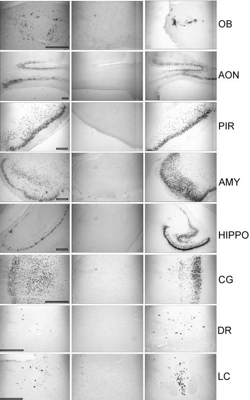
The alphaherpesvirus envelope protein Us9 is a type II viral membrane protein that is required for anterograde spread of bovine herpesvirus 5 (BHV-5) infection from the olfactory receptor neurons to the brain. In a rabbit seizure model, Us9-deleted BHV-5 failed to invade the central nervous system (CNS) following intranasal infection. However, when injected directly into the olfactory bulb, retrograde-spread infection from the olfactory bulb (OB) to the piriform cortex and other areas connected to the OB was not affected. In contrast to BHV-5, wild-type BHV-1 failed to invade the CNS following intranasal infection. In this study, we show that mature BHV-1 Us9 is a 30- to 32-kDa protein, whereas mature BHV-5 Us9 is an 18- to 20-kDa protein. In vitro, BHV-1 Us9 is expressed at 3 h postinfection (hpi), whereas BHV-5 Us9 is expressed at 6 hpi. Despite these differences, BHV-1 Us9 not only complemented for BHV-5 Us9 and rescued the anterograde-spread defect of the BHV-5 Us9-deleted virus but conferred increased neurovirulence and neuroinvasiveness in our rabbit seizure model. Rabbits infected with BHV-5 expressing BHV-1 Us9 showed severe neurological signs at 5 days postinfection, which was 1 to 2 days earlier than BHV-5 wild-type or Us9-reverted BHV-5 virus. The data underscore the importance of both Us9 genes for virion anterograde transport and neuroinvasiveness. However, Us9 is not the determinant of the differential neuropathogenesis of BHV-1 and BHV-5.


Figures

FIG. 1.
FIG. 1.
Schematic illustration of the construction of recombinant plasmids. (A) Construction of the BHV-1 Us9 deletion plasmid (pBHV-1Us9ΔEGFP), the BHV-5 Us9 deletion plasmid (pBHV-5Us9ΔEGFP), and the Us9-exchanged BHV-5 plasmid (pBHV5Us9-1). (B) Alignment showing the BHV-1 and BHV-5 Us9 upstream nucleotide sequences containing the respective putative promoter region. The gE stop (TAG) and Us9 start (ATG) codons are in boldface type. The putative TATAAA boxes are underlined. The highlighted region in the BHV-1.1 sequence shows the forward primer sequence (P9) used to amplify the BHV-1 Us9 ORF (Table 1), and the sequence above indicates the KpnI sites incorporated within the forward P9 primer sequence. The arrow (at 27 bp upstream of the Us9 start codon) in the BHV-5 Us9 sequence marks the deletion site in the BHV-5 Us9-deleted virus.
FIG. 2.
FIG. 2.
Identification of BHV-1 Us9 and characterization of BHV-1 Us9 expression by BHV-5Us9-1 (Us9-exchanged BHV-5). (A) Immunoprecipitation-immunoblotting analysis of BHV-1-, BHV-5 Us9-1-, and BHV-5-infected cell lysates were immunoprecipitated with goat anti-BHV-1 Us9 antibody. After SDS-PAGE of the immunoprecipitated samples, they were immunoblotted with rabbit anti BHV-1 Us9 antibody. (B) Autoradiograph showing phosphorylation of Us9 expressed by BHV-1-, BHV-5-, and BHV-5 Us9-1-infected MDBK cells. MDBK cells were infected with the viruses designated above for each lane and were labeled with 32P for 10 h starting at 3 hpi (BHV-1, Us9-deleted BHV-1, and BHV-5 Us9-1) or at 6 hpi (BHV-5 and BHV-5 Us9 deleted). The lysates were immunoprecipitated with goat anti-Us9 antibody as before.
FIG. 3.
FIG. 3.
(A) Autoradiograph showing a time course of Us9 expression in BHV-1, BHV-5 Us9-1, and BHV-5. Virus-infected cell proteins were labeled with [35S]methionine-cysteine at 3 or 6 hpi. Cell monolayers were washed twice with serum-free Dulbecco's modified Eagle's medium, and detergent extracts were prepared. Radiolabeled lysates were immunoprecipitated with goat anti-Us9 antibody. After labeling and washing of cell monolayers as above, 10-h chase samples (*) were further incubated for 10 h at 37°C in complete growth medium without labeled cysteine and methionine. (B) Immunoblots showing BHV-1 and BHV-5 Us9 expression in untreated (PAA−) and PAA-treated (PAA+) BHV-1-, BHV-5-, and BHV-5 Us9-1-infected cell lystaes. MDBK cells were infected with the viruses indicated above. Infected cell lysates were immunoprecipitated with goat anti-Us9 antibody. Western blots containing immunoprecipitates were immunoblotted with rabbit anti-Us9 antibody.
FIG. 4.
FIG. 4.
One-step growth curve of BHV-5 and recombinant BHV-5 Us9-1 viruses in MDBK cells. Confluent MDBK cells were infected at an MOI of 5 PFU per cell with viruses. After 1 h of adsorption at 4°C, residual input viruses were removed. The cultures were washed three times with PBS, and 5 ml of medium was added into each flask before further incubation (37°C). At indicated time intervals, replicate cultures were frozen. Virus yields were determined by titration on MDBK cells. Each data point represents the average of duplicate samples obtained from separate infections.
FIG. 5.
FIG. 5.
Localization of viral antigen in brain sections. Animals were inoculated intranasally with BHV-5 Us9-1, BHV-5 Us9Δ, and BHV-5 wild-type viruses as described in Materials and Methods. The animals were euthanized when they showed neurological signs for BHV-5 Us9-1 and BHV-5 wild-type viruses and on day 12 for BHV-5 Us9Δ virus. Representative sections of olfactory bulb (OB), anterior olfactory nucleus (AON), piriform cortex (PIR), amygdala (AMY), hippocampus (HIPPO), cingulate cortex (CG), dorsal raphe (DR), and locus coeruleus (LC) are shown. Bars, 500 μm.
FIG. 6.
FIG. 6.
Comparison of the predicted amino acid sequence of the BHV-5 Us9 with the predicted amino acid sequence of BHV-1.1 (13; GenBank accession no. 298199). The sequences were aligned with the GCG Gap program. Conserved cysteine residues are boxed. The transmembrane anchor sequences are shown by a solid boldface line. The acidic domain is in boldface, and the cluster of positively charged amino acids preceding the putative transmembrane domain is indicated by a plus sign. Four caseine kinase II phosphorylation sites are indicated (smaller dashed rectangular boxes). Putative tyrosine kinase-mediated phosphorylation sites (Y) are indicated (∧ or ∨). In addition, serine and threonine residues with potential for phosphorylation are indicated by asterisks (NetPhos 2.0 server; http:/www.cbs.dtu.dk/services/NetPhos/). Note that BHV-1 Us9 has one additional serine and threonine residue each but lacks a tyrosine phosphorylation site.

References

    1. Al-Mubarak, A., and S. I. Chowdhury. 2004. In the absence of glycoprotein I (gI), gE determines bovine herpesvirus type 5 neuroinvasiveness and neurovirulence. J. Neurovirol. 10:233-243. - PubMed
    1. Al-Mubarak, A., Y. Zhou, and S. I. Chowdhury. 2004. A glycine-rich bovine herpesvirus 5 (BHV-5) gE-specific epitope within the ectodomain is important for BHV-5 neurovirulence. J. Virol. 78:4806-4816. - PMC - PubMed
    1. Ashbaugh, S. E., K. E. Thompson, E. B. Belknap, P. C. Schultheiss, S. I. Chowdhury, and J. K. Collins. 1997. Specific detection of shedding and latency of bovine herpesvirus 1 and 5 using a nested polymerase chain reaction. J. Vet. Diagn. Investig. 9:387-394. - PubMed
    1. Belknap, E. B., J. K. Collins, V. K. Ayers, and P. C. Schulteiss. 1994. Experimental infection of neonatal calves with neurovirulent bovine herpes virus type 1.3. Vet. Pathol. 31:358-365. - PubMed
    1. Brandimarti, R., and B. Roizman. 1997. Us9, a stable lysine-less herpes simplex virus 1 protein, is ubiquitinated before packaging into virions and associates with proteasomes. Proc. Natl. Acad. Sci. USA 94:13973-13978. - PMC - PubMed

Publication types

MeSH terms

LinkOut - more resources