

DNA binding activity of the herpes simplex virus type 1 origin binding protein, UL9, can be modulated by sequences in the N terminus: correlation between transdominance and DNA binding
- PMID: 16611909
- PMCID: PMC1471996
- DOI: 10.1128/JVI.80.9.4491-4500.2006
DNA binding activity of the herpes simplex virus type 1 origin binding protein, UL9, can be modulated by sequences in the N terminus: correlation between transdominance and DNA binding
Abstract
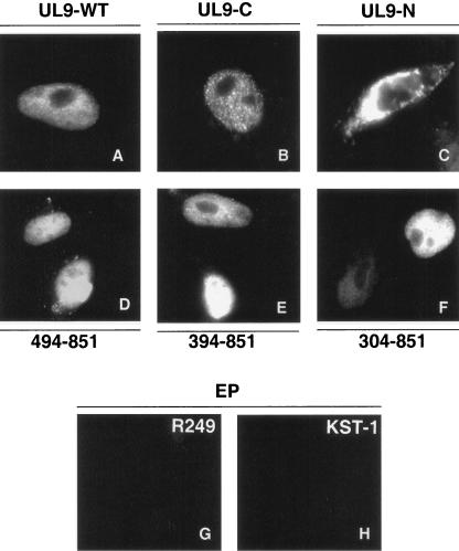
UL9, the origin binding protein of herpes simplex virus type 1, is a member of the SF2 family of helicases. Cotransfection of cells with infectious viral DNA and plasmids expressing either full-length UL9 or the C-terminal DNA binding domain alone results in the drastic inhibition of plaque formation which can be partially relieved by an insertion mutant lacking DNA binding activity. In this work, C-terminally truncated mutants which terminate at or near residue 359 were shown to potentiate plaque formation, while other C-terminal truncations were inhibitory. Thus, residues in the N-terminal region appear to regulate the inhibitory properties of UL9. To identify which residues were involved in this regulation, a series of N-terminally truncated mutants were constructed which contain the DNA binding domain and various N-terminal extensions. Mutants whose N terminus is either at residue 494 or 535 were able to bind the origin efficiently and were inhibitory to plaque formation, whereas constructs whose N terminus is at residue 304 or 394 were defective in origin binding activity and were able to relieve inhibition. Since UL9 is required for viral infection at early but not late times and is inhibitory to infection when overexpressed, we propose that the DNA binding activities of UL9 are regulated during infection. For infection to proceed, UL9 may need to switch from a DNA binding to a non-DNA binding mode, and we suggest that sequences residing in the N terminus play a role in this switch.
Figures


Similar articles
-
Existence of transdominant and potentiating mutants of UL9, the herpes simplex virus type 1 origin-binding protein, suggests that levels of UL9 protein may be regulated during infection.J Virol. 2003 Sep;77(17):9639-51. doi: 10.1128/jvi.77.17.9639-9651.2003. J Virol. 2003. PMID: 12915576 Free PMC article.
-
Direct interaction between the N- and C-terminal portions of the herpes simplex virus type 1 origin binding protein UL9 implies the formation of a head-to-tail dimer.J Virol. 2007 Dec;81(24):13659-67. doi: 10.1128/JVI.01204-07. Epub 2007 Oct 17. J Virol. 2007. PMID: 17942532 Free PMC article.
-
Helicase motif Ia is involved in single-strand DNA-binding and helicase activities of the herpes simplex virus type 1 origin-binding protein, UL9.J Virol. 2003 Feb;77(4):2477-88. doi: 10.1128/jvi.77.4.2477-2488.2003. J Virol. 2003. PMID: 12551986 Free PMC article.
-
Use of transdominant mutants of the origin-binding protein (UL9) of herpes simplex virus type 1 to define functional domains.J Virol. 1996 Nov;70(11):7859-66. doi: 10.1128/JVI.70.11.7859-7866.1996. J Virol. 1996. PMID: 8892908 Free PMC article.
-
The origin-binding domain of the herpes simplex virus type 1 UL9 protein is not required for DNA helicase activity.J Gen Virol. 1995 Dec;76 ( Pt 12):3125-30. doi: 10.1099/0022-1317-76-12-3125. J Gen Virol. 1995. PMID: 8847519
Cited by
-
The effect of amantadine on an ion channel protein from Chikungunya virus.PLoS Negl Trop Dis. 2019 Jul 24;13(7):e0007548. doi: 10.1371/journal.pntd.0007548. eCollection 2019 Jul. PLoS Negl Trop Dis. 2019. PMID: 31339886 Free PMC article.
-
A sequence within the varicella-zoster virus (VZV) OriS is a negative regulator of DNA replication and is bound by a protein complex containing the VZV ORF29 protein.J Virol. 2011 Dec;85(23):12188-200. doi: 10.1128/JVI.05501-11. Epub 2011 Sep 21. J Virol. 2011. PMID: 21937644 Free PMC article.
-
A novel 2006 Indian outbreak strain of Chikungunya virus exhibits different pattern of infection as compared to prototype strain.PLoS One. 2014 Jan 20;9(1):e85714. doi: 10.1371/journal.pone.0085714. eCollection 2014. PLoS One. 2014. PMID: 24465661 Free PMC article.
-
Cathepsin B mediates cleavage of herpes simplex virus type 1 origin binding protein (OBP) to yield OBPC-1, and cleavage is dependent upon viral DNA replication.J Virol. 2007 Sep;81(17):9175-82. doi: 10.1128/JVI.00676-07. Epub 2007 Jun 6. J Virol. 2007. PMID: 17553869 Free PMC article.
-
Herpes simplex viruses: mechanisms of DNA replication.Cold Spring Harb Perspect Biol. 2012 Sep 1;4(9):a013011. doi: 10.1101/cshperspect.a013011. Cold Spring Harb Perspect Biol. 2012. PMID: 22952399 Free PMC article. Review.
References
-
- Arbuckle, M. I., and N. D. Stow. 1993. A mutational analysis of the DNA-binding domain of the herpes simplex virus type 1 UL9 protein. J. Gen. Virol. 74(Pt 7):1349-1355. - PubMed
-
- Blumel, J., and B. Matz. 1995. Thermosensitive UL9 gene function is required for early stages of herpes simplex virus type 1 DNA synthesis. J. Gen. Virol. 76(Pt 12):3119-3124. - PubMed
-
- Challberg, M. 1996. Herpesvirus DNA replication. Cold Spring Harbor Press, Cold Spring Harbor, N.Y.
Publication types
MeSH terms
Substances
Grants and funding
LinkOut - more resources
Full Text Sources
