. 2006 May;80(9):4591-600.
doi: 10.1128/JVI.80.9.4591-4600.2006.

Human cytomegalovirus infection elicits a glycoprotein M (gM)/gN-specific virus-neutralizing antibody response

Affiliations

Human cytomegalovirus infection elicits a glycoprotein M (gM)/gN-specific virus-neutralizing antibody response

Masako Shimamura et al. J Virol. 2006 May.

Abstract

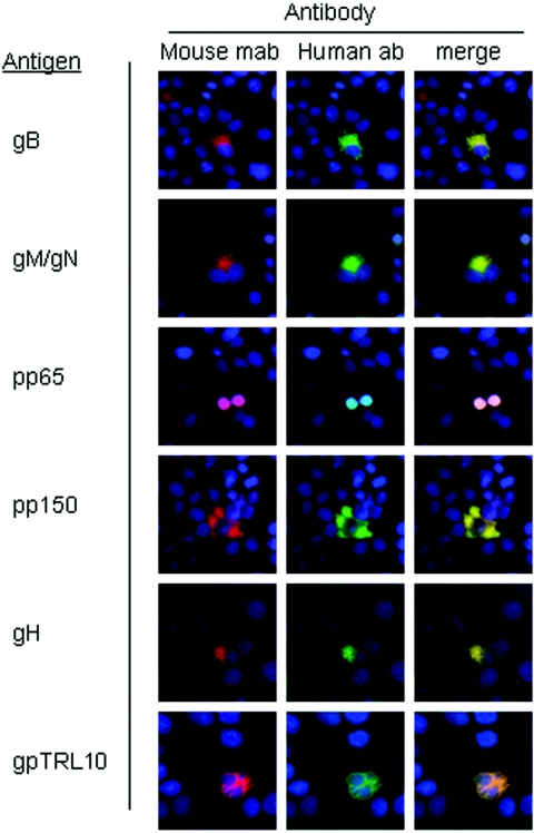
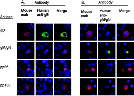
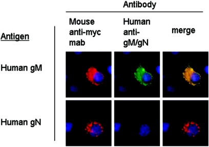
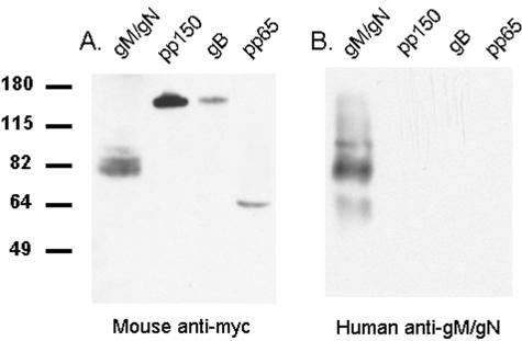
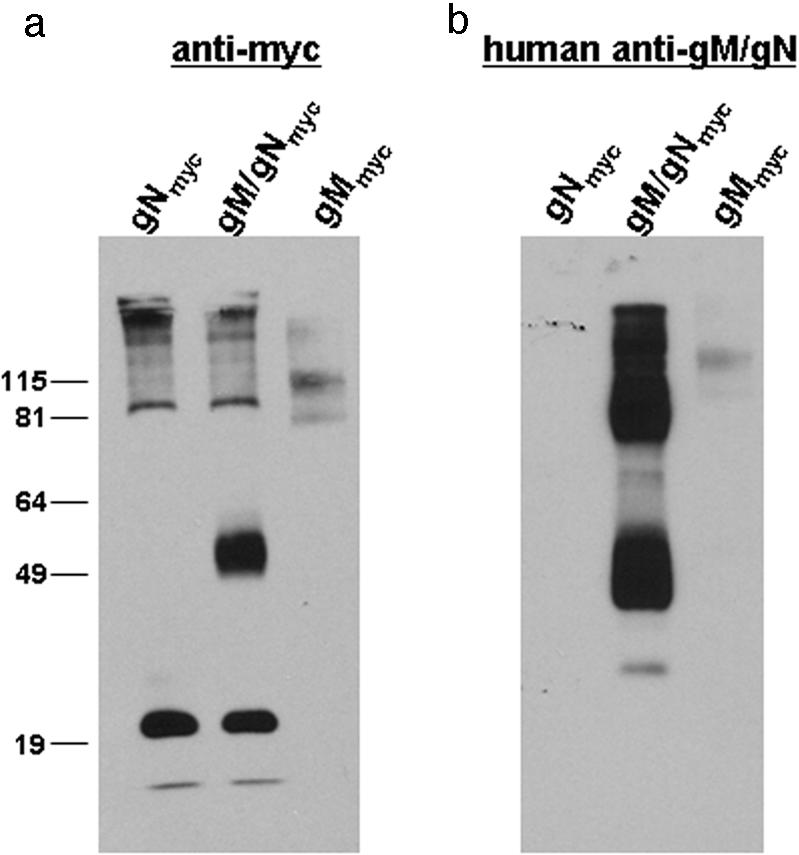
Human cytomegalovirus (HCMV) is a ubiquitous human pathogen that infects 40 to 90% of adult human populations. HCMV infections are often asymptomatic in healthy individuals but can cause severe organ and life-threatening disease in immunocompromised patients. The antiviral antibody response to HCMV infection is complex and is known to include virus-neutralizing antibody production against surface glycoproteins encoded by HCMV. We have investigated the human antibody response to a complex of HCMV surface glycoproteins composed of glycoprotein M (gM)/gN, the gene products of the UL100 and UL73 open reading frames. Mouse monoclonal antibodies generated against gM/gN have previously been shown to neutralize HCMV infection of human fibroblasts in vitro. To determine whether human antibodies reactive with the gM/gN complex possess virus-neutralizing properties, we isolated human antibodies reactive with gM/gN from pooled human HCMV hyperimmune globulin by affinity purification using recombinant gM/gN. The affinity-purified human anti-gM/gN antibodies reacted specifically by immunofluorescence with HCMV-infected human fibroblasts and with cells transiently expressing gM/gN, but not with cells transfected with plasmids encoding other immunogenic HCMV proteins. The anti-gM/gN antibodies also reacted specifically only with gM/gN in immunoblot assays using lysates of transfected cells expressing specific HCMV proteins. Last, human anti-gM/gN antibodies efficiently neutralized infectious HCMV in vitro with a capacity comparable to that of human anti-gB antibodies. These data indicated that gM/gN can elicit a virus-neutralizing antibody response in humans infected with HCMV and therefore should be considered a potential candidate for inclusion in prophylactic CMV vaccines.


Figures

FIG. 1.
FIG. 1.
Pooled human immunoglobulins contain antibodies reactive with HCMV virion proteins. Cos-7 cells were transfected with plasmids containing HCMV glycoproteins and phosphoproteins, and protein expression was demonstrated by reactivity with mouse monoclonal antibodies (Mouse mab) specific for each protein (Antigen) followed by a Texas Red-conjugated anti-mouse secondary antibody (red signal). Cells were coincubated with pooled human antibodies (Human ab) and detected using a FITC-conjugated anti-human secondary antibody (green signal). The appearance of yellow in each of the merged images (merge) shows colocalization of red and green signals in all rows, indicating reactivity of pooled human antibodies against each of the transfected HCMV proteins. The cell nuclei were stained with Hoechst dye (blue).
FIG. 2.
FIG. 2.
Affinity-purified human anti-gB and anti-gM/gN antibodies react specifically against gB or gM/gN. HCMV gB, gM/gN, pp65, or pp150 (Antigen) were expressed by transient transfection of Cos-7 cells with plasmids encoding each respective HCMV ORF. Protein expression was confirmed using mouse monoclonal antibodies specific for each protein (Mouse mab), followed by incubation with Texas Red-conjugated anti-mouse secondary antibodies (red signal). Cells were coincubated with affinity-purified human antibodies against either gB (Human anti-gB) (A) or gM/gN (Human anti-gM/gN) (B), and reactivity was detected using a FITC-conjugated anti-human secondary antibody (green signal). Merged images (Merge) show that human anti-gB reacts only with cells transfected with gB antigen (A) and human anti-gM/gN reacts only with cells transfected with gM/gN antigens (B). Hoechst nuclear stain (blue) shows the location of cell nuclei.
FIG. 3.
FIG. 3.
Affinity-purified human anti-gM/gN antibodies recognize transfected gM alone but not gN. Cos-7 cells were transfected with plasmids expressing either myc-tagged HCMV gM or gN, and protein expression was detected by reactivity with a mouse anti-myc monoclonal antibody followed by a Texas Red-conjugated anti-mouse secondary antibody (Mouse anti-myc mab, red signal). Cells were coincubated with the affinity-purified human anti-gM/gN antibodies used in Fig. 2, and reactivity was detected using a FITC-conjugated anti-human secondary antibody (Human anti-gM/gN, green signal). Merged images (merge) indicate that the human anti-gM/gN antibody recognized gM when expressed alone (yellow), but it had no detectable reactivity against gN (red) when this gene was expressed in the absence of gM.
FIG. 4.
FIG. 4.
Affinity-purified human anti-gM/gN is specific for the gM/gN complex. Myc-tagged HCMV proteins gM/gN, pp150, gB, and pp65 were transiently expressed in HEK293T cells following transfection of plasmids encoding each of these HCMV ORFs. Proteins were purified by immunoprecipitation using a rabbit anti-myc antibody and Staphylococcus aureus protein A, separated on 12% SDS-polyacrylamide gels, and electrophoretically transferred to nitrocellulose membranes. The presence of transfected proteins was confirmed by incubation of one membrane with a mouse anti-myc monoclonal antibody (Mouse anti-myc) (A), followed by a horseradish peroxidase-conjugated anti-mouse secondary antibody and developed by enhanced chemiluminescence. A second membrane, prepared using the same precipitated proteins, was incubated with the affinity-purified human anti-gM/gN and developed as described above for panel A (Human anti-gM/gN) (B). The positions of molecular mass standards (in kilodaltons) are indicated to the left of the gel. The presence of transiently expressed gM/gN (82 kDa), pp150 (150 kDa), gB (160 kDa), and pp65 (65 kDa) is shown in panel A. Panel B shows only bands in the lane containing gM/gN protein but no signal in the lanes containing pp150, gB, or pp65, indicating that affinity-purified human anti-gM/gN antibodies react specifically with components of the gM/gN complex.
FIG. 5.
FIG. 5.
Myc-tagged gN or gM were transiently expressed in HEK293T cells as described in Materials and Methods. Similarly, wild-type gM was coexpressed with myc-tagged gN (gNmyc) by cotransfection of plasmids encoding each of these HCMV ORFs. The cells were lysed, and myc-tagged proteins were collected with paramagnetic beads conjugated with anti-myc antibodies (Miltenyi Biotec, Auburn, Calif.). After the proteins were washed, they were eluted and separated by SDS-PAGE in 12% gels. The proteins were transferred to a nitrocellulose membrane and probed with (a) an anti-myc MAb (9E10) or (b) affinity-purified human anti-gM/gN antibodies, and antibody binding was detected using an ECL kit (Pierce, Rockford, Ill). The positions of molecular mass standards (in kilodaltons) are indicated to the left of the gel in panel a. The most rapidly migrating bands in panel a represent the dye front of the original gel.

Similar articles

Cited by

References

    1. Andreoni, M., M. Faircloth, L. Vugler, and W. J. Britt. 1989. A rapid microneutralization assay for the measurement of neutralizing antibody reactive with human cytomegalovirus. J. Virol. Methods 23:157-167. - PubMed
    1. Arvin, A. M., P. Fast, M. Myers, S. Plotkin, and R. Rabinovich. 2004. Vaccine development to prevent cytomegalovirus disease: report from the National Vaccine Advisory Committee. Clin. Infect. Dis. 39:233-239. - PubMed
    1. Baines, J. D., and B. Roizman. 1993. The UL10 gene of herpes simplex virus 1 encodes a novel viral glycoprotein, gM, which is present in the virion and in the plasma membrane of infected cells. J. Virol. 67:1441-1452. - PMC - PubMed
    1. Britt, W. J. 1984. Neutralizing antibodies detect a disulfide-linked glycoprotein complex within the envelope of human cytomegalovirus. Virology 135:369-378. - PubMed
    1. Britt, W. J., and D. Auger. 1985. Identification of a 65,000 dalton virion envelope of human cytomegalovirus. Virus Res. 4:31-36. - PubMed

Publication types

MeSH terms