. 2006 May;26(9):3365-77.
doi: 10.1128/MCB.26.9.3365-3377.2006.

Sonic hedgehog signaling regulates Gli2 transcriptional activity by suppressing its processing and degradation

Affiliations

Sonic hedgehog signaling regulates Gli2 transcriptional activity by suppressing its processing and degradation

Yong Pan et al. Mol Cell Biol. 2006 May.

Abstract

Gli2 and Gli3 are the primary transcription factors that mediate Sonic hedgehog (Shh) signals in the mouse. Gli3 mainly acts as a transcriptional repressor, because the majority of full-length Gli3 protein is proteolytically processed. Gli2 is mostly regarded as a transcriptional activator, even though it is also suggested to have a weak repressing activity. What the molecular basis for its possible dual function is and how its activity is regulated by Shh signaling are largely unknown. Here we demonstrate that unlike the results seen with Gli3 and Cubitus Interruptus, the fly homolog of Gli, only a minor fraction of Gli2 is proteolytically processed to form a transcriptional repressor in vivo and that in addition to being processed, Gli2 full-length protein is readily degraded. The degradation of Gli2 requires the phosphorylation of a cluster of numerous serine residues in its carboxyl terminus by protein kinase A and subsequently by casein kinase 1 and glycogen synthase kinase 3. The phosphorylated Gli2 interacts directly with betaTrCP in the SCF ubiquitin-ligase complex through two binding sites, which results in Gli2 ubiquitination and subsequent degradation by the proteasome. Both processing and degradation of Gli2 are suppressed by Shh signaling in vivo. Our findings provide the first demonstration of a molecular mechanism by which the Gli2 transcriptional activity is regulated by Shh signaling.


Figures

FIG. 1.
FIG. 1.
Differential processing of Gli2 and Gli3 proteins. (A) Characterization of Gli2 antibody. Protein extracts were prepared from E10.5 mouse embryos with indicated genotypes and immunoblotted with an anti-Gli2 antibody (αGli2) raised against the antigen shown below the panel. The αGli2 antibody detected Gli2 protein only in wild-type or Gli2lzki/+ heterozygous embryos but not Gli2lzki/lzki homozygous embryos. (B) Gli2 and Gli3 are differentially processed in vivo. Protein extracts prepared from E10.5 mouse embryos were incubated with a biotinylated double-stranded oligonucleotide containing a specific (Gli-B1) or mutated (mut) Gli-binding site. Proteins bound to the oligonucleotides were pulled down by streptavidin-conjugated magnetic beads. After the reaction mixture was divided into two halves, the proteins were detected by immunoblotting with the αGli2 and αGli3 antibodies. Full-length and processed forms of Gli2 and Gli3 proteins (indicated by arrows) were pulled down only by Gli-B1 but not Mut beads. (C) A histogram shows the ratio of full-length versus processed forms of either Gli2 or Gli3 proteins. (D) The Gli2-78 is a transcriptional repressor. Chick limb bud cell micromasses were transfected with a Gli-dependent luciferase reporter, TK-Renilla (a transfection control), and effector constructs as indicated. The data represent the normalized luciferase activities from three independent experiments.
FIG. 2.
FIG. 2.
Gli2 protein phosphorylation in vitro and in vivo. (A) A diagram shows a schematic drawing of Gli2 protein and a region of amino acid sequence including S residues for PKA (bold) and primary GSK3 (circled) and CK1 (boxed) phosphorylation. Ser residues that may become the optimal sites for GSK3 and CK1 phosphorylation, once the primary GSK3 and CK1 sites are phosphorylated, are indicated with lines over the characters representing the residues. Three Gli2 mutants shown below the Gli2 sequence contain S-to-A changes in PKA sites 1 to 4 (Gli2P1-4), GSK3 sites 2 to 4 (Gli2N2-4), or CK1 sites 1 to 4 (Gli2C1-4). (B) An immunoblot showing that Gli2 protein overexpressed in primary chick limb bud cells displays a shift in its migration upon FSK or OKA treatment. (C and D) Gli2 is phosphorylated. λ Protein phosphatase (λPPase) treatment revealed that the slowly migrating species of Gli2-185 protein from either transfected cells (C) or E10.5 mouse embryos (D) represent phosphorylated forms. (E) An immunoblot showing that Gli2 migrated more slowly than its mutants Gli2P1-4, Gli2N2-4, and Gli2C1-4, which contain point mutations as shown in panel A. (F) PKA-primed phosphorylation of Gli2 in vitro. The affinity-purified GST or fusion proteins were incubated with or without PKA in the presence of nonradioactive ATP. After PKA was removed, the proteins were incubated with CK1 or GSK3 or incubated without treatment as indicated in the presence of [γ-32P]ATP and detected by autoradiography (a 3-h exposure). An arrowhead points to a weak signal for GST-Gli2PR phosphorylated by CK1 alone, which can be readily seen after a 6-h exposure (data not shown).
FIG. 3.
FIG. 3.
Phosphorylation-induced Gli2 degradation. Chick limb bud cells were transfected with Gli2, Gli3, or Gli2-1-676 as indicated (A, B, and D). The transfected cells and untransfected MEFs (C) were incubated with FSK or the control drug ddFSK overnight (A) or with OKA and CHX for various time points (B, C, and D). Protein extracts were prepared from the treated cells and directly separated using 7% SDS-PAGE for immunoblotting with the indicated antibodies. The levels of Gli2-185 and Gli3-190 proteins in panels B and C were quantified and are plotted below the gels.
FIG. 4.
FIG. 4.
βTrCP is required for Gli2 degradation. (A) Overexpression of βTrCP promotes Gli2 degradation. Following transfection with constructs as indicated above the panel, HEK293 cells were incubated without or with FSK overnight. Gli2 proteins and tubulin were detected by immunoblotting with the αGli2 and αtubulin antibodies, respectively. Note that coexpression of βTrCP abolished hyperphosphorylated species of Gli2 (indicated by a white arrowhead; compare lane 4 to lane 3) but had little or no effect on the levels of Gli2 mutants. (B) βTrCP RNAi inhibits Gli2 degradation. HEK293 cells were transfected with Gli2 alone (lane 2) or together with either βTrCP siRNA or GFP siRNA (control) (lanes 3 and 4) and were treated with OKA and CHX. Gli2 expression was determined by immunoblotting with the αGli2 antibody (upper panel). The effectiveness of βTrCP RNAi was determined by RT-PCR (two lower panels).
FIG. 5.
FIG. 5.
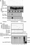
βTrCP interacts directly with phosphorylated Gli2. (A) βTrCP binds phosphorylated Gli2 in vivo. HEK293 cells were transfected with constructs as shown above the panels. A day after the transfection, the cells were incubated with FSK overnight and with MG132 for 3 h right before cells were lysed. The two upper panels show straight lysates immunoblotted using the indicated antibodies. The lysates were subjected to immunoprecipitation with anti-myc antibody followed by immunoblotting with anti-Gli2 antibody (second panel from the bottom) or to precipitation with a double-stranded oligonucleotide containing specific or nonspecific Gli-binding sites conjugated with Sepharose beads followed by immunoblotting with anti-myc antibody (bottom panel). (B) βTrCP interacts with phosphorylated Gli2 in vitro. Following the in vitro phosphorylation by the indicated kinases, GST fusion proteins (see Fig. 2A and F) were used to pull down the radiolabeled in vitro-translated myc-mβTrCP or myc-mβTrCPΔWD, which was then detected by autoradiography.
FIG. 6.
FIG. 6.
Identification of βTrCP binding sites in Gli2. (A) A Gli2 diagram (see Fig. 2 caption) representing the Gli2 mutants used for panel B. Each of the Gli2 mutants contains an A residue substitution for the corresponding S residues in the sequence. Two thick lines underline two βTrCP binding sites mapped in panel B. The alignment between βTrCP binding sites in Gli2 and the known βTrCP binding motif is shown. (B) Identification of two βTrCP binding sites in Gli2. HEK293 cells were transfected with each of the indicated Gli2 constructs along with myc-mβTrCP. Interaction between Gli2 proteins and βTrCP was determined as described for Fig. 5A.
FIG. 7.
FIG. 7.
Hh signaling inhibits Gli2 processing and degradation and induces Gli2 transcriptional activity in vivo. (A and B) Differential regulation of Gli2 and Gli3 processing by Shh signaling. Enrichment and detection of endogenous Gli2 and Gli3 proteins in Shh mutant and wild-type (WT) mouse embryos were performed as described in the Fig. 1 legend. Panel B shows the ratio of Gli2-185 to Gli2-78 (black bars), Gli3-190 to Gli3-83 (gray bars), and ratio changes between Shh mutants and wild type (Shh mutant/WT). (C and D) Stabilization of Gli2 protein by Shh signaling. The two upper panels show straight immunoblots of the Gli2-185 protein levels with α-tubulin as loading controls for E10.5 wild-type (WT) and Shh mutant (Shh/) mouse embryos and indicated cells treated with or without ShhN-conditioned medium. The two lower panels show the expression of Gli2 and actin mRNAs in the corresponding cells and mouse embryos. The relative levels of Gli2 protein and RNA were plotted in panel D. (E) ShhN treatment stabilizes endogenous Gli2-185 protein in wild-type MEFs. (F) Shh signaling induces the Gli2 transcriptional activity in vivo. Gli3Xt mutant MEFs were transfected with a Gli-dependent luciferase reporter, TK-Renilla (a transfection control), with or without a Gli1 RNAi-2 construct. Cells that received ShhN treatment are indicated. The firefly luciferase activity data were derived from three independent experiments.

References

    1. Aoto, K., T. Nishimura, K. Eto, and J. Motoyama. 2002. Mouse GLI3 regulates Fgf8 expression and apoptosis in the developing neural tube, face, and limb bud. Dev. Biol. 251:320-332. - PubMed
    1. Ausubel, Frederick M., R. Brent, Robert E. Kingston, David D. Moore, J. G. Seidman, and Kevin Struhl (ed.). 1988. Current protocols in molecular Biology. John Wiley & Sons, Inc., Hoboken, N.J.
    1. Aza-Blanc, P., H. Y. Lin, A. Ruiz i Altaba, and T. B. Kornberg. 2000. Expression of the vertebrate Gli proteins in Drosophila reveals a distribution of activator and repressor activities. Development 127:4293-4301. - PubMed
    1. Aza-Blanc, P., F. A. Ramirez-Weber, M. P. Laget, C. Schwartz, and T. B. Kornberg. 1997. Proteolysis that is inhibited by hedgehog targets Cubitus interruptus protein to the nucleus and converts it to a repressor. Cell 89:1043-1053. - PubMed
    1. Bai, C. B., W. Auerbach, J. S. Lee, D. Stephen, and A. L. Joyner. 2002. Gli2, but not Gli1, is required for initial Shh signaling and ectopic activation of the Shh pathway. Development 129:4753-4761. - PubMed

Publication types

MeSH terms