. 2006 May;26(9):3378-89.
doi: 10.1128/MCB.26.9.3378-3389.2006.

Cdc37 interacts with the glycine-rich loop of Hsp90 client kinases

Affiliations

Cdc37 interacts with the glycine-rich loop of Hsp90 client kinases

Kazuya Terasawa et al. Mol Cell Biol. 2006 May.

Abstract

Recently, we identified a client-binding site of Cdc37 that is required for its association with protein kinases. Phage display technology and liquid chromatography-tandem mass spectrometry (which identifies a total of 33 proteins) consistently identify a unique sequence, GXFG, as a Cdc37-interacting motif that occurs in the canonical glycine-rich loop (GXGXXG) of protein kinases, regardless of their dependence on Hsp90 or Cdc37. The glycine-rich motif of Raf-1 (GSGSFG) is necessary for its association with Cdc37; nevertheless, the N lobe of Raf-1 (which includes the GSGSFG motif) on its own cannot interact with Cdc37. Chimeric mutants of Cdk2 and Cdk4, which differ sharply in their affinities toward Cdc37, show that their C-terminal portions may determine this difference. In addition, a nonclient kinase, the catalytic subunit of cyclic AMP-dependent protein kinase, interacts with Cdc37 but only when a threonine residue in the activation segment of its C lobe is unphosphorylated. Thus, although a region in the C termini of protein kinases may be crucial for accomplishing and maintaining their interaction with Cdc37, we conclude that the N-terminal glycine-rich loop of protein kinases is essential for physically associating with Cdc37.


Figures

FIG. 1.
FIG. 1.
Cdc37(181-378) interacts directly with the kinase domain of Raf-1. The Myc-tagged kinase domain of Raf-1 and either FLAG-Cdc37 (FL) or FLAG-Cdc37(181-378) (181-378) were expressed in COS7 cells. The cell lysates (whole, lanes 1 and 2) were subjected to immunoprecipitation (IP) with anti-FLAG (IP: α-FLAG, lanes 3 and 4) or anti-Myc (IP: α-Myc, lanes 5 and 6) antibody, followed by immunoblotting (WB) with the indicated antibodies. The asterisk indicates nonspecific bands.
FIG. 2.
FIG. 2.
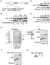
The glycine-rich loop of Raf-1 interacts with Cdc37. (A) Schematic of the primary structures of the EGFP-Myc fusion proteins showing the peptide sequences of wild-type (wt) and mutant (ASA) Raf-1; the altered amino acids are marked by a box. M indicates the N-terminal methionine. (B) EGFP-Myc alone (empty) and EGFP-Myc fused to the wild-type or mutant Raf-1 peptide were coexpressed with FLAG-Cdc37(181-378) in COS7 cells. Cell extracts (whole, lanes 1 to 3) were subjected to immunoprecipitation with anti-FLAG (IP: α-FLAG, lanes 4 to 6) or anti-Myc (IP: α-Myc, lanes 7 to 9) antibody, followed by immunoblotting with both anti-FLAG and anti-Myc antibodies. The asterisk indicates nonspecific bands. (C and D) Wild-type and mutant forms of the Myc-tagged Raf-1 kinase domain (Myc-Knd) and either FLAG-Cdc37 (C) or FLAG-Cdc37(181-378) (D) were expressed in COS7 cells. The cell lysates (whole) were subjected to immunoprecipitation with anti-FLAG or anti-Myc antibody, followed by immunoblotting with both anti-FLAG and anti-Myc antibodies. (E) FLAG-Cdc37(181-378) (181-378) was expressed in COS7 cells; the cell lysates (whole, lane 7) and purified proteins (IP, lane 8) were immunoblotted with the indicated antibodies. The purified FLAG-Cdc37(181-378) protein was subjected to SDS-PAGE and Coomassie staining (lane 1). Myc-GST (empty) and wild-type (wt) and mutant (ASA) forms of Myc-GST fused to either the kinase domain of Raf-1 (Knd) or its N lobe were separated by SDS-PAGE and then subjected to either Coomassie staining (lanes 2 to 6) or immunoblotting with the indicated antibodies (lanes 9 to 13). Asterisks indicate unknown proteins that are present in all preparations of the Myc-GST fusion proteins. Molecular mass markers are shown in kDa on the left of the panel. (F) Myc-GST, the Myc-GST-fused kinase domain of Raf-1, and the Myc-GST-fused N lobe of Raf-1, which were adsorbed to glutathione Sepharose, were incubated with FLAG-Cdc37(181-378) and then subjected to a GST pull-down assay. Samples were analyzed by immunoblotting with the indicated antibodies. (G) Wild-type and mutant forms of the His-tagged Raf-1 peptide adsorbed to Ni-NTA agarose were incubated with recombinant GST-Cdc37(181-378) proteins, and then proteins bound to the peptide-adsorbed resin were analyzed by immunoblotting with anti-Cdc37 antibody.
FIG. 3.
FIG. 3.
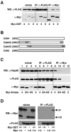
Full-length Cdc37 does not bind to the N-lobe or glycine-rich loop of Raf-1. (A) Myc-tagged Raf-1 kinase domain (Knd) and Myc-tagged GST fused to the N-lobe of Raf-1 were coexpressed with either FLAG-Cdc37 or FLAG-Cdc37(181-378) (FL or 181-378, respectively) in COS7 cells (whole, lanes 1 to 4), and the indicated immunoprecipitates (IP: α-FLAG, lanes 5 to 8; IP: α-Myc, lanes 9 to 12) were analyzed by immunoblotting with both anti-FLAG and anti-Myc antibodies. (B) FLAG-Cdc37 or FLAG-Cdc37(181-378) was coexpressed in COS7 cells with EGFP-Myc either alone (e) or fused to the Raf-1 peptide (p) shown in Fig. 2A. Cell extracts were prepared (whole) and subjected to immunoprecipitation with anti-FLAG (IP: α-FLAG) or anti-Myc (IP: α-Myc) antibody, followed by immunoblotting as indicated. Asterisks indicates nonspecific bands.
FIG. 4.
FIG. 4.
Chimeric mutants of Cdk2 and Cdk4. (A) FLAG-Cdc37(181-378) was coexpressed in COS7 cells with Myc-tagged GST (Myc-GST) either alone (e) or fused to the N lobe of Cdk4 or Cdk2 (4 or 2, respectively) and subjected to immunoprecipitation with anti-FLAG (IP: α-FLAG) or anti-Myc (IP: α-Myc) antibody, followed by immunoblotting with anti-FLAG and anti-Myc antibodies. (B) Schematic of Cdk4, Cdk2, and two chimeric mutants. Numbers in parentheses indicate the total number of amino acids; those in boxes indicate the original residue numbers. (C) FLAG-Cdc37 was coexpressed in COS7 cells with the indicated Myc-tagged kinases; Cdk4, Cdk2, and the two chimeric mutants, Cdk2/4 and Cdk4/2, are indicated as 4, 2, 2/4, and 4/2, respectively. Cell extracts (whole, lanes 1 to 4) were subjected to immunoprecipitation (IP: α-FLAG, lanes 5 to 8; IP: α-Myc, lanes 9 to 12) and immunoblotting with anti-Hsp90, anti-FLAG, and anti-Myc antibodies. (D) Myc-GST fused to the N-terminal portion of the Raf-1 kinase domain (I to IV) and/or the Myc-tagged C-terminal portion of the Raf-1 kinase domain (V to XI) was expressed in COS7 cells in the presence or absence of FLAG-Cdc37 as indicated. Cell extracts (whole) were treated as described for panel C.
FIG. 5.
FIG. 5.
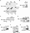
Unphosphorylated PKA binds to Cdc37. (A) Myc-tagged PKA (Myc-PKA) was coexpressed with FLAG-Cdc37, FLAG-Cdc37(1-180), or FLAG-Cdc37(181-378) in COS7 cells (FL, 1-180, or 181-378, respectively). Cell extracts (whole, lanes 1 to 3) were immunoprecipitated with anti-FLAG (IP: α-FLAG, lanes 4 to 6) or anti-Myc (IP: α-Myc, lanes 7 to 9) antibody and subjected to immunoblotting with both anti-FLAG and anti-Myc antibodies. The two short horizontal lines indicate a doublet band. (B) Myc-PKA and FLAG-Cdc37(181-378) were coexpressed in COS7 cells, and the indicated immunoprecipitates (IP: α-FLAG, lane 1; IP: α-Myc, lane 2) were analyzed by immunoblotting with antiphosphorylated (at Thr197) PKA (α-pPKA) and anti-Myc (to detect Myc-PKA) antibodies. (C) Myc-PKA was expressed in COS7 cells either alone (control, lane 1 to 3) or with FLAG-Cdc37(181-378) [FLAG-Cdc37(181-378), lanes 4 to 6] and purified by immunoprecipitation with anti-Myc or anti-FLAG antibody, respectively. The purified PKA (input, lanes 1 and 4) was either mock treated (without phosphatase [PPase −], lanes 2 and 5) or treated with phosphatase (PPase +, lanes 3 and 6) and subjected to immunoblotting with anti-Myc antibody. (D) Wild-type and mutant forms of Myc-PKA (wt and TA, respectively) were coexpressed with FLAG-Cdc37 in COS7 cells, and the immunoprecipitates obtained were analyzed by immunoblotting with both anti-FLAG and anti-Myc antibodies. (E) FLAG-tagged PKA was translated from mRNAs with or without a stop codon (+ or −, respectively) in rabbit reticulocyte lysate for 20 min at 30°C. Reaction mixtures were immunoprecipitated with anti-FLAG antibody and subjected to immunoblotting with both anti-phosphorylated PKA and anti-FLAG antibodies. (F and G) After protein synthesis, as described for panel E, PKA released from the ribosome (F, RNase at 0 min) or the ribosome-bound PKA (G, puromycin at 0 min) was incubated for a further 30 min at 30°C in the presence of either 0.2 mg/ml of RNase to terminate translation (F, RNase at 30 min) or 1 mM puromycin to release translation products from the ribosome (G, puromycin at 30 min). Immunoprecipitates obtained with anti-FLAG antibody were subjected to immunoblotting with the indicated antibodies.

Similar articles

Cited by

References

    1. Basso, A. D., D. B. Solit, G. Chiosis, B. Giri, P. Tsichlis, and N. Rosen. 2002. Akt forms an intracellular complex with heat shock protein 90 (Hsp90) and Cdc37 and is destabilized by inhibitors of Hsp90 function. J. Biol. Chem. 277:39858-39866. - PubMed
    1. Bukau, B., E. Deuerling, C. Pfund, and E. A. Craig. 2000. Getting newly synthesized proteins into shape. Cell 101:119-122. - PubMed
    1. Dai, K., R. Kobayashi, and D. Beach. 1996. Physical interaction of mammalian CDC37 with CDK4. J. Biol. Chem. 271:22030-22034. - PubMed
    1. Deuerling, E., and B. Bukau. 2004. Chaperone-assisted folding of newly synthesized proteins in the cytosol. Crit. Rev. Biochem. Mol. Biol. 39:261-277. - PubMed
    1. Farrell, A., and D. Morgan. 2000. Cdc37 promotes the stability of protein kinases Cdc28 and Cak1. Mol. Cell. Biol. 20:749-754. - PMC - PubMed

Publication types

MeSH terms