. 2006 May;26(9):3390-400.
doi: 10.1128/MCB.26.9.3390-3400.2006.

Role for Upf2p phosphorylation in Saccharomyces cerevisiae nonsense-mediated mRNA decay

Affiliations

Role for Upf2p phosphorylation in Saccharomyces cerevisiae nonsense-mediated mRNA decay

Weirong Wang et al. Mol Cell Biol. 2006 May.

Abstract

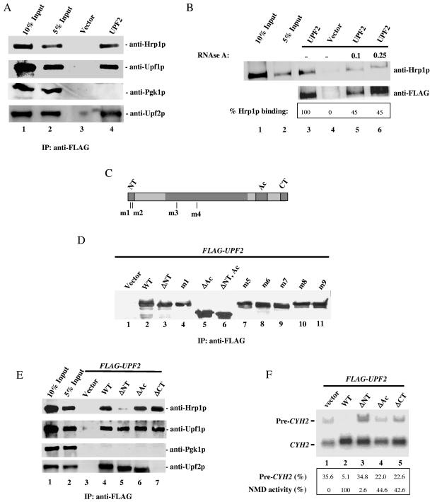
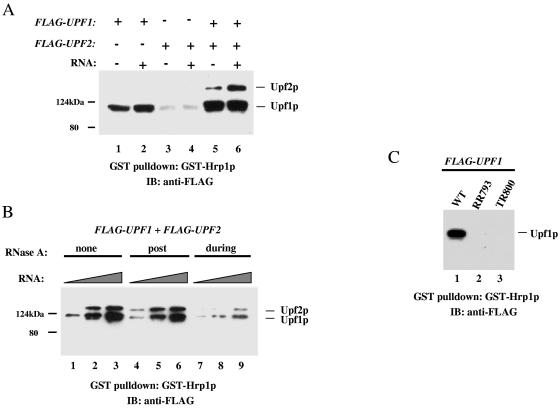
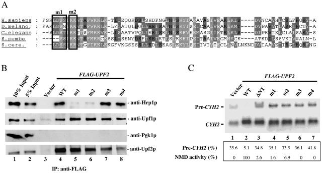
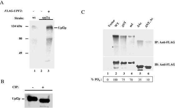
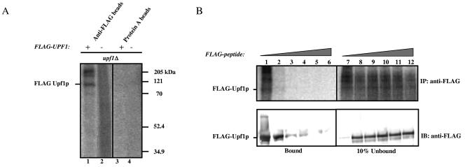
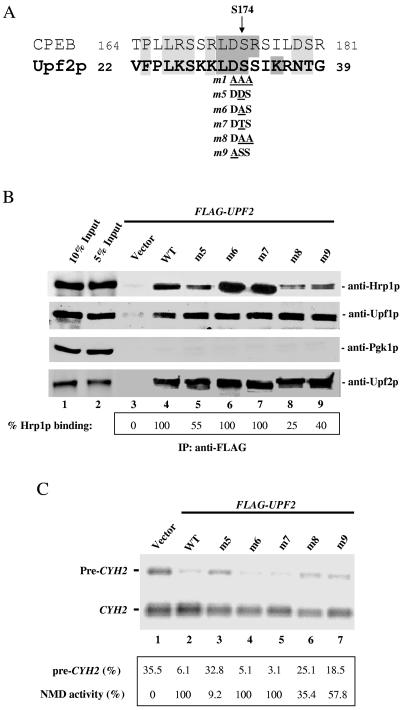
Premature termination (nonsense) codons trigger rapid mRNA decay by the nonsense-mediated mRNA decay (NMD) pathway. Two conserved proteins essential for NMD, UPF1 and UPF2, are phosphorylated in higher eukaryotes. The phosphorylation and dephosphorylation of UPF1 appear to be crucial for NMD, as blockade of either event in Caenorhabditis elegans and mammals largely prevents NMD. The universality of this phosphorylation/dephosphorylation cycle pathway has been questioned, however, because the well-studied Saccharomyces cerevisiae NMD pathway has not been shown to be regulated by phosphorylation. Here, we used in vitro and in vivo biochemical techniques to show that both S. cerevisiae Upf1p and Upf2p are phosphoproteins. We provide evidence that the phosphorylation of the N-terminal region of Upf2p is crucial for its interaction with Hrp1p, an RNA-binding protein that we previously showed is essential for NMD. We identify specific amino acids in Upf2p's N-terminal domain, including phosphorylated serines, which dictate both its interaction with Hrp1p and its ability to elicit NMD. Our results indicate that phosphorylation of UPF1 and UPF2 is a conserved event in eukaryotes and for the first time provide evidence that Upf2p phosphorylation is crucial for NMD.


Figures

FIG. 1.
FIG. 1.
Hrp1p and Upf2p interact. (A) Coimmunoprecipitation of Hrp1p and Upf2p. Cytoplasmic extracts from strains transformed with FLAG-UPF2 or vector only were immunoprecipitated (IP) with anti-FLAG monoclonal antibody and immunoblotted with anti-Hrp1p, anti-Upf1p (positive control), or anti-Pgk1p (negative control) antibodies. The anti-Upf2p antibody was used to demonstrate the amount of Upf2p immunoprecipitated in the assay. Lanes 1 and 2 contain 10% and 5% of the total extract used for immunoprecipitation, respectively. (B) The assay was performed as described for panel A except that cytoplasmic extracts were treated with RNase A (μg/μl concentration indicated) for 20 minutes at room temperature prior to immunoprecipitation with anti-FLAG antibody. High concentrations of RNase A were used to ensure that the RNA present in the cytoplasmic extracts was completely degraded. (C) Schematic diagram of Upf2p, showing the NT, Ac, and CT domains. The positions of the mutations in the m1 to m4 mutants are indicated. (D) Western blot analysis performed as described for panel A, demonstrating that the mutant Upf2p proteins shown are expressed at a level similar to that of wild-type Upf2p (WT). (E) Coimmunoprecipitation of mutant Upf2p proteins and Hrp1p. Cytoplasmic extracts from yeast upf2Δ strains expressing the indicated FLAG-UPF2 constructs were immunoprecipitated as described for panel A. The anti-Upf2p antibody used in these experiments recognizes a specific peptide located in the C-terminal domain of Upf2p. These amino acids are not present in the ΔCT mutant protein used in these coimmunoprecipitation studies (see lane 7, anti-Upf2p). (F) NMD activity of Upf2p mutants determined using Northern blot analysis of total cellular RNA from the upf2Δ strains in panel E. The migration of the nonsense codon-containing CYH2 pre-mRNA (pre-CYH2) and CYH2 mature mRNA (CYH2) bands is indicated. The ratio of pre-CYH2 to CYH2 mRNA gives the magnitude of NMD (quantified by phosphorimaging).
FIG. 2.
FIG. 2.
Upf2p-Hrp1p interactions promoted by Upf1p and RNA. (A) GST pull-down experiment in which immobilized GST-Hrp1p is incubated with FLAG-Upf1p and/or FLAG-Upf2p and/or poly(U) RNA. Bound proteins were detected with anti-FLAG antibody. The weak band with a size similar to that of FLAG-Upf1p in lanes 3 and 4 is GST-Hrp1p, which is weakly recognized by the anti-FLAG antibody. (B) Experiment performed as described for panel A except that RNase A (0.25 μg/μl) was added either during or after (post) complex formation (see Materials and Methods). The amount of poly(U) RNA added was 2, 20, or 200 ng. (C) Immobilized GST-Hrp1p was incubated with wild-type UPF1 or an RNA-binding-deficient UPF1 mutant (RR793 or TR800), and their interactions were examined as described for panel A.
FIG. 3.
FIG. 3.
Specific N-terminal amino acids of Upf2p crucial for NMD and its interaction with Hrp1p. (A) Alignment of the N-terminal region of UPF2 homologues from S. cerevisiae, Schizosaccharomyces pombe, C. elegans, D. melanogaster, and Homo sapiens. Black boxes represent the positions of amino acid substitutions in the m1 and m2 mutants (Fig. 6A and the text indicate the substitutions made). (B) Coimmunoprecipitation of mutant Upf2p proteins and Hrp1p, performed as described for Fig. 1A. The Western blot was first probed with an anti-Hrp1p antibody, followed by anti-Upf1p (positive control) and anti-Pgk1p (negative control) antibodies. The mutant proteins were expressed at levels virtually identical to those of wild-type Upf2p, as determined by Western blot analysis (Fig. 1D and data not shown). In addition, Western blot analysis showed that the expression of Hrp1p does not vary in these different strains (data not shown). (C) NMD activity of the Upf2p mutants shown, determined using Northern blot analysis of total cellular RNA (performed as for Fig. 1F) from the upf2Δ strains used in panel B.
FIG. 4.
FIG. 4.
Upf2p is phosphorylated in vivo. (A) A wild-type strain (lane 1), a upf2Δ strain (lane 2), and a upf2Δ strain expressing FLAG-Upf2p (lane 3) were labeled with [32P]orthophosphate and immunoprecipitated with anti-Upf2p polyclonal antibody. Labeled proteins were detected by phosphorimaging. (B) Immunopurified FLAG-Upf2p was incubated with or without calf intestinal phosphatase (CIP) and analyzed by Western blotting with anti-Upf2p polyclonal antibody. (C) In vivo labeling experiment, performed as described for panel A, with yeast upf2Δ strains expressing the indicated upf2 constructs. The assay was performed as described for panel A, except that the cytoplasmic extracts were immunoprecipitated with anti-FLAG antibody and detected by autoradiography (upper panel) or by Western blotting with anti-FLAG antibody (lower panel). The asterisk indicates an N-terminally truncated Upf2p product.
FIG. 5.
FIG. 5.
Yeast Upf1p is phosphorylated in vivo. (A) In vivo labeling experiment performed as described for Fig. 4A using a yeast upf1Δ strain that expresses FLAG-Upf1p and a negative control that does not. (B) Increasing concentrations of FLAG peptide (0 to 400 ng/μl) were added to immunoprecipitations performed as described for panel A to test the specificity of the affinity matrix against FLAG-Upf1p (upper panel). The immunoprecipitated proteins were transferred to a polyvinylidene fluoride membrane and probed with anti-FLAG monoclonal antibody (lower panel).
FIG. 6.
FIG. 6.
The N-terminal domain of Upf2p harbors a phosphorylation site. (A) Sequence alignment of the phosphorylation site of Xenopus CPEB protein and the N-terminal region phosphorylated in S. cerevisiae Upf2p. (B) Coimmunoprecipitation of mutant Upf2p proteins with Hrp1p, Upf1p, and Pgk1p, performed as described for Fig. 1A. The percentage of Hrp1p interacting with the FLAG-tagged Upf2p mutants (relative to wild-type [WT]) was calculated as described in Materials and Methods. (C) NMD activity of the Upf2p mutants shown, determined using Northern blot analysis of total cellular RNA (performed as described for Fig. 1D) from the upf2Δ strains used in panel B.

Similar articles

Cited by

References

    1. Amrani, N., R. Ganesan, S. Kervestin, D. A. Mangus, S. Ghosh, and A. Jacobson. 2004. A faux 3′-UTR promotes aberrant termination and triggers nonsense-mediated mRNA decay. Nature 432:112-118. - PubMed
    1. Anders, K. R., A. Grimson, and P. Anderson. 2003. SMG-5, required for C. elegans nonsense-mediated mRNA decay, associates with SMG-2 and protein phosphatase 2A. EMBO J. 22:641-650. - PMC - PubMed
    1. Baker, K. E., and R. Parker. 2004. Nonsense-mediated mRNA decay: terminating erroneous gene expression. Curr. Opin. Cell Biol. 16:293-299. - PubMed
    1. Bhattacharya, A., K. Czaplinski, P. Trifillis, F. He, A. Jacobson, and S. W. Peltz. 2000. Characterization of the biochemical properties of the human Upf1 gene product that is involved in nonsense-mediated mRNA decay. RNA 6:1226-1235. - PMC - PubMed
    1. Chiu, S. Y., G. Serin, O. Ohara, and L. E. Maquat. 2003. Characterization of human Smg5/7a: a protein with similarities to Caenorhabditis elegans SMG5 and SMG7 that functions in the dephosphorylation of Upf1. RNA 9:77-87. - PMC - PubMed

Publication types

MeSH terms