. 2006 May;26(9):3414-31.
doi: 10.1128/MCB.26.9.3414-3431.2006.

Protein kinase C delta activates topoisomerase IIalpha to induce apoptotic cell death in response to DNA damage

Affiliations

Protein kinase C delta activates topoisomerase IIalpha to induce apoptotic cell death in response to DNA damage

Kiyotsugu Yoshida et al. Mol Cell Biol. 2006 May.

Abstract

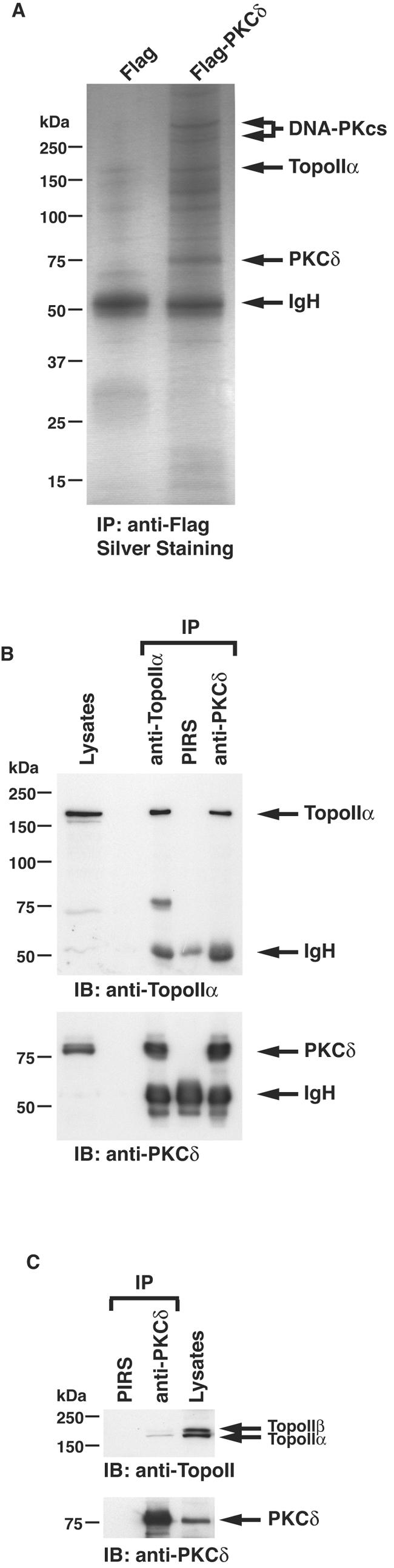
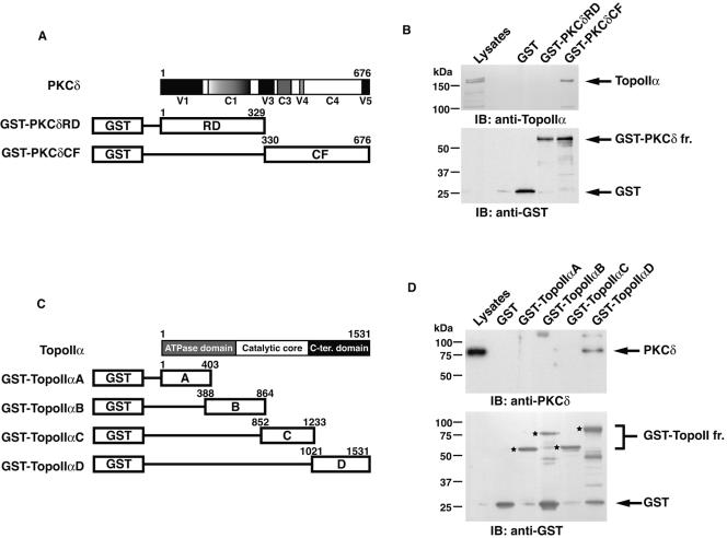
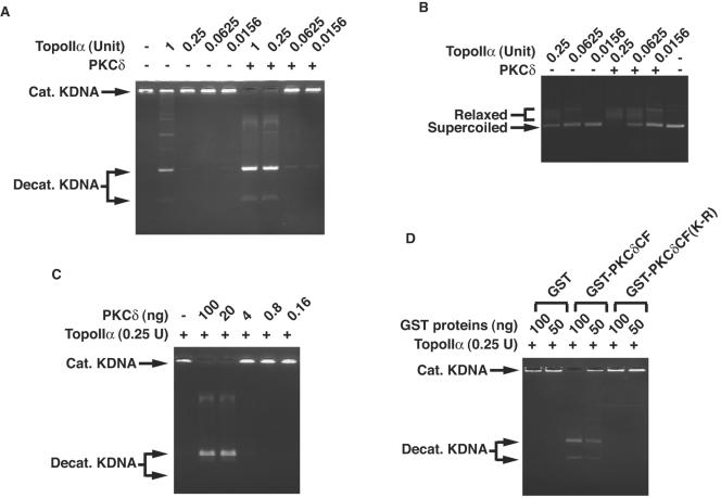
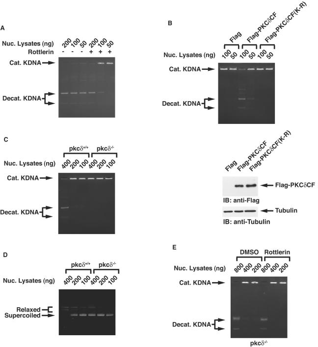
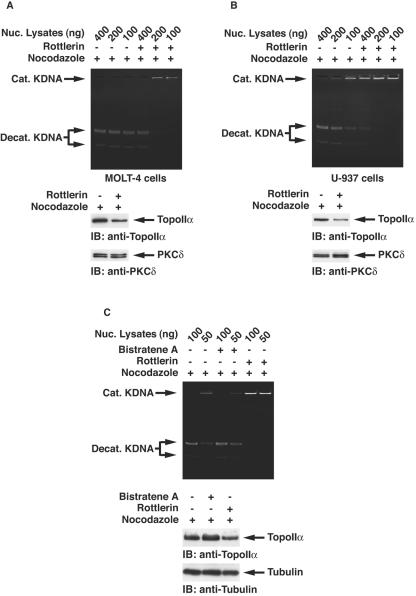
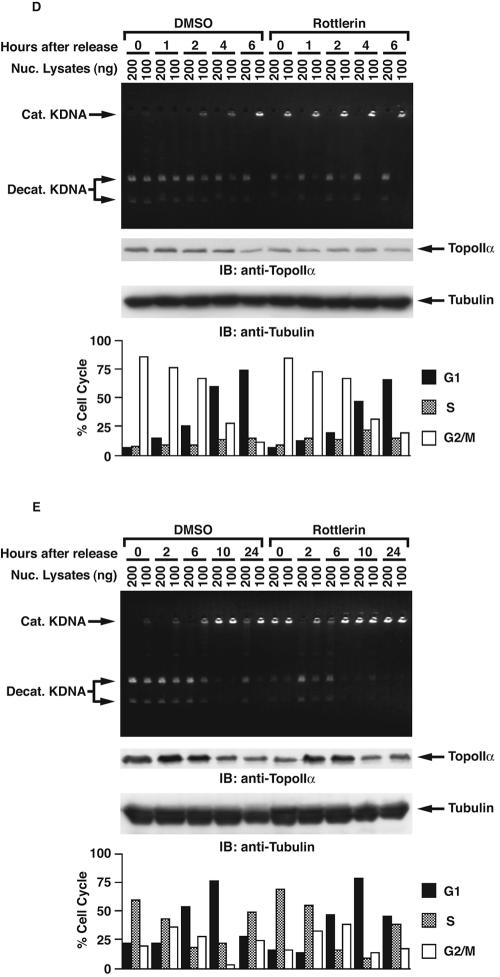
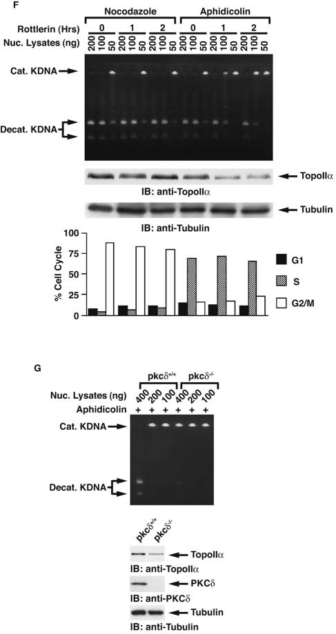
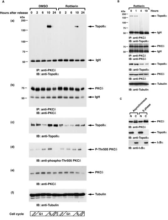
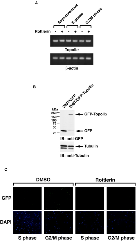
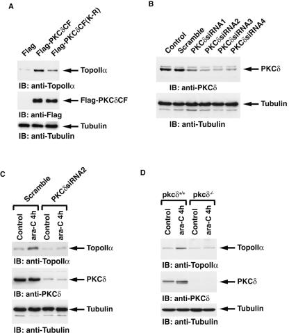
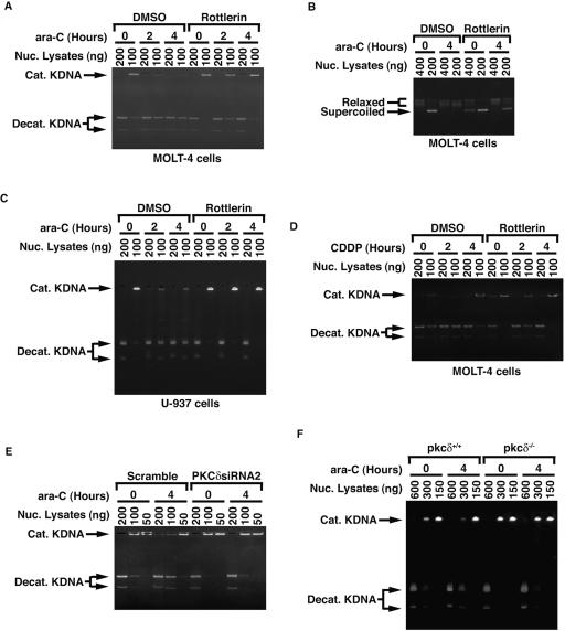
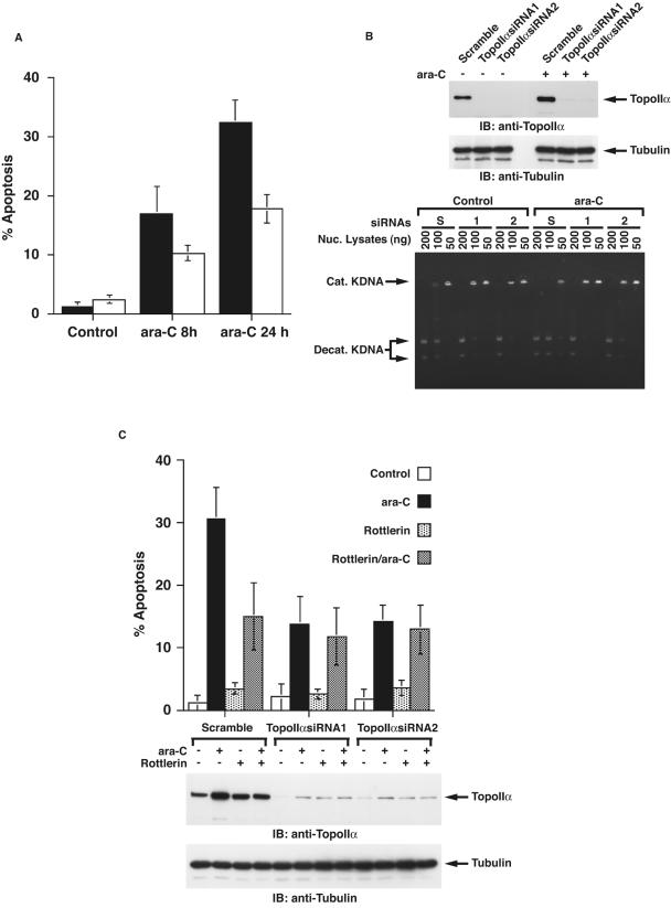
DNA topoisomerase II is an essential nuclear enzyme that modulates DNA processes by altering the topological state of double-stranded DNA. This enzyme is required for chromosome condensation and segregation; however, the regulatory mechanism of its activation is largely unknown. Here we demonstrate that topoisomerase IIalpha is activated in response to genotoxic stress. Concomitant with the activation, the expression of topoisomerase IIalpha is increased following DNA damage. The results also demonstrate that the proapoptotic kinase protein kinase C delta (PKCdelta) interacts with topoisomerase IIalpha. This association is in an S-phase-specific manner and is required for stabilization and catalytic activation of topoisomerase IIalpha in response to DNA damage. Conversely, inhibition of PKCdelta activity attenuates DNA damage-induced activation of topoisomerase IIalpha. Finally, aberrant activation of topoisomerase IIalpha by PKCdelta is associated with induction of apoptosis upon exposure to genotoxic agents. These findings indicate that PKCdelta regulates topoisomerase IIalpha and thereby cell fate in the genotoxic stress response.


Figures

FIG. 1.
FIG. 1.
Association of PKCδ with topoisomerase IIα (TopoIIα). (A) Cell lysates from 293T cells transfected with Flag vector or Flag-PKCδ were immunoprecipitated (IP) with anti-Flag. Immunoprecipi- tates were resolved by SDS-PAGE and analyzed by silver staining. The polypeptides identified by mass spectrometric analyses are indicated to the right of the blot (IgH, immunoglobulin heavy chain). (B) Nuclear lysates from MOLT-4 cells were subjected to immunoprecipitation (IP) with preimmune rabbit serum (PIRS), anti-topoisomerase IIα (anti-TopoIIα), or anti-PKCδ. Cell lysates and immunoprecipitates were analyzed by immunoblotting (IB) with anti-topoisomerase IIα or anti-PKCδ. The finding that the binding stoichiometry of PKCδ with topoisomerase IIα was relatively high is mainly due to using nuclear lysates for immunoprecipitation. (C) Nuclear lysates from MOLT-4 cells were subjected to immunoprecipitation with PIRS or anti-PKCδ. Cell lysates and immunoprecipitates were analyzed by immunoblotting with anti-topoisomerase IIα and IIβ (anti-TopoII) or anti-PKCδ.
FIG. 2.
FIG. 2.
PKCδ directly interacts with and phosphorylates topoisomerase IIα. (A) Schematic representation of PKCδ. RD, regulatory domain; CF, catalytic fragment. (B) MOLT-4 cell lysates were incubated with GST, GST-PKCδRD, or GST-PKCδCF bound to glutathione beads. The adsorbates were analyzed by immunoblotting (IB) with anti-topoisomerase IIα (anti-TopoIIα) or anti-GST. GST-PKCδ fr., GST-PKCδ fragment. (C) Schematic representation of topoisomerase IIα. C-ter. domain, C-terminal domain. (D) MOLT-4 cell lysates were incubated with GST or GST-topoisomerase IIα fragments (GST-TopoIIα fr.) bound to glutathione beads. The adsorbates were analyzed by immunoblotting (IB) with anti-PKCδ or anti-GST. Each one of the bands specific for GST-topoisomerase IIα fragments is indicated by an asterisk. (E) 293T cells were transfected with GFP-TopoIIα or a truncated form of GFP-TopoIIα that encodes amino acid residues 1 to 1145 (GFP-TopoIIαΔC). Lysates were subjected to immunoprecipitation (IP) with anti-PKCδ or PIRS followed by immunoblot (IB) analysis with anti-GFP or anti-PKCδ. IgH, immunoglobulin heavy chain. (F) Recombinant topoisomerase IIα was incubated with glutathione beads containing GST or GST-PKCδ. The adsorbates were subjected to immunoblot (IB) analysis with anti-topoisomerase IIα (anti-TopoIIα) or anti-GST. (G) GST-PKCδ was incubated with or without recombinant topoisomerase IIα and [γ-32P]ATP. The reaction products were analyzed by SDS-PAGE and autoradiography or by Coomassie brilliant blue R-250 (CBB) staining.
FIG. 2.
FIG. 2.
PKCδ directly interacts with and phosphorylates topoisomerase IIα. (A) Schematic representation of PKCδ. RD, regulatory domain; CF, catalytic fragment. (B) MOLT-4 cell lysates were incubated with GST, GST-PKCδRD, or GST-PKCδCF bound to glutathione beads. The adsorbates were analyzed by immunoblotting (IB) with anti-topoisomerase IIα (anti-TopoIIα) or anti-GST. GST-PKCδ fr., GST-PKCδ fragment. (C) Schematic representation of topoisomerase IIα. C-ter. domain, C-terminal domain. (D) MOLT-4 cell lysates were incubated with GST or GST-topoisomerase IIα fragments (GST-TopoIIα fr.) bound to glutathione beads. The adsorbates were analyzed by immunoblotting (IB) with anti-PKCδ or anti-GST. Each one of the bands specific for GST-topoisomerase IIα fragments is indicated by an asterisk. (E) 293T cells were transfected with GFP-TopoIIα or a truncated form of GFP-TopoIIα that encodes amino acid residues 1 to 1145 (GFP-TopoIIαΔC). Lysates were subjected to immunoprecipitation (IP) with anti-PKCδ or PIRS followed by immunoblot (IB) analysis with anti-GFP or anti-PKCδ. IgH, immunoglobulin heavy chain. (F) Recombinant topoisomerase IIα was incubated with glutathione beads containing GST or GST-PKCδ. The adsorbates were subjected to immunoblot (IB) analysis with anti-topoisomerase IIα (anti-TopoIIα) or anti-GST. (G) GST-PKCδ was incubated with or without recombinant topoisomerase IIα and [γ-32P]ATP. The reaction products were analyzed by SDS-PAGE and autoradiography or by Coomassie brilliant blue R-250 (CBB) staining.
FIG. 3.
FIG. 3.
Topoisomerase IIα is activated by PKCδ in vitro. (A) The indicated amount of purified topoisomerase IIα (TopoIIα) was incubated with (+) or without (−) kinase-active recombinant PKCδ (50 ng). Decatenation assays using reaction mixtures containing kinetoplast DNA were performed, and the reaction products were analyzed on a 1% agarose gel. Cat. KDNA, catenated KDNA; Decat. KDNA, decatenated KDNA. (B) Purified topoisomerase IIα was incubated with or without recombinant PKCδ. Relaxation assays using reaction mixtures containing pBluescript were performed, and the reaction products were analyzed on a 1% agarose gel. (C) Purified topoisomerase IIα (0.25 U) was incubated with or without (−) the indicated amount of kinase-active recombinant PKCδ. Decatenation assays using reaction mixtures containing KDNA were performed, and the reaction products were analyzed on a 1% agarose gel. (D) Purified topoisomerase IIα (0.25 U) was incubated with recombinant GST, GST-PKCδCF, or GST-PKCδCF(K-R). Decatenation assays using reaction mixtures containing KDNA were performed, and the reaction products were analyzed on a 1% agarose gel.
FIG. 4.
FIG. 4.
Topoisomerase IIα is activated by PKCδ in cells. (A) MOLT-4 cells were incubated in the presence (+) or absence (−) of rottlerin. Decatenation assays using nuclear lysates (Nuc. Lysates) were performed, and the reaction products were resolved on a 1% agarose gel. Cat. KDNA, catenated KDNA; Decat. KDNA, decatenated KDNA. (B) 293T cells were transfected with Flag vector, Flag-PKCδCF, or Flag-PKCδCF(K-R). Nuclear lysates were analyzed by decatenation assays (top blot). Cell lysates were subjected to immunoblot (IB) analysis with anti-Flag or antitubulin. (C and D) Nuclear lysates from pkcδ+/+ and pkcδ−/− MEFs were analyzed by the decatenation (C) and DNA relaxation (D) assays. (E) pkcδ−/− MEFs were left untreated or treated with rottlerin for 1 h. Nuclear lysates were analyzed by the decatenation assays. DMSO, dimethyl sulfoxide.
FIG. 5.
FIG. 5.
PKCδ is involved in cell cycle-dependent activation of topoisomerase IIα. (A and B) MOLT-4 (A) and U-937 (B) cells were treated with nocodazole (+) in the presence (+) or absence (−) of rottlerin. Topoisomerase IIα activity was analyzed by decatenation assays (top blot). Lysates were analyzed by immunoblotting (IB) with anti-topoisomerase IIα (anti-TopoIIα) or anti-PKCδ. Nuc. Lysates, nuclear lysates; Cat. KDNA, catenated KDNA; Decat. KDNA, decatenated KDNA. (C) MOLT-4 cells were treated with nocodazole in the presence or absence of bistratene A or rottlerin. Nuclear lysates were analyzed by decatenation assays (top blot). Lysates were analyzed by immunoblotting with anti-topoisomerase IIα (anti-TopoIIα) or antitubulin (bottom blot). (D) MOLT-4 cells were treated with nocodazole for 16 h in the presence or absence of rottlerin and then released by nocodazole removal and harvested at the indicated times. Nuclear (Nuc.) lysates were prepared, and topoisomerase IIα activity was analyzed by decatenation assays (top blot). Lysates were analyzed by immunoblotting (IB) with anti-topoisomerase IIα or antitubulin (bottom blot). The cell cycle was monitored by using a FACscan and represented as the percentage of population in each cell cycle phase in the graph at the bottom of panel D. (E) MOLT-4 cells were treated with aphidicolin for 16 h in the presence or absence of rottlerin and then released by aphidicolin removal and harvested at the indicated times. Nuclear lysates were prepared, and topoisomerase IIα activity was analyzed by decatenation assays (top blot). Lysates were analyzed by immunoblotting with anti-topoisomerase IIα or antitubulin (bottom blot). The cell cycle was monitored by using a FACscan and represented as the percentage of population in each cell cycle phase in the graph. (F) MOLT-4 cells were treated with nocodazole or aphidicolin for 16 h and then with rottlerin for the indicated times. Topoisomerase IIα activity was analyzed by decatenation assays (top blot). Lysates were analyzed by immunoblotting with anti-topoisomerase IIα or antitubulin (bottom blot). The cell cycle was monitored by using a FACscan and represented as the percentage of population in each cell cycle phase in the graph. (G) pkcδ+/+ and pkcδ−/− MEFs were treated with aphidicolin for 16 h. Nuclear lysates were analyzed by the decatenation assays (top blot). Lysates were analyzed by immunoblotting with anti-topoisomerase IIα, anti-PKCδ, or antitubulin.
FIG. 5.
FIG. 5.
PKCδ is involved in cell cycle-dependent activation of topoisomerase IIα. (A and B) MOLT-4 (A) and U-937 (B) cells were treated with nocodazole (+) in the presence (+) or absence (−) of rottlerin. Topoisomerase IIα activity was analyzed by decatenation assays (top blot). Lysates were analyzed by immunoblotting (IB) with anti-topoisomerase IIα (anti-TopoIIα) or anti-PKCδ. Nuc. Lysates, nuclear lysates; Cat. KDNA, catenated KDNA; Decat. KDNA, decatenated KDNA. (C) MOLT-4 cells were treated with nocodazole in the presence or absence of bistratene A or rottlerin. Nuclear lysates were analyzed by decatenation assays (top blot). Lysates were analyzed by immunoblotting with anti-topoisomerase IIα (anti-TopoIIα) or antitubulin (bottom blot). (D) MOLT-4 cells were treated with nocodazole for 16 h in the presence or absence of rottlerin and then released by nocodazole removal and harvested at the indicated times. Nuclear (Nuc.) lysates were prepared, and topoisomerase IIα activity was analyzed by decatenation assays (top blot). Lysates were analyzed by immunoblotting (IB) with anti-topoisomerase IIα or antitubulin (bottom blot). The cell cycle was monitored by using a FACscan and represented as the percentage of population in each cell cycle phase in the graph at the bottom of panel D. (E) MOLT-4 cells were treated with aphidicolin for 16 h in the presence or absence of rottlerin and then released by aphidicolin removal and harvested at the indicated times. Nuclear lysates were prepared, and topoisomerase IIα activity was analyzed by decatenation assays (top blot). Lysates were analyzed by immunoblotting with anti-topoisomerase IIα or antitubulin (bottom blot). The cell cycle was monitored by using a FACscan and represented as the percentage of population in each cell cycle phase in the graph. (F) MOLT-4 cells were treated with nocodazole or aphidicolin for 16 h and then with rottlerin for the indicated times. Topoisomerase IIα activity was analyzed by decatenation assays (top blot). Lysates were analyzed by immunoblotting with anti-topoisomerase IIα or antitubulin (bottom blot). The cell cycle was monitored by using a FACscan and represented as the percentage of population in each cell cycle phase in the graph. (G) pkcδ+/+ and pkcδ−/− MEFs were treated with aphidicolin for 16 h. Nuclear lysates were analyzed by the decatenation assays (top blot). Lysates were analyzed by immunoblotting with anti-topoisomerase IIα, anti-PKCδ, or antitubulin.
FIG. 6.
FIG. 6.
S-phase-specific interaction of PKCδ with topoisomerase IIα. (A) MOLT-4 cells were synchronized in G2/M phase by treatment with nocodazole in the presence or absence of rottlerin and then released into the cell cycle by its removal. Cells were harvested at the indicated times, and lysates were analyzed by immunoprecipitation (IP) with anti-PKCδ followed by immunoblotting (IB) with anti-topoisomerase IIα (anti-TopoIIα) (a) or anti-PKCδ (b). Cell lysates were also analyzed by immunoblotting with anti-topoisomerase IIα (c), anti-phospho-Thr505 PKCδ (d), anti-PKCδ (e), or antitubulin (f). The cell cycle was monitored by using a FACscan. DMSO, dimethyl sulfoxide; IgH, immunoglobulin heavy chain; P-Thr505 PKCδ, phospho-Thr505 PKCδ. (B) MOLT-4 cells were left untreated or treated with rottlerin for the indicated times. Cell lysates were subjected to immunoprecipitation with anti-PKCδ followed by immunoblot analysis with anti-topoisomerase IIα (anti-TopoIIα) or anti-PKCδ. Lysates were also analyzed by immunoblotting with anti-topoisomerase IIα, anti-PKCδ, or antitubulin. (C) MOLT-4 cells were left untreated or treated with aphidicolin to synchronize cells in S phase. Lysates from nuclear (N) and cytoplasmic (C) fractions were subjected to immunoblot (IB) analysis with anti-PKCδ, anti-topoisomerase IIα (anti-TopoIIα), or anti-IκBα.
FIG. 7.
FIG. 7.
PKCδ regulates a posttranslational modification of topoisomerase IIα. (A) MOLT-4 cells were left untreated or treated with aphidicolin or nocodazole for 16 h in the presence (+) or absence (−) of rottlerin. Total RNA was subjected to RT-PCR analysis using primer sets for topoisomerase IIα (TopoIIα) or β-actin. (B) 293T cells were stably transfected with GFP vector (293T/GFP) or GFP-topoisomerase IIα (293T/GFP-TopoIIα). Cell lysates were analyzed by immunoblotting (IB) with anti-GFP or antitubulin. (C) 293T/GFP-TopoIIα cells were treated with aphidicolin or nocodazole for 16 h in the presence or absence of rottlerin. After the cells were washed with phosphate-buffered saline twice, cells were mounted with Vectashield mounting medium containing 4′,6-diamidino-2-phenylindole (DAPI; Vector Laboratories) and analyzed with a Nikon Eclipse TE2000-U microscope. The cell cycle was determined by using a FACscan. DMSO, dimethyl sulfoxide.
FIG. 8.
FIG. 8.
Inhibition of PKCδ by rottlerin abrogates DNA damage-induced expression of topoisomerase IIα. (A and B) MOLT-4 cells were pretreated with or without rottlerin for 1 h followed by treatment with ara-C (A) or cisplatin (CDDP) (B) for the indicated periods. Cell lysates were subjected to immunoblot (IB) analysis with anti-topoisomerase IIα (anti-TopoIIα) or anti-PKCδ. DMSO, dimethyl sulfoxide. (C) U-937 cells were treated and analyzed as described above for panel A. (D) MOLT-4 cells were left untreated or treated with ara-C for 4 h in the presence or absence of rottlerin. Total RNA was subjected to RT-PCR analysis using primer sets for topoisomerase IIα (TopoIIα) or β-actin. (E) 293T/GFP-TopoIIα cells were left untreated or treated with rottlerin for 1 h followed by treatment with ara-C for 4 h. Cell lysates were subjected to immunoblot analysis with anti-topoisomerase IIα or anti-PKCδ.
FIG. 9.
FIG. 9.
PKCδ-dependent induction of topoisomerase IIα expression in response to DNA damage. (A) 293T cells were transfected with Flag vector, Flag-PKCδCF, or Flag-PKCδCF(K-R). Cell lysates were analyzed by immunoblotting (IB) with anti-topoisomerase IIα (anti-TopoIIα), anti-Flag, or antitubulin. (B) U2-OS cells were left untransfected (Control) or transfected with the indicated siRNAs. Cell lysates were subjected to immunoblot (IB) analysis with anti-PKCδ or antitubulin. (C) U2-OS cells transfected with scrambled siRNA or PKCδsiRNA2 were left untreated or treated with ara-C for 4 h. Cell lysates were analyzed by immunoblotting with anti-topoisomerase IIα, anti-PKCδ, or antitubulin. (D) pkcδ+/+ and pkcδ−/− MEFs were left untreated or treated with ara-C for 4 h. Cell lysates were analyzed by immunoblotting with anti-topoisomerase IIα, anti-PKCδ, or antitubulin.
FIG. 10.
FIG. 10.
PKCδ-dependent activation of topoisomerase IIα in response to DNA damage. (A and B) MOLT-4 cells were pretreated with or without rottlerin for 1 h followed by treatment with ara-C. Nuclear lysates (Nuc. Lysates) were analyzed by decatenation (A) and DNA relaxation (B) assays. DMSO, dimethyl sulfoxide; Cat. KDNA, catenated KDNA; Decat. KDNA, decatenated KDNA. (C) U-937 cells were treated as described above for panel A, and nuclear lysates were analyzed by the decatenation assays. (D) MOLT-4 cells were left untreated or treated with rottlerin for 1 h followed by the treatment with CDDP for the indicated times. Topoisomerase IIα activity was analyzed by decatenation assays. (E) U2-OS cells transfected with scrambled siRNA or PKCδsiRNA2 were left untreated or treated with ara-C for 4 h. Nuclear lysates were analyzed by the decatenation assays. (F) pkcδ+/+ and pkcδ−/− MEFs were left untreated or treated with ara-C for 4 h. Nuclear lysates were analyzed by the decatenation assays.
FIG. 11.
FIG. 11.
Topoisomerase IIα is required for PKCδ-induced apoptotic cell death in response to genotoxic stress. (A) MOLT-4 cells were treated with ara-C for the indicated times in the presence (open bar) or absence (closed bar) of ICRF-193. The percentages of apoptotic cells were determined by TUNEL assays. The results are represented as means ± standard deviations (error bars) obtained from four fields of 100 to 300 cells (each field) and three independent experiments. (B) U2-OS cells were transfected with the indicated siRNAs for 48 h and then left untreated (−) or treated (+) with ara-C for 4 h. Cell lysates were subjected to immunoblot (IB) analysis with anti-topoisomerase IIα (anti-TopoIIα) or antitubulin. Nuclear lysates were analyzed by decatenation assays (bottom blot). The cells had been transfected with scrambled siRNA (lanes S), topoisomerase IIα siRNA1 (lanes 1), or topoisomerase IIα siRNA2 (lanes 2). Nuc. Lysates, nuclear lysates; Cat. KDNA, catenated KDNA; Decat. KDNA, decatenated KDNA. (C) U2-OS cells transfected with the indicated siRNAs were left untreated (−) or treated (+) with rottlerin for 1 h followed by treatment with ara-C (+) for 24 h. The percentages of apoptotic cells were determined by TUNEL assays. The results are represented as means ± standard deviations (error bars) obtained from four fields of 100 to 300 cells, each performed over three independent experiments. Cell lysates were also analyzed by immunoblotting (IB) with anti-topoisomerase IIα (anti-TopoIIα) or antitubulin.

Similar articles

Cited by

References

    1. Akimitsu, N., K. Kamura, S. Tone, A. Sakaguchi, A. Kikuchi, H. Hamamoto, and K. Sekimizu. 2003. Induction of apoptosis by depletion of DNA topoisomerase IIα in mammalian cells. Biochem. Biophys. Res. Commun. 307:301-307. - PubMed
    1. Bharti, A., S. K. Kraeft, M. Gounder, P. Pandey, S. Jin, Z. M. Yuan, S. P. Lees-Miller, R. Weichselbaum, D. Weaver, L. B. Chen, D. Kufe, and S. Kharbanda. 1998. Inactivation of DNA-dependent protein kinase by protein kinase Cδ: implications for apoptosis. Mol. Cell. Biol. 18:6719-6728. - PMC - PubMed
    1. Blass, M., I. Kronfeld, G. Kazimirsky, P. M. Blumberg, and C. Brodie. 2002. Tyrosine phosphorylation of protein kinase Cδ is essential for its apoptotic effect in response to etoposide. Mol. Cell. Biol. 22:182-195. - PMC - PubMed
    1. Castedo, M., J. L. Perfettini, T. Roumier, K. Andreau, R. Medema, and G. Kroemer. 2004. Cell death by mitotic catastrophe: a molecular definition. Oncogene 23:2825-2837. - PubMed
    1. Chen, A. Y., and L. F. Liu. 1994. DNA topoisomerases: essential enzymes and lethal targets. Annu. Rev. Pharmacol. Toxicol. 34:191-218. - PubMed

Publication types

LinkOut - more resources