. 2007 Jan;56(1):48-59.
doi: 10.1007/s00262-006-0160-8. Epub 2006 Apr 13.

Tumor-derived CD4(+)CD25(+) regulatory T cell suppression of dendritic cell function involves TGF-beta and IL-10

Affiliations

Tumor-derived CD4(+)CD25(+) regulatory T cell suppression of dendritic cell function involves TGF-beta and IL-10

Nicolas Larmonier et al. Cancer Immunol Immunother. 2007 Jan.

Abstract

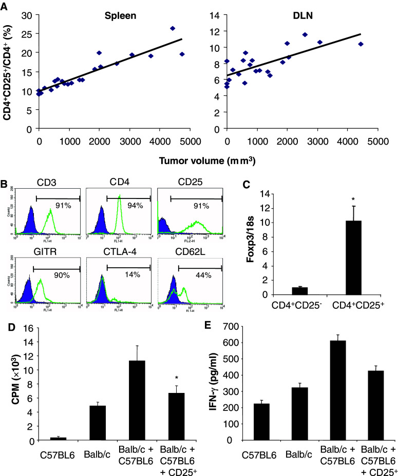
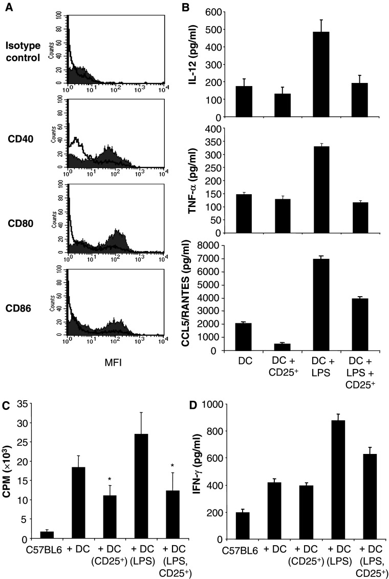
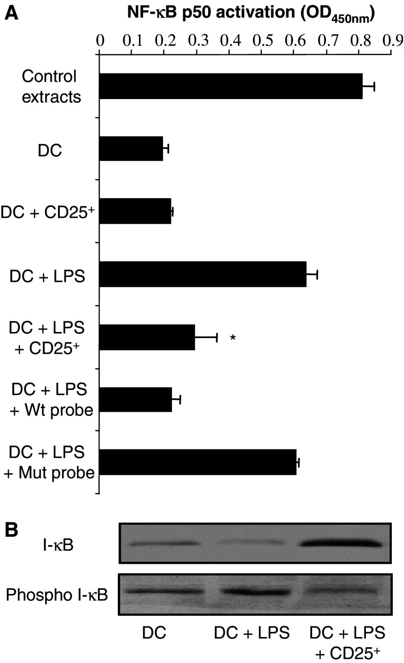
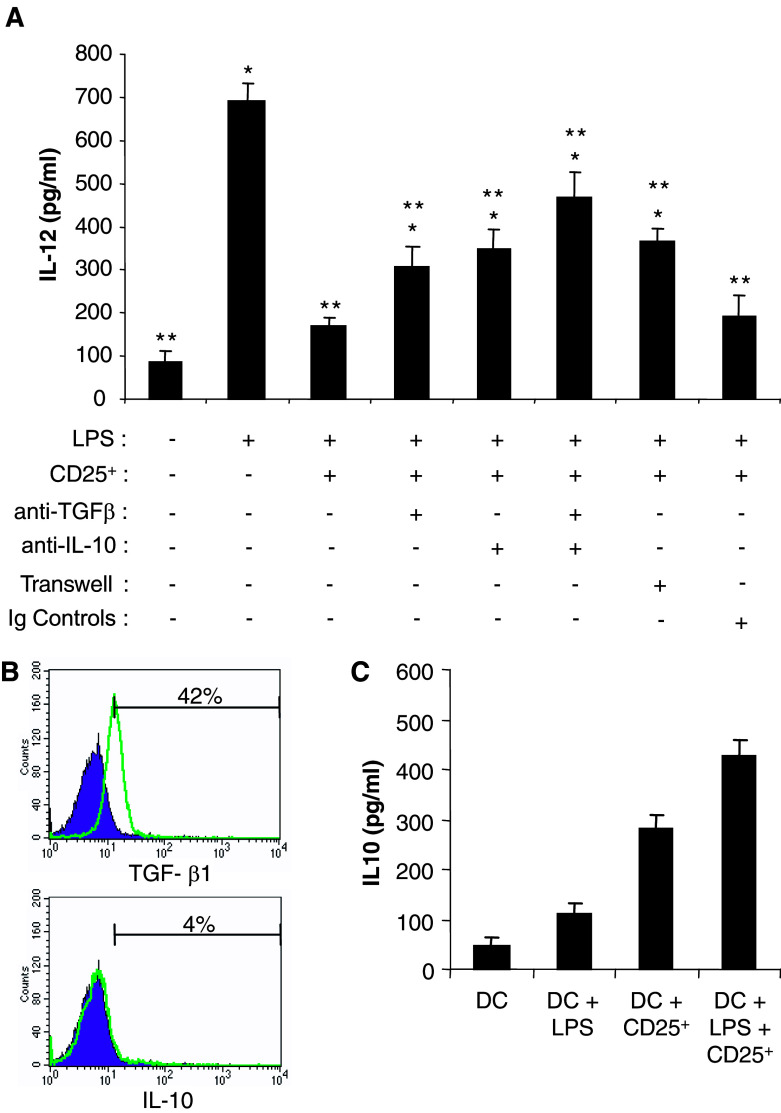
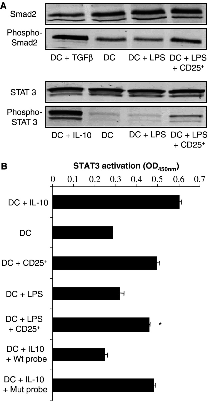
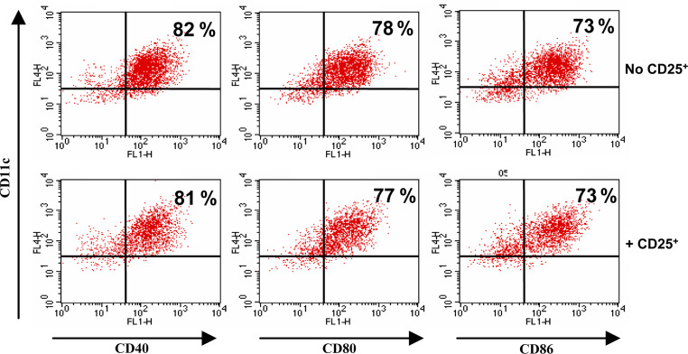
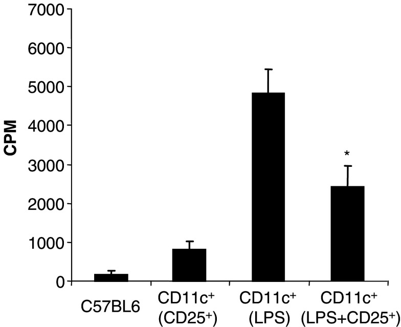
CD4(+)CD25(+) regulatory T cells have been characterized as a critical population of immunosuppressive cells. They play a crucial role in cancer progression by inhibiting the effector function of CD4(+) or CD8(+) T lymphocytes. However, whether regulatory T lymphocytes that expand during tumor progression can modulate dendritic cell function is unclear. To address this issue, we have evaluated the inhibitory potential of CD4(+)CD25(+) regulatory T cells from mice bearing a BCR-ABL(+) leukemia on bone marrow-derived dendritic cells. We present data demonstrating that CD4(+)CD25(+)FoxP3(+) regulatory T cells from tumor-bearing animals impede dendritic cell function by down-regulating the activation of the transcription factor NF-kappaB. The expression of the co-stimulatory molecules CD80, CD86 and CD40, the production of TNF-alpha, IL-12, and CCL5/RANTES by the suppressed DC is strongly down-regulated. The suppression mechanism requires TGF-beta and IL-10 and is associated with induction of the Smad signaling pathway and activation of the STAT3 transcription factor.


Figures

Fig. 1
Fig. 1
Characteristics of CD4+CD25+FoxP3+ regulatory T cells in mice bearing established 12B1 tumors. a Increase in the ratio of CD4+CD25+ to total CD4+ during tumor progression. Total splenocytes (Spleen) or draining lymph node (DLN) cells from mice bearing established 12B1 tumors at different stages were analyzed. The ratio CD4+CD25+/CD4+ was determined for each individual mouse and reported as a function of the tumor volume (r 2=0.9270, P<0.00005 in the spleen and r 2=0.7364, P<0.0005 in the draining lymph nodes). b Phenotype of CD4+CD25+ T lymphocytes purified using magnetic cell sorting from the spleen of mice bearing established 12B1 tumors (2,000–3,000 mm3). The percentage of positive cells is indicated. c Foxp3 mRNA levels are increased in CD4+CD25+ T cells purified from the spleen of mice bearing established tumors. Expression levels (average ± SD) relative to 18S ribosomal RNA are shown (*P<0.01). d Inhibition of the proliferation of allogeneic splenocytes by CD4+CD25+ T cells from tumor-bearing mice. Responder BALB/c splenocytes (Balb/c) were stimulated with C57BL6 splenocytes (C57BL6) in presence or absence of CD4+CD25+ T cells (CD25+) isolated from the spleen of mice bearing 12B1 tumors. The data are shown as mean ± SD of quadruplicate wells of 3[H]thymidine incorporation. e CD4+CD25+ T cells from tumor-bearing mice inhibit IFN-γ production. The culture supernatants of the experiment described in d were collected and IFN-γ concentration was determined by ELISA. The results are shown as the mean of duplicate wells. Asterisk is a significant difference when compared to control without CD4+CD25+ regulatory T cells (P<0.01)
Fig. 2
Fig. 2
CD4+CD25+ regulatory T cells from tumor-bearing mice suppress DC. a CD4+CD25+ regulatory T cells (CD25+) from tumor-bearing mice inhibit the expression of the co-stimulatory molecules by DC. Day 4 bone marrow-derived DC were cultured in the presence or absence of CD4+CD25+ regulatory T cells isolated from tumor-bearing mice (tumor volume = 2,000–3,000 mm3), and subjected to LPS treatment. DC were then purified, stained and analyzed by FACS. Shaded histogram, DC + LPS; non-shaded histogram, DC + LPS + CD25+. b CD4+CD25+ regulatory T cells inhibit cytokine production by DC. CD4+CD25+ regulatory T cells-DC co-culture supernatants were collected and the production of IL-12, TNF-α and CCL5/RANTES was analyzed by ELISA. Control DC were cultured in medium alone. Results are the mean ± SD of triplicate wells. c The capability of DC to induce the proliferation of allo-splenocytes is hampered by CD4+CD25+ regulatory T cells from tumor-bearing mice. DC purified from the co-culture with CD4+CD25+ regulatory T cells were used as stimulator in an allogeneic MLR. C57BL6 splenocytes were the responder cells and were cultured alone (C57BL6), with DC (+DC), with DC pre-incubated with regulatory T cells (+DC(CD25+)), or LPS (+DC(LPS)), or both (+DC(LPS,CD25+)). The data are shown as mean ± SD of quadruplicate wells of 3H-thymidine incorporation. d CD4+CD25+ regulatory T cells isolated from 12B1 tumor-bearing mice inhibit the potency of DC to induce IFN-γ production by allogeneic splenocytes. The same experiments as described in c were performed. At the end of the MLR, the supernatants were collected and ELISA were carried out. Results are the mean ± SD of duplicate wells. Asterisk is a significant difference when compared to the corresponding control without CD4+CD25+ regulatory T cells (P<0.01)
Fig. 3
Fig. 3
Inhibition of LPS-induced activation of nuclear factor-κB in DC by CD4+CD25+ regulatory T cells from tumor-bearing mice. a CD4+CD25+ regulatory T cells (CD25+) inhibit the DNA binding activity of NF-κB. CD11c+ cells were isolated at the end of the co-culture with or without CD4+CD25+ T lymphocytes, in the presence or absence of LPS. Nuclear extracts were performed and the DNA binding activity of NF-κB P50 to a consensus DNA oligonucleotide probe was assessed as described in Materials and methods. Positive controls (control extracts), provided by the manufacturer, correspond to TPA-treated Jurkat cells. To confirm DNA-binding specificity of the transcription factor, wild type (Wt) or mutated (Mut) NF-κB consensus oligonucleotides were added to the assay. The data are shown as mean ± SD of duplicate wells of NF-κB P50 activation determined as the OD value at 450 nm as indicated by the manufacturer. Asterisk is a significant difference when compared to the corresponding control without CD4+CD25+ regulatory T cells (P<0.01). b CD4+CD25+ regulatory T cells inhibit I-κB phosphorylation. Day 4 DC were cultured and CD11c+ cells were recovered as previously mentioned. Western blot analysis was performed for phospho-I-κB or I-κB. a, b Negative controls consisted of DC cultured alone, and positive controls of DC cultured with LPS (1 μg/ml)
Fig. 4
Fig. 4
DC suppression by CD4+CD25+ regulatory T cells depends on TGF-β and IL-10. a The effects of CD4+CD25+ T cells (CD25+) are partially abrogated by anti-TGF-β1,2,3 or anti-IL-10 Ab. Day 4 DC were cultured with CD4+CD25+ regulatory T cells from tumor-bearing mice, with or without LPS, anti-TGF-β1,2,3, anti-IL-10 Ab, both anti-IL-10 and anti-TGF-β1,2,3 Ab, or isotype control antibodies as indicated. DC were also cultured separated from CD4+CD25+ T cells by a 0.4 μm pore size Transwell insert. IL-12 production was determined by ELISA in the culture supernatants. Results are the mean ± SD of duplicate wells. Asterisk is a significant difference when compared to LPS + CD4+CD25+ regulatory T cell group (P<0.05). Double asterisk is a significant difference when compared to LPS group (P<0.05). b Membrane expression of TGF-β1 and IL-10 by CD4+CD25+ regulatory T cells from tumor-bearing animal analyzed by flow cytometry. Representative histograms of three experiments are presented. Percentage of positive cells is indicated. c IL-10 secretion in the DC-CD4+CD25+ regulatory T cells co-cultures. Day 4 DC were cultured with CD4+CD25+ regulatory T cells and were treated with LPS as mentioned above. The indicated culture supernatants were tested for IL-10 concentration by ELISA. Results are the mean ± SD of duplicate wells
Fig. 5
Fig. 5
DC suppression by CD4+CD25+ regulatory T cells is associated with the phosphorylation of Smad 2 and the activation of STAT3. a Detection of Smad2 and STAT3 phosphorylation in DC after co-culture with CD4+CD25+ regulatory T cells (CD25+). Day 4 DC were cultured in the presence or absence of CD4+CD25+ regulatory T cells, and treated with LPS. CD11c+ cells were selected and western blot analysis was carried out for phospho-Smad2, Smad2, phospho-STAT3, or STAT3. Positive controls for Smad2 or STAT3 phosphorylation consisted of DC treated with TGF-β (90 min, 20 pg/ml) or IL-10 (20 min, 10 ng/ml), respectively. Negative controls consisted of DC cultured alone. b CD4+CD25+ regulatory T cells enhance the DNA binding activity of STAT3 in DC. CD11c+ cells were obtained after co-culture with CD4+CD25+ regulatory T cells as outlined in (a). Nuclear extracts were performed and the DNA binding activity of STAT3 to a consensus DNA probe was assessed. To confirm the DNA-binding specificity of the transcription factor, wild type (Wt) or mutated (Mut) consensus oligonucleotides were added. Negative controls consisted of DC cultured alone, and positive control of DC treated with IL-10 (20 min, 10 ng/ml). The data are shown as mean ± SD of duplicate wells for which the OD value was determined at 450 nm as indicated by the manufacturer. Asterisk is a significant difference when compared to the corresponding control without CD4+CD25+ regulatory T cells (P<0.01)
Fig. 6
Fig. 6
Activated DC are not susceptible to CD4+CD25+ regulatory T cell-mediated suppression. Day 4 DC were first activated with LPS (1 μg/ml) and then co-cultured (+CD25+) or not (No CD25+) with CD4+CD25+ regulatory T cells from tumor-bearing animals. CD11c+ cells were then purified, stained for the indicated markers and analyzed by flow cytometry. Percentage of positive cells is indicated
Fig. 7
Fig. 7
CD4+CD25+ regulatory T lymphocytes from tumor-bearing mice modulate DC function in vivo. Mice (three per group) received PBS, or LPS (10 μg), or CD4+CD25+ T cells isolated from tumor-bearing mice (2×10⁶ cells) or LPS and CD4+CD25+ T cells injection (sc). Two days later, the draining lymph nodes were collected and CD11c+ cells were positively selected, treated with mitomycin C and used as stimulators in a MLR, with C57BL6 splenocytes (C57BL6) as responders. The data are shown as mean ± SD of triplicate wells of 3[H]thymidine incorporation. Asterisk is a significant difference when compared to the corresponding control without CD4+CD25+ regulatory T cells (P<0.01)

References

    1. Sakaguchi S. Naturally arising Foxp3-expressing CD25(+)CD4(+) regulatory T cells in immunological tolerance to self and non-self. Nat Immunol. 2005;6:345–352. doi: 10.1038/ni1178. - DOI - PubMed
    1. Shevach EM. CD4+ CD25+ suppressor T cells: more questions than answers. Nat Rev Immunol. 2002;2:389–400. - PubMed
    1. Asano M, Toda M, Sakaguchi N, et al. Autoimmune disease as a consequence of developmental abnormality of a T cell subpopulation. J Exp Med. 1996;184:387–396. doi: 10.1084/jem.184.2.387. - DOI - PMC - PubMed
    1. Sakaguchi S, Sakaguchi N, Asano M, et al. Immunologic self-tolerance maintained by activated T cells expressing IL-2 receptor alpha-chains (CD25). Breakdown of a single mechanism of self-tolerance causes various autoimmune diseases. J Immunol. 1995;155:1151–1164. - PubMed
    1. Belkaid Y, Piccirillo CA, Mendez S, et al. CD4+CD25+ regulatory T cells control Leishmania major persistence and immunity. Nature. 2002;420:502–507. doi: 10.1038/nature01152. - DOI - PubMed

Publication types

MeSH terms