. 2006;8(3):R70.
doi: 10.1186/ar1938. Epub 2006 Apr 13.

The role played by cell-substrate interactions in the pathogenesis of osteoclast-mediated peri-implant osteolysis

Affiliations

The role played by cell-substrate interactions in the pathogenesis of osteoclast-mediated peri-implant osteolysis

Zhenxin Shen et al. Arthritis Res Ther. 2006.

Abstract

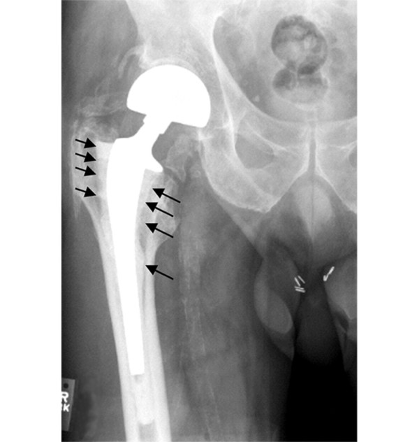
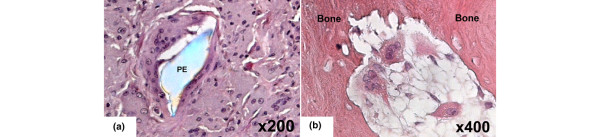
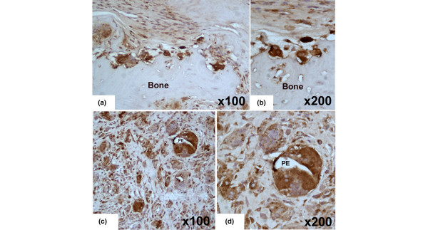
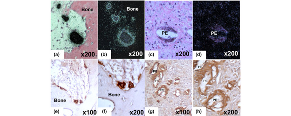
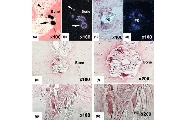
Prosthetic wear debris-induced peri-implant osteolysis is a major cause of aseptic loosening after total joint replacement. In this condition, wear particles released from the implant components induce a granulomatous inflammatory reaction at the interface between implant and adjacent bone, leading to progressive bone resorption and loss of fixation. The present study was undertaken to characterize definitively the phenotype of osteoclast-like cells associated with regions of peri-implant focal bone resorption and to compare the phenotypic features of these cells with those of mononucleated and multinucleated cells associated with polyethylene wear particles. Peri-implant tissues were obtained from patients undergoing hip revision surgery for aseptic loosening after total joint replacement. Cells were examined for the expression of several markers associated with the osteoclast phenotype using immunohistochemistry, histochemistry, and/or in situ hybridization. CD68 protein, a marker expressed by multiple macrophage lineage cell types, was detected in mononucleated and multinucleated cells associated with polyethylene particles and the bone surface. Cathepsin K and tartrate-resistant acid phosphatase were expressed highly in both mononucleated and multinucleated cells associated with the bone surface. Levels of expression were much lower in cells associated with polyethylene particles. High levels of beta3 integrin protein were detected in cells in contact with bone. Multinucleated cells associated with polyethylene particles exhibited faint positive staining. Calcitonin receptor mRNA expression was detected solely in multinucleated cells present in resorption lacunae on the bone surface and was absent in cells associated with polyethylene particles. Our findings provide further evidence that cells expressing the full repertoire of osteoclast phenotypic markers are involved in the pathogenesis of peri-implant osteolysis after total joint replacement. They also demonstrate that foreign body giant cells, although believed to be phenotypically and functionally distinct from osteoclasts, express many osteoclast-associated genes and gene products. However, the levels and patterns of expression of these genes in the two cell types differ. We speculate that, in addition to the role of cytokines and growth factors, the substrate with which these cells interact plays a critical role in their differential phenotypic and functional properties.


Figures

Figure 1
Figure 1
Preoperative radiograph from a study patient before revision hip arthroplasty for aseptic loosening. Arrows denote the area of extensive peri-implant osteolysis along the femoral shaft.
Figure 2
Figure 2
Sections of human peri-implant tissues stained by hematoxylin and eosin. (a) Multinucleated cell associated with a polyethylene (PE) wear particle. (b) Multinucleated cells line the bone surface at site of bone resorption.
Figure 3
Figure 3
CD68 detection in sections of human peri-implant tissues using immunohistochemistry with rabbit polyclonal antibody. (a, b) CD68 is detected in the multinuclear and mononuclear cells located in the soft tissues and multinuclear cells on the bone surface. (c, d) CD68 is also detected in multinuclear and mononuclear cells associated with polyethylene (PE) particles.
Figure 4
Figure 4
Detection of cathepsin K mRNA and protein in sections of human peri-implantation tissues. The techniques used were in situ hybridization with a 35S-labeled anti-sense RNA probe ((a, c) bright field and (b, d) dark field) and (e-h) immunohistochemistry with a rabbit polyclonal antibody to human cathepsin K. Multinuclear cells on the bone surface and some mononuclear cells in the peri-implant tissues adjacent to the bone express high levels of cathepsin K mRNA (panels a [bright field] and b [dark field]). Low levels of cathepsin K mRNA were detected in multinuclear cells associated with polyethylene (PE) particles (panels c [bright field] and d [dark field]). Multinuclear cells on the bone surface (panels e and f) and mononuclear and multinuclear cells associated with PE particles (panels g and h) stain positively for cathepsin K protein. PE particles are easily identified by their strong bi-refringence with polarizing light microscopy.
Figure 5
Figure 5
Detection of TRAP mRNA and TRAP enzymatic activity in sections of human peri-implant tissues. The techniques used were in situ hybridization with a 35S-labeled antisense RNA probe ((a, c) bright field and (b, d) and dark field) and (e-h) histochemistry. TRAP mRNA expression is detected in mononuclear (arrows) and multinuclear cells (arrowheads) on the bone surface adjacent to the peri-implant granuloma (panels a and b). Cells associated with polyethylene (PE) particles also express TRAP mRNA (panels c and d). The enzymatic activity is evident as purple staining seen in similar cells (panels e-h). TRAP, tartrate-resistant acid phosphatase.
Figure 6
Figure 6
Detection of β3 integrin in sections of human peri-implant by immunohistochemistry using a rabbit polyclonal antibody. (a, b) β3 Integrin is evident in the membrane of mononuclear and multinuclear cells adjacent to bone. (d, e) Weak staining is evident in cells associated with polyethylene (PE) particles. Normal IgG was used as a negative control (panel c).
Figure 7
Figure 7
Detection of CTR mRNA in sections of human peri-implant tissues. The techniques used were in situ hybridization with a 35S-labeled antisense RNA probe ((a, c) bright field and (b, d) dark field). Expression of CTR mRNA is evident only in multinuclear cells in contact with the bone surface (panels a and b). Cells associated with polyethylene (PE) particles do not express detectable CTR mRNA (panels c and d).

Similar articles

Cited by

References

    1. Takagi M, Santavirta S, Ida H, Ishii M, Takei I, Niissalo S, Ogino T, Konttinen YT. High-turnover periprosthetic bone remodeling and immature bone formation around loose cemented total hip joints. J Bone Miner Res. 2001;16:79–88. doi: 10.1359/jbmr.2001.16.1.79. - DOI - PubMed
    1. Shinar DM, Schmidt A, Halperin D, Rodan GA, Weinreb M. Expression of alpha v and beta 3 integrin subunits in rat osteoclasts in situ. J Bone Miner Res. 1993;8:403–414. - PubMed
    1. Davies J, Warwick J, Totty N, Philp R, Helfrich M, Horton M. The osteoclast functional antigen, implicated in the regulation of bone resorption, is biochemically related to the vitronectin receptor. J Cell Biol. 1989;109:1817–1826. doi: 10.1083/jcb.109.4.1817. - DOI - PMC - PubMed
    1. Nesbitt S, Nesbit A, Helfrich M, Horton M. Biochemical characterization of human osteoclast integrins. Osteoclasts express alpha v beta 3, alpha 2 beta 1, and alpha v beta 1 integrins. J Biol Chem. 1993;268:16737–16745. - PubMed
    1. Teitelbaum S. Bone resorption by osteoclasts. Science. 2000;289:1504–1508. doi: 10.1126/science.289.5484.1504. - DOI - PubMed

Publication types

MeSH terms

LinkOut - more resources